Purtscher-Like Retinopathy Associated with Atypical Hemolytic Uremic Syndrome
- PMID: 29326853
- PMCID: PMC5758771
- DOI: 10.4274/tjo.66502
Purtscher-Like Retinopathy Associated with Atypical Hemolytic Uremic Syndrome
Abstract
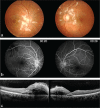
A 25-year-old woman presented with acute bilateral blurred vision and history of headache, dizziness, and syncope for three days. Her visual acuity was 20/60 in both eyes. Fundoscopy revealed multiple bilateral peripapillary yellow-white patches like cotton wool spots, intraretinal hemorrhages and macular edema. The patient was diagnosed with Purtscher-like retinopathy based on the retinal findings and lack of trauma history. She was urgently admitted to the nephrology clinic due to thrombotic microangiopathy findings (hemoglobinemia, thrombocytopenia, and acute renal failure). After excluding thrombotic microangiopathy, the patient was diagnosed with atypical hemolytic uremic syndrome (aHUS) with the clinical and laboratory findings. Eculizumab treatment was added to hemodialysis and plasmapheresis therapy. Three months after starting treatment, retinal lesions regressed and visual acuity increased to 20/20 in both eyes. To the best of our knowledge, this is the first reported case of Purtscher-like retinopathy associated with aHUS.
Keywords: Atypical hemolytic uremic syndrome; Purtscher retinopathy; Purtscher-like retinopathy; eculizumab; thrombotic microangiopathy.
Conflict of interest statement
Conflict of Interest: No conflict of interest was declared by the authors.
Figures


References
-
- Dragon-Durey MA, Sethi SK, Bagga A, Blanc C, Blouin J, Ranchin B, André JL, Takagi N, Cheong HI, Hari P, Le Quintrec M, Niaudet P, Loirat C, Fridman WH, Frémeaux-Bacchi V. Clinical features of anti-factor H autoantibody-associated hemolytic uremic syndrome. J Am Soc Nephrol. 2010;21:2180–2187. - PMC - PubMed
-
- Zheng X, Gorovoy IR, Mao J, Jin J, Chen X, Cui QN. Recurrent ocular involvement in pediatric atypical hemolytic uremic syndrome. J Pediatr Ophthalmol Strabismus. 2014;51:62–65. - PubMed
LinkOut - more resources
Full Text Sources
Other Literature Sources
