

Aggressive neuroendocrine tumor of the ovary with multiple metastases treated with everolimus: A case report
- PMID: 29326972
- PMCID: PMC5760241
- DOI: 10.1016/j.gore.2018.01.002
Aggressive neuroendocrine tumor of the ovary with multiple metastases treated with everolimus: A case report
Abstract
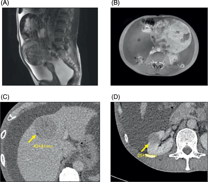
•Neuroendocrine tumors (NETs) frequently occur in the lungs or the gastrointestinal tract; they are uncommon in the ovary.•The mammalian target of rapamycin (mTOR) pathway has been reported as a treatment for advanced NETs.•We describe a patient with an aggressive primary ovarian NET, successfully treated with everolimus (an mTOR inhibitor).
Keywords: BEP, bleomycin, etoposide, and cisplatin; CA-125, carbohydrate antigen 125; CD56, cluster of differentiation 56; CDX2, caudal-type homeobox transcription factor 2; CT, computed tomography; Carcinoid; EMA, epithelial membrane antigen; Everolimus; FIGO, International Federation of Gynecology and Obstetrics; MRI, magnetic resonance imaging; Multiple metastases; NETs, neuroendocrine tumors; Neuroendocrine tumor; Ovary; mTOR, mammalian target of rapamycin.
Figures


References
-
- Cives M., Strosberg J. Treatment strategies for metastatic neuroendocrine tumors of the gastrointestinal tract. Curr. Treat. Options in Oncol. 2017;18 - PubMed
-
- Desouki M.M., Lioyd J., Xu H., Cao D., Barner R., Zhao C. CDX2 may be a useful marker to distinguish primary ovarian carcinoid from gastrointestinal metastatic carcinoids to the ovary. Hum. Pathol. 2013;44:2536–2541. - PubMed
-
- Gardner G.J., Reidy-Lagunes D., Gehrig P.A. Neuroendocrine tumors of the gynecologic tract: a Society of Gynecologic Oncology (SGO) clinical document. Gynecol. Oncol. 2011;122:190–198. - PubMed
-
- Jamar F., Fiasse R., Leners N., Pauwels S. Somatostatin receptor imaging with indium-111-pentetreotide in gastroenteropancreatic neuroendocrine tumors: safety, efficacy and impact on patient management. J. Nucl. Med. 1995;36:542–549. - PubMed
Publication types
LinkOut - more resources
Full Text Sources
Other Literature Sources
Research Materials
Miscellaneous