Review
. 2018 Apr;91(1084):20170533.
doi: 10.1259/bjr.20170533. Epub 2018 Feb 6.

The different faces of renal angiomyolipomas on radiologic imaging: a pictorial review

Affiliations
Review

The different faces of renal angiomyolipomas on radiologic imaging: a pictorial review

Shanigarn Thiravit et al. Br J Radiol. 2018 Apr.

Abstract

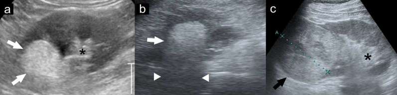
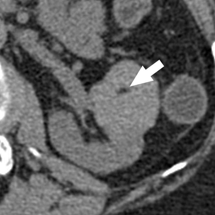
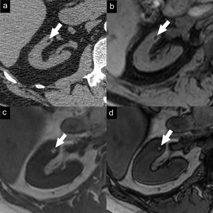
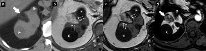
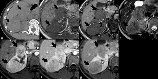
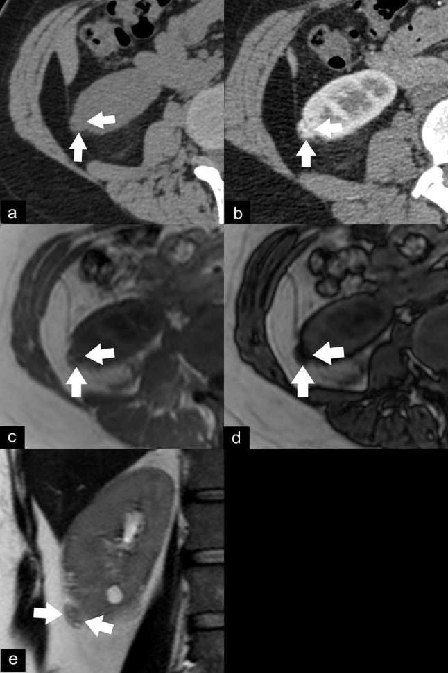
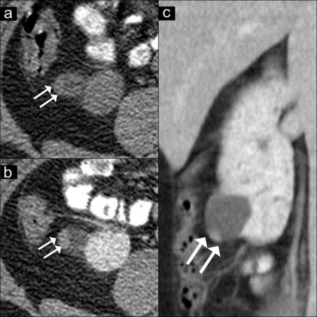
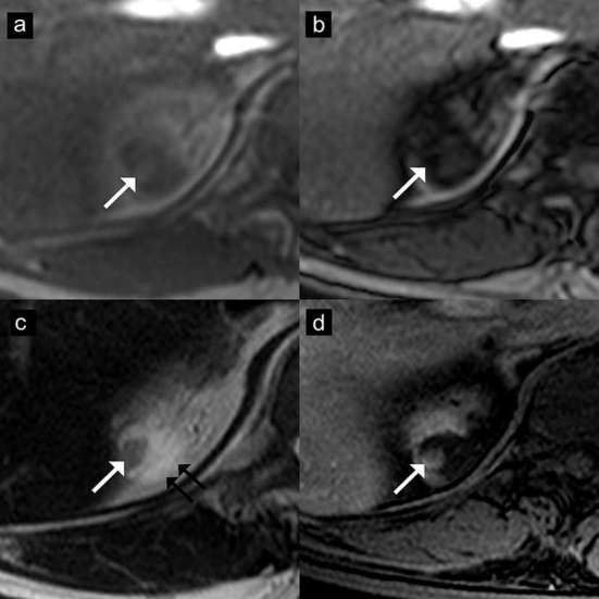
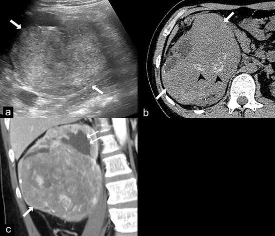
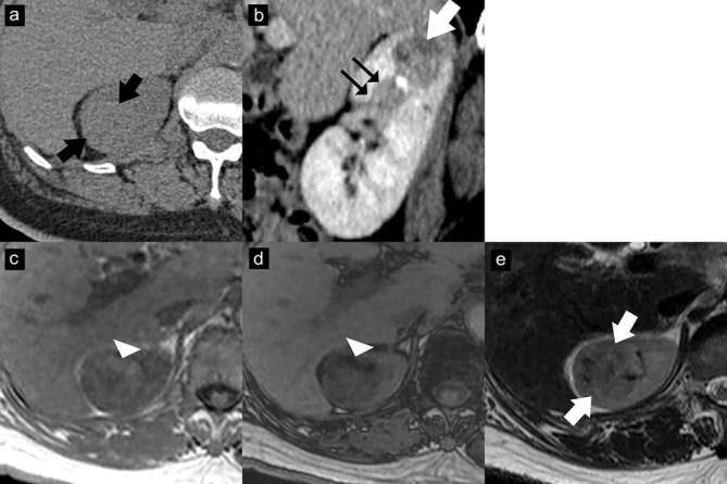
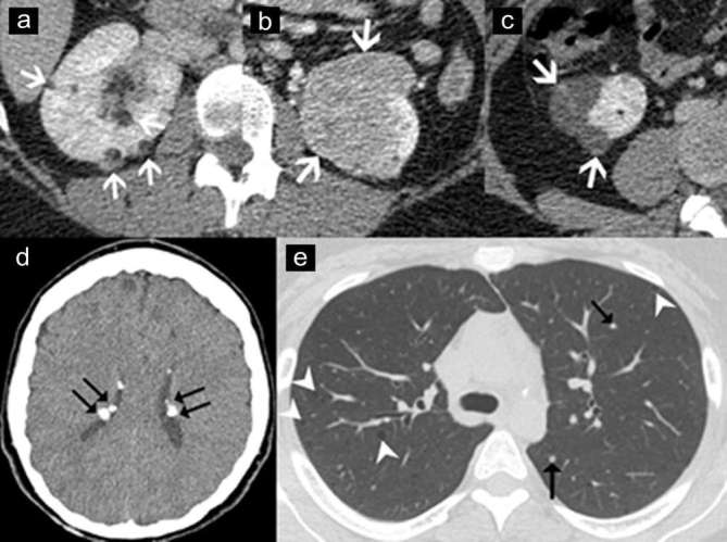
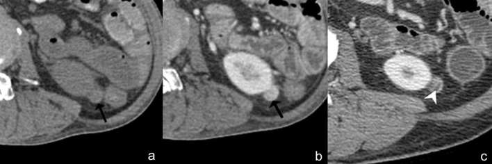
Renal angiomyolipoma (AML) is an uncommon renal tumour, generally composed of mature adipose tissue, dysmorphic blood vessels and smooth muscle. Identification of intratumoral fat on unenhanced CT images is the most reliable finding for establishing the diagnosis of renal AML. However, AMLs sometimes exhibit atypical findings, including cystic as well as solid forms; some of these variants overlap with the appearance of other renal tumours. A rare type of AML, the epithelioid type, possesses malignant potential. The aim of this pictorial review is to gather the different imaging features of AMLs including the classic and fat-poor types, AMLs with epithelial cysts, epithelioid AML, AML associated with tuberous sclerosis, haemorrhagic AML and large AMLs mimicking retroperitoneal liposarcomas. The diagnostic clues that help to distinguish AMLs from other renal tumours are also described in the review.


Figures

Figure 1.
Figure 1.
Ultrasound findings of classic angiomyolipoma and renal cell carcinoma. The angiomyolipoma (a, b: white arrows) is highly hyperechoic, equal to the echogenicity of renal sinus fat (asterisk) with posterior shadowing (arrowheads), while the renal cell carcinoma (c: black arrow) is hyperechoic, but less than renal sinus fat (asterisk) and has a hypoechoic rim and internal cysts (open arrows).
Figure 2.
Figure 2.
Classic angiomyolipoma with a small amount of fat. Axial unenhanced CT image with 1.5 mm sections nicely demonstrates a small amount (arrow) of internal fat attenuation (−48 HU). Note a tiny renal cyst located in the posterior cortex. HU, Hounsfield unit.
Figure 3.
Figure 3.
Small classic angiomyolipoma. Axial unenhanced CT image (a) with 1.5 mm sections demonstrates a small fat-containing renal mass (arrow). Axial gradient-echo fat-suppressed T1 weighted MR image (b) demonstrates a signal drop in the mass due to adipose tissue. Axial T1 weighted dual-echo in-phase (c) and opposed-phase (d) MR images show a black line between the mass and the renal parenchyma on opposed-phase images (India-ink artefact).
Figure 4.
Figure 4.
Classic angiomyolipoma. Axial unenhanced CT image (a) demonstrates a typical fat-containing angiomyolipoma (arrow). Axial T1 weighted dual-echo in-phase (b) and opposed-phase (c) MR images show an India-ink artefact at the border of the mass and the renal parenchyma on opposed-phase images (double arrows). The mass (arrow) shows early enhancement in the corticomedullary phase (d) of the axial gradient-echo fat-suppressed T1 weighted MR image after gadolinium administration. Note two renal cysts adjacent to the AML (asterisks).
Figure 5.
Figure 5.
Fat-poor angiomyolipoma, hyperattenuating type. Axial unenhanced CT image (a) demonstrates two right renal masses (arrows) with hyperattenuation (51 and 52 HU) and no visible fat density. On MRI, the masses show no signal drop on axial T1 weighted dual-echo in-phase (b) and opposed-phase (c) images, and show hypointensity on axial T2 weighted image (d). Axial gradient-echo fat-suppressed T1 weighted MR images after gadolinium administration show masses with early enhancement on the corticomedullary phase and washout on the nephrographic phase (the percentage change in signal intensity from corticomedullary (e), nephrographic (f) and excretory phases (g) relative to precontrast phase was 203, 174 and 147% for the mass in the posterior cortex, and 213, 171 and 151% for the mass in the anterior cortex, respectively). The mass in the posterior cortex was proven AML by percutaneous biopsy. HU, Hounsfield unit.
Figure 6.
Figure 6.
Fat-poor angiomyolipoma, isoattenuating type. Axial unenhanced CT image (a) demonstrates a cortical renal mass (arrows) with isoattenuation (27 HU) and no visible fat density. Axial CECT image demonstrates the mass enhancing in the corticomedullary phase (b). Axial T1 weighted dual-echo in-phase (c) and opposed-phase (d) images show signal drop within the mass on opposed-phase images. The mass appears slightly hypointense on coronal T2 weighted image (e). This type of angiomyolipoma is difficult to differentiate from other renal cortical tumours. This mass developed obvious fat components on follow-up 3 year later (not shown). HU, Hounsfield unit.
Figure 7.
Figure 7.
AMLEC on CT images. Axial unenhanced CT image (a) show a renal mass with peripheral slight hyperattenuation (36 HU) (double arrows). Axial (b) and coronal (c) contrast-enhanced CT images show a cyst with an enhancing peripheral solid nodule (arrows), concerning as a Bosniak category 4 lesion. A partial nephrectomy was performed. The final diagnosis of AMLEC was proven by histopathology. AMLEC, angiomyolipoma with epithelial cysts; HU, Hounsfield unit.
Figure 8.
Figure 8.
Angiomyolipoma with epithelial cysts (AMLEC) on MR images. Axial T1 weighted dual-echo in-phase (a) and opposed-phase (b) images show a mass with peripheral signal drop on opposed-phase image (arrows). This lesion shows hypointensity in the solid portion (arrow) and hyperintensity in the cystic portion (double arrows) on axial T2 weighted image (c). The solid part also shows homogeneous enhancement (arrow) on the axial contrast-enhanced T1 weighted image (d).The final diagnosis of AMLEC was proven by histopathology.
Figure 9.
Figure 9.
Epithelioid angiomyolipoma (EAML). Ultrasound (a) demonstrates a large heterogeneous hyperechoic mass in the right kidney (arrows). Axial unenhanced CT image (b) shows a 14 cm renal mass (arrows) with internal calcifications (arrow heads) and no gross fat component. Coronal contrast-enhanced CT image (c) shows the heterogeneous enhancing mass (arrow) in the lower pole of the right kidney which compresses the renal pelvis, causing mild dilatation of the upper pole calyces (double arrows). Renal cell carcinoma was suspected and a radical nephrectomy was performed. The final diagnosis of EAML was proven by histopathology and immunohistochemistry.
Figure 10.
Figure 10.
EAML with segmental renal vein invasion. Axial unenhanced CT image (a) shows an isoattenuating renal mass (37 HU) without calcification or gross fat component (arrows). Contrast-enhanced CT image with sagittal multiplanar reconstruction in the nephrographic phase (b) shows a heterogeneously enhancing mass (arrow) in the upper pole with extension into a segmental renal vein (double arrows). Axial T1 weighted dual-echo in-phase (c) and opposed-phase (d) images show signal drop in the anterior part of the mass on the opposed-phase image, representing intracytoplasmic lipid (arrowhead). This lesion shows iso-to-hypointensity (arrows) on the axial T2 weighted image (e). The mass was resected due to segmental renal vein invasion. The final diagnosis of EAML was proven by histopathology and immunohistochemistry. EAML, epithelioid angiomyolipoma; HU, Hounsfield unit
Figure 11.
Figure 11.
Angiomyolipoma (AML) associated with tuberous sclerosis. Axial contrast-enhanced CT images of a 40 year-old-woman with tuberous sclerosis, demonstrating multiple typical AMLs (a: arrows), a fat-poor AML (b: arrows), and angiomyolipomas with epithelial cysts (c: arrows). Axial CT image of brain (d) in the same patient shows multiple calcified subependymal tubers (double arrows). Axial CT image of the chest (e) demonstrates multiple well-defined lung cysts (arrowheads) and multiple tiny nodules (arrows), which are consistent with lymphangiomyomatosis and multifocal micronodular pneumocyte hyperplasia, respectively.
Figure 12.
Figure 12.
Angiomyolipoma with internal haemorrhage. Axial unenhanced (a) and contrast-enhanced CT (b) images show an isoattenuating renal mass (28 HU) with homogeneous enhancement, no calcification or fat component (arrow). Based on these findings, the differential diagnosis includes renal cell carcinoma, oncocytoma or isoattenuating angiomyolipoma. However, typical angiomyolipoma with internal haemorrhage can be diagnosed due to the presence of intratumoral fat (arrowhead) on the previous image (c). HU, Hounsfield unit.
Figure 13.
Figure 13.
Exophytic renal angiomyolipoma can be differentiated from other retroperitoneal fat-containing tumours by demonstrating renal parenchyma draping around the mass (the claw sign: arrows) on ultrasound (a) and contrast-enhanced CT (b) images. Contrast-enhanced CT image (b) also demonstrates feeding vessels (arrow head) supplying the mass from kidneys.

References

    1. Jinzaki M, Silverman SG, Akita H, Nagashima Y, Mikami S, Oya M. Renal angiomyolipoma: a radiological classification and update on recent developments in diagnosis and management. Abdom Imaging 2014; 39: 588–604. doi: 10.1007/s00261-014-0083-3 - DOI - PMC - PubMed
    1. Kapoor A, Girard L, Lattouf JB, Pei Y, Rendon R, Card P, et al. . Evolving strategies in the treatment of tuberous sclerosis complex-associated angiomyolipomas (TSC-AML). Urology 2016; 89: 19–26. doi: 10.1016/j.urology.2015.12.009 - DOI - PubMed
    1. Song S, Park BK, Park JJ. New radiologic classification of renal angiomyolipomas. Eur J Radiol 2016; 85: 1835–42. doi: 10.1016/j.ejrad.2016.08.012 - DOI - PubMed
    1. Farrelly C, Delaney H, McDermott R, Malone D. Do all non-calcified echogenic renal lesions found on ultrasound need further evaluation with CT? Abdom Imaging 2008; 33: 44–7. doi: 10.1007/s00261-007-9306-1 - DOI - PubMed
    1. Schieda N, Kielar AZ, Al Dandan O, McInnes MD, Flood TA. Ten uncommon and unusual variants of renal angiomyolipoma (AML): radiologic-pathologic correlation. Clin Radiol 2015; 70: 206–20. doi: 10.1016/j.crad.2014.10.001 - DOI - PubMed

MeSH terms