Review
. 2018 Mar;41(3):1165-1176.
doi: 10.3892/ijmm.2018.3364. Epub 2018 Jan 4.

Paediatric Virology and its interaction between basic science and clinical practice (Review)

Affiliations
Review

Paediatric Virology and its interaction between basic science and clinical practice (Review)

Ioannis N Mammas et al. Int J Mol Med. 2018 Mar.

Abstract

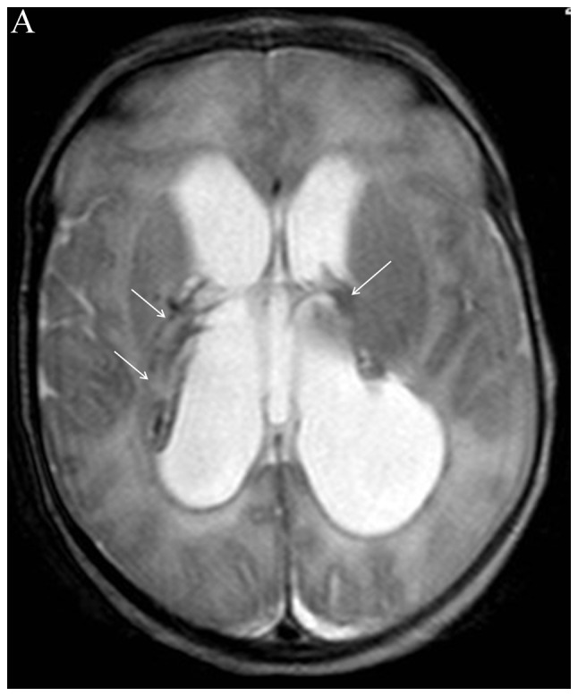
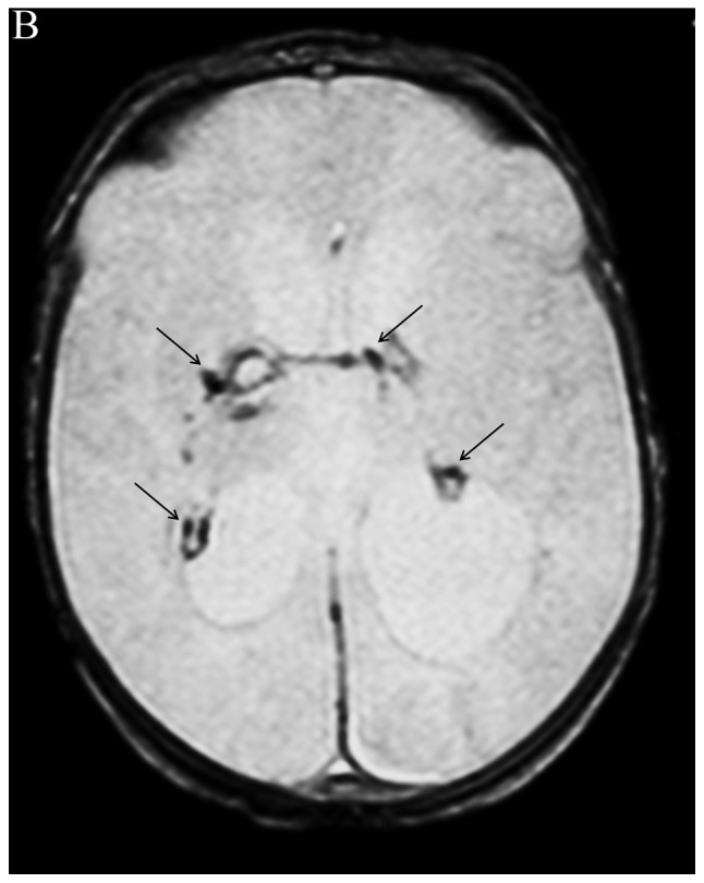
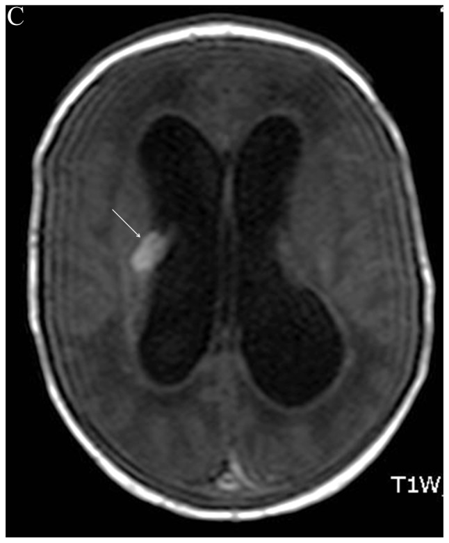
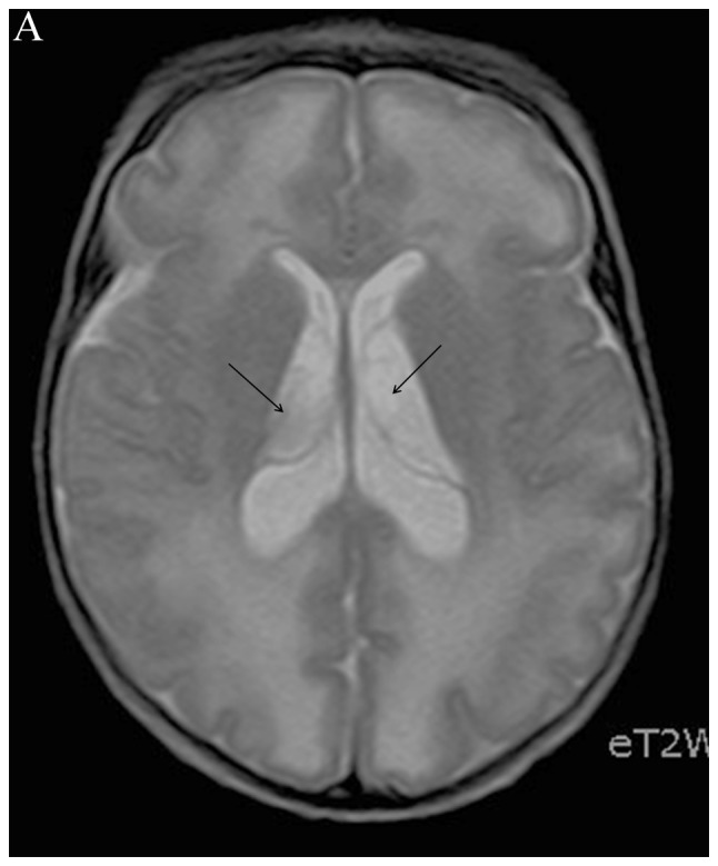
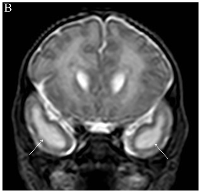
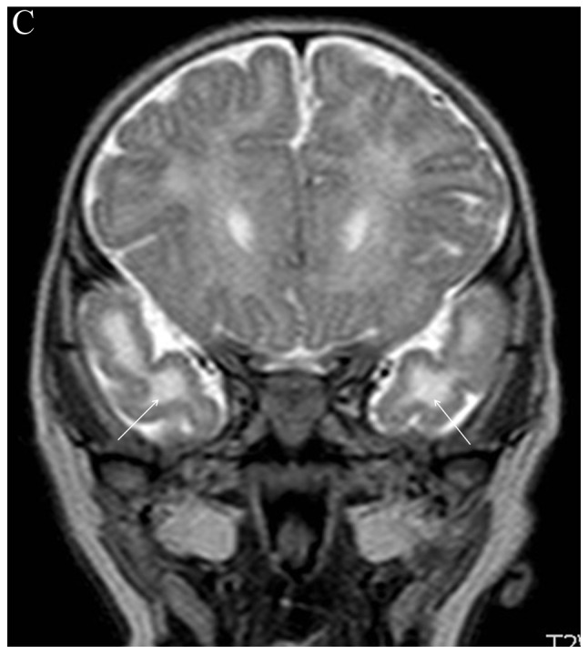
The 3rd Workshop on Paediatric Virology, which took place on October 7th, 2017 in Athens, Greece, highlighted the role of breast feeding in the prevention of viral infections during the first years of life. Moreover, it focused on the long-term outcomes of respiratory syncytial virus and rhinovirus infections in prematurely born infants and emphasised the necessity for the development of relevant preventative strategies. Other topics that were covered included the vaccination policy in relation to the migration crisis, mother‑to‑child transmission of hepatitis B and C viruses, vaccination against human papilloma viruses in boys and advances on intranasal live‑attenuated vaccination against influenza. Emphasis was also given to the role of probiotics in the management of viral infections in childhood, the potential association between viral infections and the pathogenesis of asthma, fetal and neonatal brain imaging and the paediatric intensive care of children with central nervous system viral infections. Moreover, an interesting overview of the viral causes of perinatal mortality in ancient Greece was given, where recent archaeological findings from the Athenian Agora's bone well were presented. Finally, different continuing medical educational options in Paediatric Virology were analysed and evaluated. The present review provides an update of the key topics discussed during the workshop.


Conflict of interest statement

Competing interests

Demetrios Spandidos is the Editor-in-Chief for the journal, but had no personal involvement in the reviewing process, or any influence in terms of adjudicating on the final decision, for this article.

Figures

Figure 1
Figure 1
Brain magnetic resonance imaging (MRI) in a term neonate with post-natal enterovirus infection. (A) axial T2-Weighted image through the basal ganglia - white arrows indicate the presence of residual bilateral thalamocaudate groove hemorrhages. (B) Foci of hemosiderin deposits are more conspicuous on axial T2* image, obvious not only in the thalamocaudate grooves, but also along the choroid plexuses (black arrows). (C) This abnormal area (white arrow) is also evident with increased signal intensity on axial T1-Weighted image through the same level.
Figure 1
Figure 1
Brain magnetic resonance imaging (MRI) in a term neonate with post-natal enterovirus infection. (A) axial T2-Weighted image through the basal ganglia - white arrows indicate the presence of residual bilateral thalamocaudate groove hemorrhages. (B) Foci of hemosiderin deposits are more conspicuous on axial T2* image, obvious not only in the thalamocaudate grooves, but also along the choroid plexuses (black arrows). (C) This abnormal area (white arrow) is also evident with increased signal intensity on axial T1-Weighted image through the same level.
Figure 2
Figure 2
Brain magnetic resonance imaging (MRI) in a neonate with history of third trimester cytomegalovirus (CMV) infection. (A) axial T2-Weighted image through the lateral ventricles - black arrows indicate the presence of intraventricular cysts. (B) Coronal T2-Weighted image reveals signal abnormality in the temporal lobes bilaterally (white arrows). (C) On the 3-month follow-up, same to section b demonstrates persistent signal abnormality in the temporal lobes and additional white matter volume loss (white arrows).
Figure 2
Figure 2
Brain magnetic resonance imaging (MRI) in a neonate with history of third trimester cytomegalovirus (CMV) infection. (A) axial T2-Weighted image through the lateral ventricles - black arrows indicate the presence of intraventricular cysts. (B) Coronal T2-Weighted image reveals signal abnormality in the temporal lobes bilaterally (white arrows). (C) On the 3-month follow-up, same to section b demonstrates persistent signal abnormality in the temporal lobes and additional white matter volume loss (white arrows).

References

    1. Greenough A, Osborne J, Sutherland S, editors. Congenital, Perinatal, and Neonatal Infections. Churchill Livingstone; Edinburgh: 1992.
    1. Shulman ST. The history of pediatric infectious diseases. Pediatr Res. 2004;55:163–176. doi: 10.1203/01.PDR.0000101756.93542.09. - DOI - PMC - PubMed
    1. Starr M. Paediatric infectious diseases: The last 50 years. J Paediatr Child Health. 2015;51:12–15. doi: 10.1111/jpc.12795. - DOI - PubMed
    1. Mammas IN, Greenough A, Theodoridou M, Kramvis A, Christaki I, Koutsaftiki C, Koutsaki M, Portaliou DM, Kostagianni G, Panagopoulou P, Sourvinos G, Spandidos DA. Current views and advances on Paediatric Virology: An update for paediatric trainees. Exp Ther Med. 2016;11:6–14. doi: 10.3892/etm.2015.2890. - DOI - PMC - PubMed
    1. Mammas IN, Greenough A, Theodoridou M, Spandidos DA. Paediatric Virology: A new paediatric subspecialty? A proposal at the Workshop on Paediatric Virology, Athens, October 10,2015. Exp Ther Med. 2016;11:3–5. doi: 10.3892/etm.2015.2889. - DOI - PMC - PubMed