. 2018 Mar;17(3):4474-4482.
doi: 10.3892/mmr.2018.8413. Epub 2018 Jan 9.

The human umbilical cord stem cells improve the viability of OA degenerated chondrocytes

Affiliations

The human umbilical cord stem cells improve the viability of OA degenerated chondrocytes

Hao Wang et al. Mol Med Rep. 2018 Mar.

Abstract

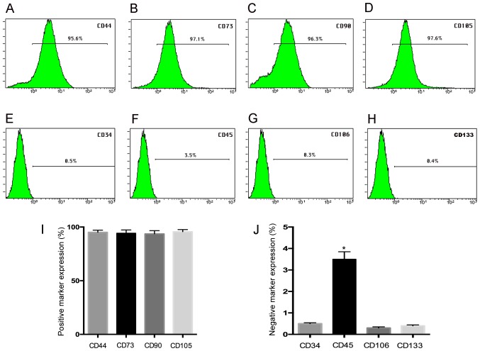
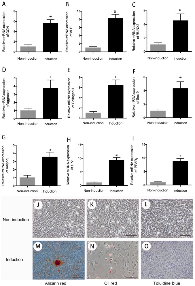
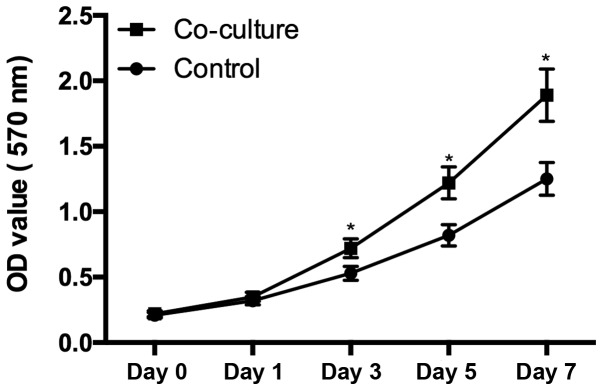
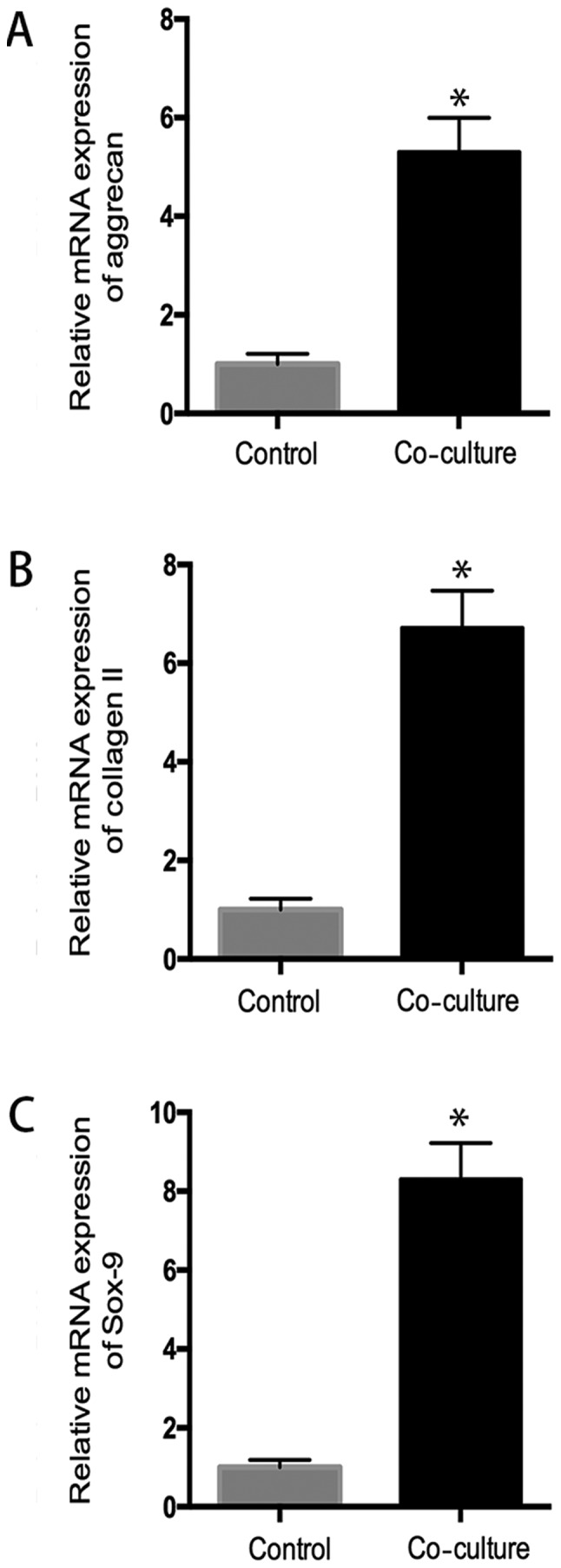
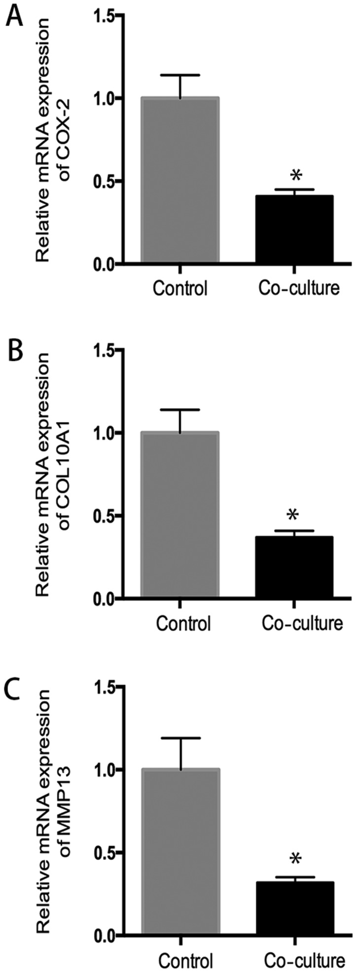
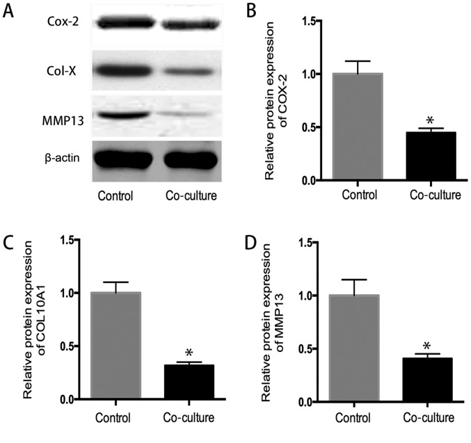
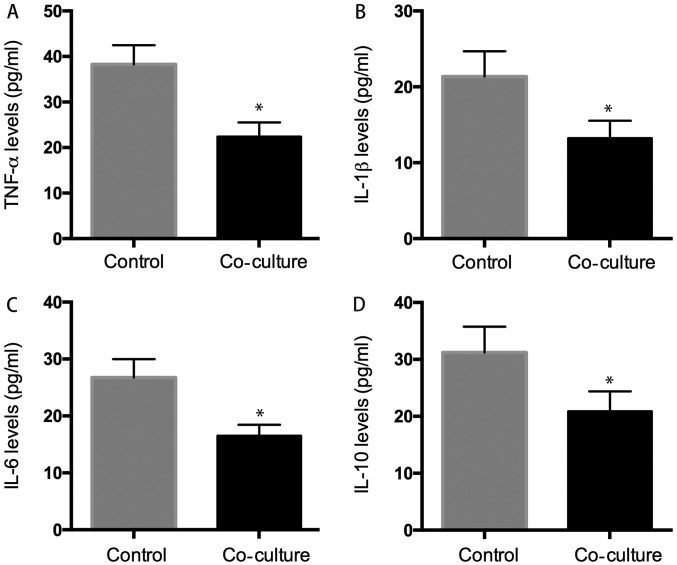
Osteoarthritis (OA) affects a large number of patients; however, human umbilical cord stem cells exhibit therapeutic potential for treating OA. The aim of the present study was to explore the interaction between human umbilical cord stem cells and degenerated chondrocytes, and the therapeutic potential of human umbilical cord stem cells on degenerated chondrocytes. Human umbilical cord‑derived mesenchymal stem cells (hUC‑MSCs) were harvested from human umbilical cords, and flow cytometry was used to analyze the surface antigen markers, in addition, chondrogenic, osteogenic and adipogenic differentiation on the cells was investigated. OA cells at P3 were cocultured with hUC‑MSCs in a separated co‑culture system, and reverse transcription‑polymerase chain reaction and western blot were used to evaluate the mRNA, and protein expression of collagen type II (Col2), SRY‑box 9 (sox‑9) and aggrecan. The level of inflammatory cytokines, tumor necrosis factor‑α, interleukin (IL)‑1β, IL‑6, IL‑10, were analyzed by ELISA in the supernatant. hUC‑MSCs grow in a fibroblastic shape with stable proliferation. hUC‑MSCs expressed cluster of differentiation 44 (CD44), CD73, CD90, CD105; while did not express CD34, CD45, CD106, CD133. After multi‑induction, hUC‑MSCs were able to differatiate into adipogenic, osteogenic and chondrogenic lineage. hUC‑MSCs inhibited the expression of matrix metalloproteinase‑13, collagen type X α1 chain and cyclooxygenase‑2 in OA chondrocytes, and enhanced the proliferation of OA chondrocytes, while OA chondrocytes stimulated the production of Col2, sox‑9 and aggrecan and promoted hUC‑MSCs differentiate into chondrocytes. Flow cytometry analysis demonstrated hUC‑MSCs have a predominant expression of stem cell markers, while the hematopoietic and endothelial markers were absent. Osteogenic, chondrogenic and adipogenic differentiation was observed in certain induction conditions. hUC‑MSCs improved the proliferation of OA chondrocytes and downregulated the expression of inflammatory cytokines, while OA chondrocytes promoted MSCs to differentiate into chondrocytes. Taken together, the co‑culture of hUC‑MSCs and OA chondrocytes may provide a therapeutic potential in OA treatment.

Keywords: human umbilical cord-derived mesenchymal stem cells; osteoarthritis; degenerated chondrocytes; differentiation; co-culture.


Figures

Figure 1.
Figure 1.
Surface marker expression by FACS analysis. The hUC-MSC at passage 3 demonstrated a high expression of (A) CD44 (95.6%), (B) CD73 (97.1%), (C) CD90 (96.3%), (D) CD105 (97.6%) by FACS analysis, but they express (E) CD34 (0.5%) and (F) CD45 (3.5%), (G) CD106 (0.3%), and (H) CD133 (0.4%) negatively; (I and J) the histogram gives a clear picture of the expression ratio. *P<0.05 when compared to control group.
Figure 2.
Figure 2.
Cell differentiation. After 3 weeks of multi-differentiation induction, cells in the experiment group have higher expression of osteogenic genes, (A) OCN, (B) ALP, (C) RUNX2, chondrogenic genes, (D) aggrecan, (E) collagen II, (F) sox-9, and adipogenic genes, (G) adipoq, (H) aP2, (I) PPARγ than that in control group (P<0.05). There is no positive staining in control group (J-L), wheread, positive Alizarin Red staining was observed in osteogenic differentiation group for 21 days (M); Oil Red O-positive staining of lipid droplets were detected in induction group after 3 weeks of chondrogenic differentiation (N); Cells in chondrogenic induction group showed remarkable changes in cellular morphology and were stain positive for Toluidine Blue (O). *P<0.05 when compared to control group. Scale bar, 100 µm.
Figure 3.
Figure 3.
Cell proliferation. There is a significant difference of the chondrocytes proliferation between the cells cultured in the supernatant from hUC-MSC and cells culture alone (P<0.05), cells in coculture group demonstrated a high proliferation rate from day 3. *P<0.05 when compared to control group.
Figure 4.
Figure 4.
Chondrocytes promotes the chondrogenesis of hUC-MSC. RT-PCR analysis revealed increased compared with control group, aggrecan mRNA expression was increased 5.3-fold (A) and collagen II mRNA expression was increased 6.7-fold (B), as well as sox-9 mRNA expression was increased 8.2-fold (C) in co-culture system, indicating chondrocytes promoted the chondrogenic differentiation of hUC-MSC. *P<0.05 when compared to control group.
Figure 5.
Figure 5.
Effects of hUC-MSC on expression of genes related to inflammation and matrix degradation. In co-culture system, hUC-MSC downregulated (A) COX-2, (B) collagen 10A1, and (C) MMP13 gene expressions, compared to control group (P<0.05), indicating that hUC-MSC can have an inhibitory effect in genes expression related to inflammation and matrix degradation in human osteoarthritic chondrocytes. *P<0.05 when compared to control group.
Figure 6.
Figure 6.
(A) The western blot analysis. The protein expression of (B) COX-2, (C) collagen 10A1 and (D) MMP13 in co-culture system was lower than that in control group (P<0.05), which confirmed the previously mRNA expression results. *P<0.05 when compared to control group.
Figure 7.
Figure 7.
ELISA analysis. We evaluated whether hUC-MSC affected inflammatory cytokines secretion of osteoarthritic chondrocytes, the results indicated that the level of inflammatory cytokines (TNF-α, IL-1β, IL-6, IL-10) was 22.38±3.2 pg/ml (A), 13.23±2.3 pg/ml (B), 16.53±1.9 pg/ml (C), 20.89±3.5 pg/ml (D) in co-culture group decreased significant than that was 38.28±4.3 pg/ml (A), 21.37±3.3 pg/ml (B), 26.75±3.6 pg/ml (C), 31.23±4.5 pg/ml (D) in control group. *P<0.05 when compared to control group.

References

    1. Fransen M, Bridgett L, March L, Hoy D, Penserga E, Brooks P. The epidemiology of osteoarthritis in Asia. Int J Rheum Dis. 2011;14:113–121. doi: 10.1111/j.1756-185X.2011.01608.x. - DOI - PubMed
    1. van der Kraan PM, van den Berg WB. Chondrocyte hypertrophy and osteoarthritis: Role in initiation and progression of cartilage degeneration? Osteoarthritis Cartilage. 2012;20:223–232. doi: 10.1016/j.joca.2011.12.003. - DOI - PubMed
    1. Zhang Y, Jordan JM. Epidemiology of osteoarthritis. Clin Geriatr Med. 2010;26:355–369. doi: 10.1016/j.cger.2010.03.001. - DOI - PMC - PubMed
    1. Neogi T, Zhang Y. Epidemiology of osteoarthritis. Rheum Dis Clin North Am. 2013;39:1–19. doi: 10.1016/j.rdc.2012.10.004. - DOI - PMC - PubMed
    1. Cross M, Smith E, Hoy D, Nolte S, Ackerman I, Fransen M, Bridgett L, Williams S, Guillemin F, Hill CL, et al. The global burden of hip and knee osteoarthritis: Estimates from the global burden of disease 2010 study. Ann Rheum Dis. 2014;73:1323–1330. doi: 10.1136/annrheumdis-2013-204763. - DOI - PubMed

MeSH terms