. 2018 Jan 11;172(1-2):262-274.e11.
doi: 10.1016/j.cell.2017.12.022.

Retrovirus-like Gag Protein Arc1 Binds RNA and Traffics across Synaptic Boutons

Affiliations

Retrovirus-like Gag Protein Arc1 Binds RNA and Traffics across Synaptic Boutons

James Ashley et al. Cell. .

Abstract

Arc/Arg3.1 is required for synaptic plasticity and cognition, and mutations in this gene are linked to autism and schizophrenia. Arc bears a domain resembling retroviral/retrotransposon Gag-like proteins, which multimerize into a capsid that packages viral RNA. The significance of such a domain in a plasticity molecule is uncertain. Here, we report that the Drosophila Arc1 protein forms capsid-like structures that bind darc1 mRNA in neurons and is loaded into extracellular vesicles that are transferred from motorneurons to muscles. This loading and transfer depends on the darc1-mRNA 3' untranslated region, which contains retrotransposon-like sequences. Disrupting transfer blocks synaptic plasticity, suggesting that transfer of dArc1 complexed with its mRNA is required for this function. Notably, cultured cells also release extracellular vesicles containing the Gag region of the Copia retrotransposon complexed with its own mRNA. Taken together, our results point to a trans-synaptic mRNA transport mechanism involving retrovirus-like capsids and extracellular vesicles.

Keywords: Arc/Arg3.1; Gag domain; RNA trafficking; RNA-binding protein; exosomes; extracellular vesicles; plasticity; retrotransposon; synapse; trans-synaptic RNA transport.


Figures

Figure 1
Figure 1. darc1 mRNA is enriched in EVs
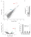
(A) Comparison of RNA expression levels in EV versus cellular RNA from S2 cells determined by deep sequencing, and enrichment of darc1 (red) in the EV fraction. Note that darc2 mRNA (blue) levels are not statistically different from cellular levels. (B) Volcano Plot of RNA-Seq data from 4 replicates, where the x-axis represents fold change in transcripts from EVs vs. total cellular mRNA levels (a positive score represents enrichment, a negative score represents depletion). The y-axis represents statistical confidence for each x-axis point. Green circles are transcripts that are significantly enriched. darc1 is encircled by a red marker. (C) Normalized quantitative PCR confirming that darc1 is enriched in the exosome fraction compared to the total cell. N= 3 biological replicates. (D) Raw number of RNA-seq reads for darc1 from 4 biological RNA-Seq replicates.
Figure 2
Figure 2. darc1 mRNA and protein are present at the Drosophila NMJ, and are transferred from pre- to postsynaptic sites
(A–D) Confocal slices of NMJ branches in preparations double labeled with anti-HRP and either (A,B) a darc1 FISH probe, or (C,D) anti-dArc1 in (A,C) wild type and (B) darc1esm113 and (D) darc1E8/darc1esm113 mutants. (EL) Confocal slices of NMJ branches in preparations double labeled with anti-HRP and either (E–H) a darc1 FISH probe, or (I–L) anti-dArc1, in (E,I) C380-Gal4/+ control, upon expression of (F,J) dArc1-RNAi-neuron (C380-Gal4>UAS-RNAi1), (G,K) Rab11-DN-neuron (C380-Gal4>UAS-Rab11DN), and (H,L) dArc1-RNAi1-muscle (C57-Gal4> dArc1-RNAi1). (M–R) Quantification of (M,O,Q) normalized darc1 FISH signal and (N,P,R) normalized dArc1 immunocytochemical signal at the NMJ in the indicated genotypes. (S) Normalized ratio of postsynaptic to presynaptic signal in the indicated genotypes (T) Western blot of body wall muscle protein extracts derived from the indicated genotypes probed sequentially with anti-dArc1 (top) and Tubulin (Tub; bottom). Numbers at the left of the blots represent molecular weight in kilodaltons. Arrow points to an unspecific band labeled by the dArc1 antibody. Tub=tubulin. (U) Diagram of darc1 mRNA showing the 5′ UTR (blue), the open reading frame (ORF; red), and the 3′ UTR (green). Black lines underneath represent different portions of the darc1 transcript. Orange bars represent regions of the darc1 mRNA resembling Gypsy-like Gag sequences. Note that the entire ORF encodes a Gypsy-like Gag protein. Calibration bar is 6 μm; N=(from left to right; animals/arbors) M(6/11, 6/10), N(12/16, 6/10, 6/10), O(8/14, 8/13, 8/15, 8/16), P(21/44, 12/24, 9/14, 18/29, 9/17, 9/14), Q(8/14, 8/13), R(15/28, 15/28), S(15/28,15/28); Data are represented as mean and error bars represent SEM; statistical analysis was conducted using student’s t-test for M and one-way ANOVA with Tukey Post Hoc test for the rest of the graphs. *= p < 0.05; **= p < 0.001; ***= p < 0.0001.
Figure 3
Figure 3. darc1 mRNA and protein transfer across synaptic boutons depends on darc1 3UTR (see also Supplemental Fig. 1)
(A–H,L–M) Single confocal slices of NMJ branches (A–D) from darc1 mutant larvae expressing dArc1 transgenes either (A,D) containing, or (B,C) lacking the 3′UTR, double labeled with antibodies to dArc1 and HRP. Transgenes were expressed in (A–C) neurons (C380-Gal4>transgene)or (D) muscles (C57-Gal4>transgene). (EH) in preparations double labeled with GFP and HRP from larvae expressing (E) darc1 3′UTR-GFP-neuron (C380> darc1 3′UTR-GFP), (F) darc1 3′UTR A-fragment-GFP-neuron (C380-Gal4> darc1 3′UTR A-fragment-GFP, (G) UGR-GFP (C380>UAS-UGR-GFP), (H) GFP neuron (C380-Gal4>UAS-GFP). (L,M) in preparations double labeled with GFP and HRP from darc1 3′UTR-GFP-neuron (C380> darc1 3′UTR-GFP) in a (L) wild type and (M) darc1E8/darc1esm113 mutant background. (IK) Quantification of (I) dArc1 immunoreactive and (J,K) GFP immunoreactive signal in the indicated genotypes. (L) Diagram of darc1 mRNA showing the 5′ (blue), the ORF encoding (red), and 3′ (green) UTR. The black lines underneath represent different portions of the darc1 transcript testing the darc1 localization signal. Calibration bar is 8 μm; N=(from left to right; animals/arbors) I(10/13, 10/13, 10/23, 9/17, 9/15, 9/13, 9/19, 9/17, 9/15), J(9/18, 15/27, 9/18, 9/16), K(10/29, 10/25). Data are represented as mean and error bars represent SEM; statistical analysis was conducted using one-way ANOVA with Tukey post hoc test for I,J, and Student’s t-test for K. *= p < 0.05; **= p < 0.001; ***= p < 0.0001.
Figure 4
Figure 4. Enrichment of Copia-retrotransposon RNA and protein in S2 cell EVs and darc1 mRNA association withdarc1 protein
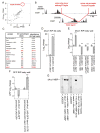
(A) Enrichment of copia mRNA in the S2 EV fraction. (B) Long (L) and short (S) copia isoforms, predicted to be generated by alternative RNA splicing, and enrichment of copiaS, encoding the Gag region, in EVs. (C) Selected proteins showing enrichment in S2 cell EVs and their abundance in the EV vs cellular fractions, highlighting dArc1, dArc2 and Copia. (D,E) Immunoprecipitation of darc1 RNA using anti-dArc1 antibodies from extracts of (D) S2 cells and (E) body wall muscles. (F) Immunoprecipitation of GFP RNA using anti-dArc1 antibodies from extracts of body wall muscles with neurons expressing either GFP alone or GFP upstream of the darc1 3′UTR. (G) Biotinylated RNA pull down of dArc1, using biotinylated darc1 3′UTR RNA or control RNA. Both pull down dArc1 protein, while beads or RNA alone do not. N=3 biological repeats for D,E,F; data are represented as mean and error bars represent SEM; statistical analysis was conducted using student’s T-test; *= p < 0.05; **= p < 0.001; ***= p < 0.0001.
Figure 5
Figure 5. Purified dArc1 protein assembles into capsid-like structures and these structures are contained in EVs
(A,B) Negatively stained capsid-like structures (white arrow) formed by purified dArc1 protein shown at (A) low and (B) high magnification. (C,D) Capsid-like structures (white arrow) observed after EV lysis, shown at (A) low and (B) high magnification. (E–G) Anti-dArc1 ImmunoEM labeling of capsid-like structures (black arrows) derived from lysed EVs, shown at (E) low and (F,G) high magnification. White arrows point to unlabeled capsid like structures. Calibration bar is 120 nm for A,C,E and 40 nm for B,D,F,G.
Figure 6
Figure 6. dArc1 influences developmental and activity-dependent plasticity at the NMJ
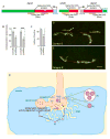
(A–D) Merged confocal Z-stacks of NMJ arbors labeled with antibodies against HRP and DLG in (A) C380-Gal4/+ control, (B) darc1esm113 mutant, (C) darc1E8/darc1esm113 mutant, and (D) motorneuron expression of dArc1-RNAi1. (E–H) High magnification view of single confocal slices through NMJ branches in the same genotypes as (A–D). (I,J,O) Quantification of third instar larval (I) synaptic boutons, (J) ghost boutons, and (O) activity induced ghost boutons in the indicated genotypes and conditions. (K–M) Single confocal slices of NMJs in preparations with antibodies against HRP and DLG in (K,L) unstimulated NMJs and (M,N) after stimulating NMJs with a spaced stimulation protocol in the indicated genotypes. Calibration bar is 46 μm in AH and 6 μm in K–N; N=(from left to right; animals/arbors) I,J(14/27, 8/15, 6/12, 6/12, 6/10, 6/12, 12/21, 6/12, 6/12, 12/21, 8/16, 6/10, 6/10, 14/27, 10/20, 9/16, 6/10), O(12/19,12/22,6/12,6/11,7/14,7/12). Data are represented as mean and error bars represent SEM. Statistical analysis was conducted using one-way ANOVA with Tukey post hoc test for I,J and Student’s t-test for O. *= p < 0.05; **= p < 0.001; ***= p < 0.0001.
Figure 7
Figure 7. Consequences of the UGR for darc1 expression and NMJ morphology, and proposed model of dArc1 transfer (see also Supplemental Fig. 2)
(A) Diagram of the darc1 genomic region in OR showing the UGR element between the darc1 and darc2 genes. (B,C) Quantification in the indicated genotypes of (B) anti-dArc1 signal and (C) number of synaptic boutons. (D,E) Merged confocal Z-stacks of NMJ arbors labeled with antibodies to HRP and DLG in (D) Canton-S and (E) Oregon-R. (F) Diagram depicting a larval NMJ, in which exosome-like vesicles (EV) containing dArc1 protein and transcript are packaged then released from non-synaptic sites. It is still unclear whether these EVs contain multiple enveloped capsid-like particles (a) or a single capsid-like structure (b). These capsid-like particles (c) are taken up by the postsynaptic muscle, either through EV fusion with the muscle membrane, or endocytosis and further fusion with the endosome membrane. We propose that this transfer serves to stimulate synaptic maturation, as downregulation of presynaptic dArc1 leads to accumulation of ghost boutons. (MVB = multivesicular body, SV = synaptic vesicle, AZ = active zone, SSR = subsynaptic reticulum) Calibration bar is 26 μm; N=(from left to right; animals/arbors) B(12/12,12/24), C(6/10,6/12); data are represented as mean and error bars represent SEM. Statistical analysis was conducted using student’s T-test. *= p < 0.05; **= p < 0.001; ***= p < 0.0001.

Comment in

References

    1. Abrusan G, Szilagyi A, Zhang Y, Papp B. Turning gold into ‘junk’: transposable elements utilize central proteins of cellular networks. Nucleic Acids Res. 2013a;41:3190–3200. doi:3110.1093/nar/gkt3011. Epub 2013 Jan 3121. - PMC - PubMed
    1. Abrusan G, Szilagyi A, Zhang Y, Papp B. Turning gold into ‘junk’: transposable elements utilize central proteins of cellular networks. Nucleic Acids Res. 2013b;41:3190–3200. - PMC - PubMed
    1. Alenquer M, Amorim MJ. Exosome Biogenesis, Regulation, and Function in Viral Infection. Viruses. 2015;7:5066–5083. doi:5010.3390/v7092862. - PMC - PubMed
    1. Alhowikan AM. Activity-Regulated Cytoskeleton-Associated Protein Dysfunction May Contribute to Memory Disorder and Earlier Detection of Autism Spectrum Disorders. Med Princ Pract. 2016;25:350–354. doi:310.1159/000445351. Epub 000442016 Mar 000445311. - PMC - PubMed
    1. Ataman B, Ashley J, Gorczyca D, Gorczyca M, Mathew D, Wichmann C, Sigrist SJ, Budnik V. Nuclear trafficking of Drosophila Frizzled-2 during synapse development requires the PDZ protein dGRIP. Proc Natl Acad Sci U S A. 2006;103:7841–7846. - PMC - PubMed

Publication types

MeSH terms