. 2018 Jan 11;172(1-2):305-317.e10.
doi: 10.1016/j.cell.2017.12.023.

Transmembrane Pickets Connect Cyto- and Pericellular Skeletons Forming Barriers to Receptor Engagement

Affiliations

Transmembrane Pickets Connect Cyto- and Pericellular Skeletons Forming Barriers to Receptor Engagement

Spencer A Freeman et al. Cell. .

Abstract

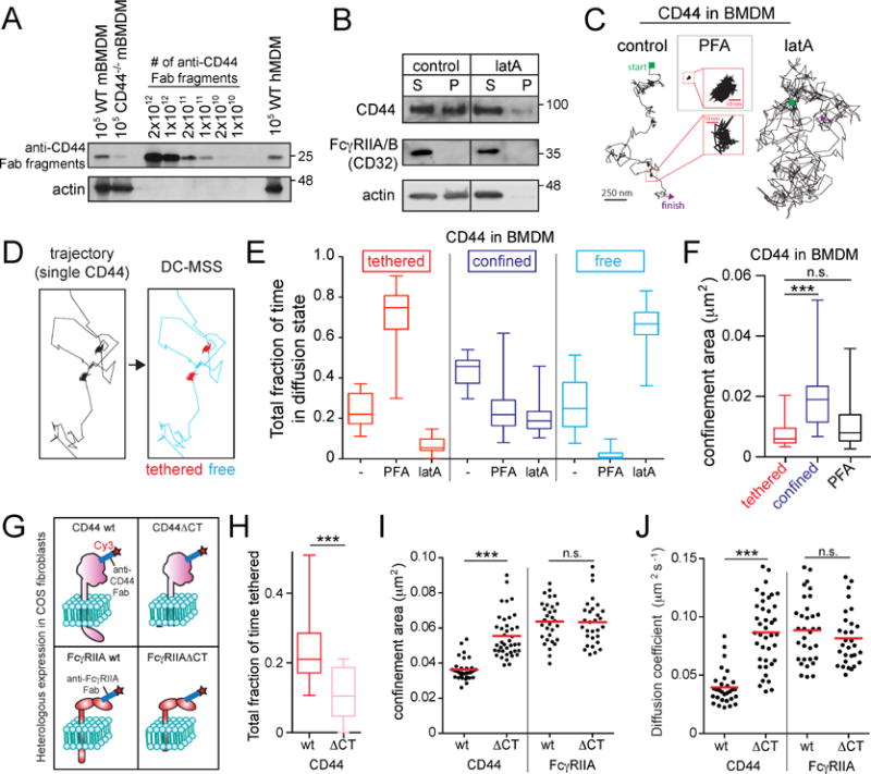
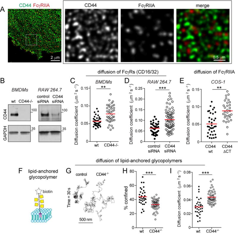
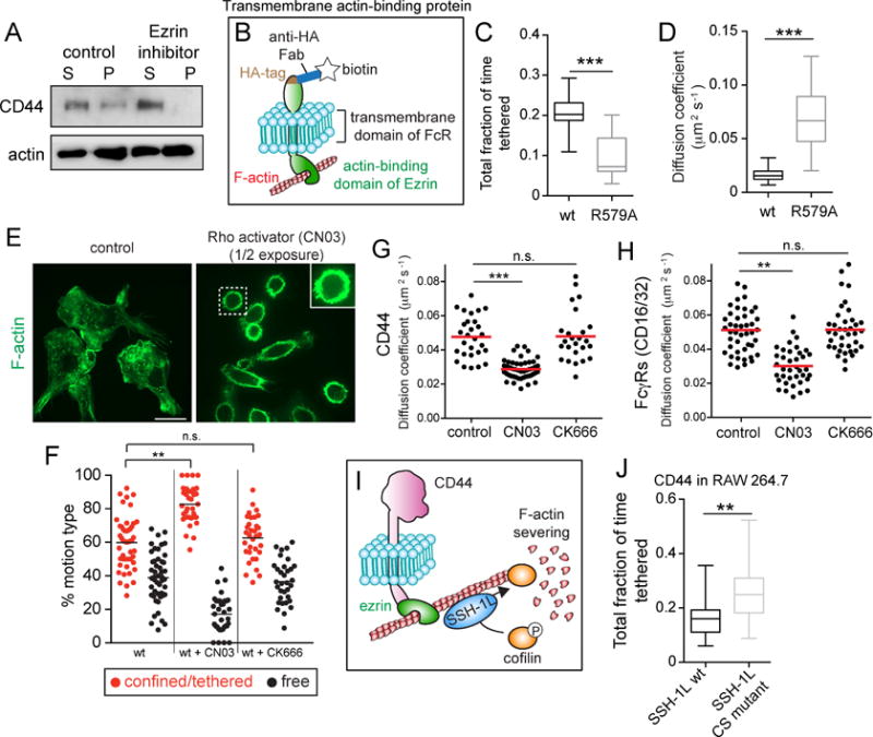
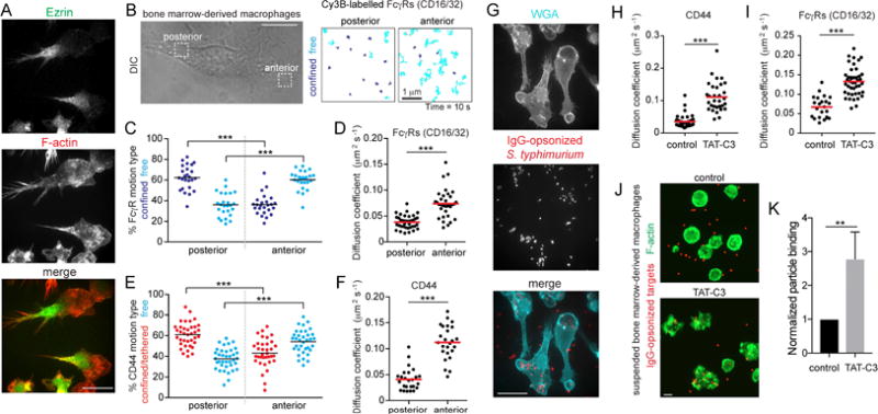
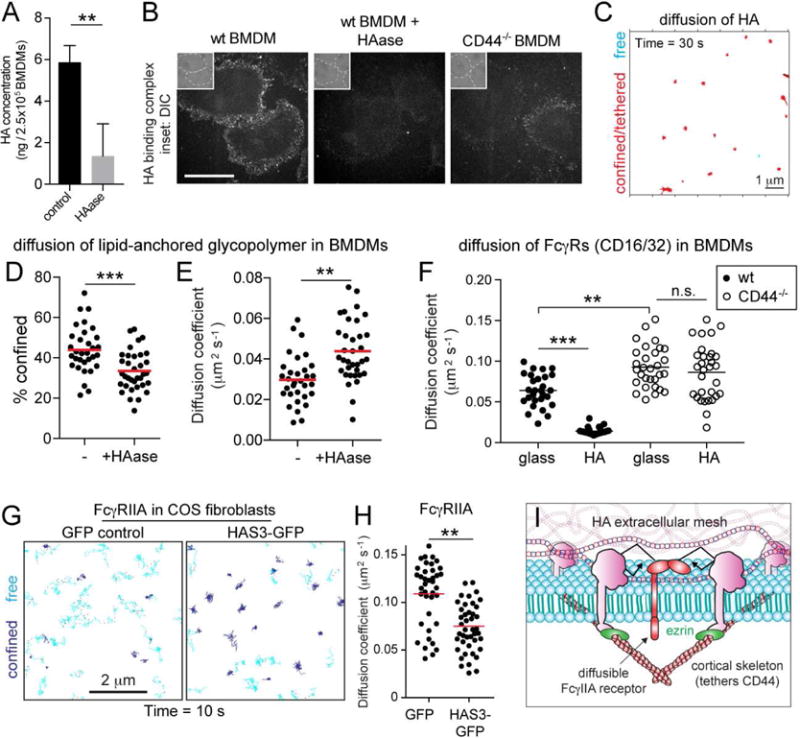
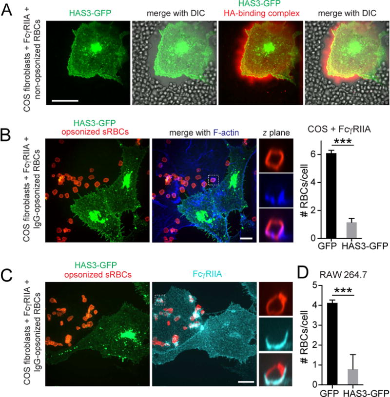
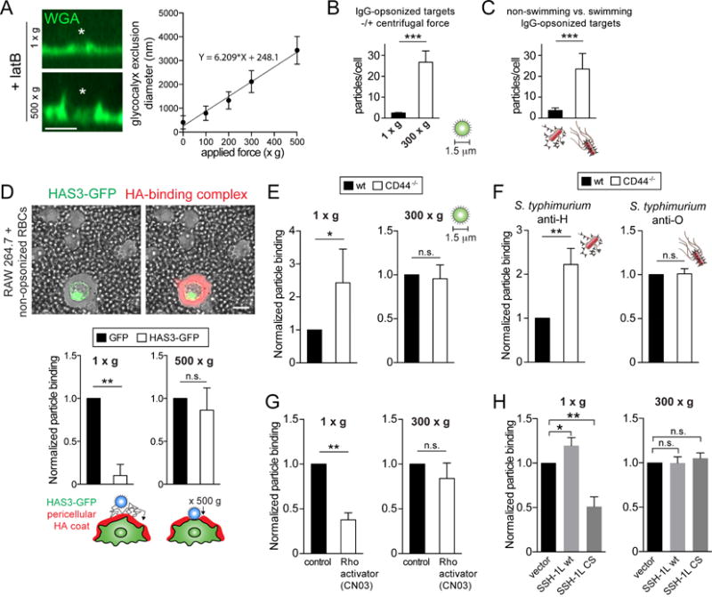
Phagocytic receptors must diffuse laterally to become activated upon clustering by multivalent targets. Receptor diffusion, however, can be obstructed by transmembrane proteins ("pickets") that are immobilized by interacting with the cortical cytoskeleton. The molecular identity of these pickets and their role in phagocytosis have not been defined. We used single-molecule tracking to study the interaction between Fcγ receptors and CD44, an abundant transmembrane protein capable of indirect association with F-actin, hence likely to serve as a picket. CD44 tethers reversibly to formin-induced actin filaments, curtailing receptor diffusion. Such linear filaments predominate in the trailing end of polarized macrophages, where receptor mobility was minimal. Conversely, receptors were most mobile at the leading edge, where Arp2/3-driven actin branching predominates. CD44 binds hyaluronan, anchoring a pericellular coat that also limits receptor displacement and obstructs access to phagocytic targets. Force must be applied to traverse the pericellular barrier, enabling receptors to engage their targets.

Keywords: Arp2/3; CD44; Fc receptor; diffusion barrier; ezrin; formin; glycocalyx; hyaluronan; phagocytosis; single particle tracking.


Figures

Figure 1
Figure 1. CD44 undergoes “stop-and-go” motion by tethering to the submembrane cytoskeleton
A) Murine bone marrow-derived macrophages (mBMDM) or human monocyte-derived macrophages (hMDM) were incubated with saturating amounts of biotinylated anti-CD44 Fab, subjected to electrophoresis and blotting, and compared to known quantities of Fab. Actin used as loading control. B) BMDM were untreated (control) or treated with 1 μM latrunculin A (latA) for 5 min, extracted in actin-stabilizing buffer and centrifuged. Pellet (P) and supernatant (S) analyzed by immunoblotting. Representative of 5 experiments. C) Single CD44 particles were visualized on the dorsal side of BMDMs using anti-CD44 Fab labeled with Qdots and tracked for 30 s at 33 Hz under control conditions, or following treatment with LatA (5 min) or PFA (10 min). D–F) Trajectories were analyzed using DC-MSS, that recognized segments of trajectories as tethered (red), confined-mobile (blue), or free (cyan). See also supplementary Figure 1. E) Fraction of time CD44 displays each motion type, determined for >25 cells recorded for 30 s at 33 Hz from 3 experiments. Here and elsewhere boxplots represent pooled distribution of single cells, illustrating the median (central line), interquartile ranges (lower and upper regions of box), and full range (whiskers). F) CD44 confinement area determined using DC-MSS. G–J) Full-length CD44 (wt), CD44 with its cytoplasmic tail deleted (CD44ΔCT), FcγRIIA wt or FcγRIIAΔCT were heterologously expressed in COS-1 cells. Single CD44 or FcγRIIA particles visualized using Fab labeled with Qdots (H) or Cy3B (I–J, shown diagrammatically in G). H) Fraction of time spent in tethered mode by CD44wt and CD44ΔCT. I) Confinement area of CD44 or FcγRIIA determined by traditional MSS for >30 cells recorded 5 s at 10 Hz from 3 experiments. Here and elsewhere dots represent the median for each cell, while horizontal line is the overall mean. J) Diffusion coefficient determined for the same recordings as in (H).
Figure 2
Figure 2. CD44 curtails diffusion of Fcγ receptors and lipid anchored glycopolymers
A) Human macrophages stained for CD44 (green) and FcγRIIA (red) and imaged by STED. B) CD44 expression in wildtype or CD44−/−BMDM, or RAW macrophages. C–D) Comparison of diffusion coefficient of Fcγ receptors in wt or CD44−/−BMDM, or RAW cells treated with scrambled or CD44 siRNA (D). Fcγ receptors labelled with Cy3B-tagged Fab and tracked over 5 s at 10 Hz. Median diffusion coefficients determined for >30 cells from 3 experiments. E) Diffusion coefficient of Fcγ receptors in COS-1 cells transfected with full-length (wt) or truncated (ΔCT) CD44. F) Schematic of synthetic glycopolymer used in panels C–E. It consists of repeating units of N-acetyl galactosamine, is labelled with biotin and inserts into the outer leaflet of the membrane via a dipalmitoyl terminus. G–I) Qdots were affixed to single glycopolymers and their motion tracked in wt and CD44−/−BMDMs over 30 s at 33 Hz. G) Representative trajectories. H) Fraction of particles undergoing confined diffusion for >30 cells from 3 experiments. I) Median diffusion coefficients for the recordings in (D).
Figure 3
Figure 3. The actin-binding domain of ezrin is necessary and sufficient to tether CD44, which is released by cofilin-mediated severing of actin filaments
A) BMDMs treated with or without 10 μM of ezrin inhibitor NSC668394 were extracted in actin-stabilizing buffer and centrifuged. Pellet (P) and supernatant (S) were analyzed by immunoblotting. Representative of 3 experiments. B) Schematic of the construct used in C,D and F. A transmembrane actin-binding protein was engineered by fusing the actin-binding domain of ezrin to the transmembrane domain of Fc receptor, tagged with hemagglutinin (HA) used to attach biotinylated anti-HA Fab for single-particle tracking. An otherwise identical construct bearing a R579A mutation was used as an inactive control. C) Fraction of time spent tethered by the unmodified (wt) and mutant (R579A) constructs, determined for >25 cells from 3 experiments. D) Diffusion coefficient estimates for the recordings in I. E) BMDMs treated with or without Rho activator (CN03) were fixed and stained for F-actin. Right panel image was acquired using ½ the exposure time used for the left; exposure time for inset in right panel was same as for the left panel. F) Cells expressing the actin-binding chimera were treated with 1.0 μg/mL CN03 for 2–3 h or 10 μM CK666 for 20 min. Fraction of time spent undergoing restricted or free motion determined for >35 cells from 3 experiments. Tethered and confined modes were not separated. G–H) BMDMs were treated with CN03 or CK666 as above before labeling single particles with Cy3-labeled Fab against CD44 or FcγRs. Particles tracked for 10 s at 10 Hz and median diffusion coefficients for >25 cells from 3 experiments are plotted for CD44 (G) and FcγRs (H). I) Schematic illustrating the regulation of actin filament stability by cofilin. Slingshot (SSH-1L) localizes to actin filaments and dephosphorylates cofilin to enable its binding and severing of the filament. J) CD44 mobility assessed in RAW cells transfected with wt or phosphatase-dead (CS) SSH-1L. Single CD44 molecules visualized using Qdots tracked for 30 s at 33 Hz and the fraction of time spent in the tethered state calculated for >25 cells from 3 experiments.
Figure 4
Figure 4. Polarization of cytoskeletal networks orchestrates picket and receptor diffusion
A) BMDMs fixed and stained for ezrin (top) and F-actin (middle); merged images at bottom. Bar = 40 μm. B) Polarized BMDMs were selected to visualize CD44 or FcγRs with Cy3B-labelled Fab. Particles tracked for 10 s at 10 Hz at the front and back of the cell. Scale bar = 10 μm. Trajectories are color-coded as confined/tethered (blue) or free (cyan). C–F) Motion type (C and E) and diffusion coefficient (D and F) of FcγRs (C–D) or CD44 (E–F) determined for >25 cells from 3 experiments. G) BMDMs were challenged with ΔinvA S. typhimurium expressing dsRed and opsonized against O-antigens for 5 min and stained with wheat-germ agglutinin (WGA, cyan). H–I) BMDMs were treated with the Rho inhibitor (TAT-C3) before single-particle tracking for 10 s at 10 Hz; diffusion coefficient of CD44 (H) or FcγRs (I) was determined for >23 cells from 3 experiments. J–K) BMDMs treated in suspension with TAT-C3 for 2 h before challenge with 1.5 μm IgG-opsonized latex beads (red). Cells were fixed and stained for F-actin (green; panel J). Bar = 10 μm. Particle binding presented as means ± SE of >15 fields containing >5 cells each from 3 experiments (K).
Figure 5
Figure 5. An immobile pericellular exoskeleton of hyaluronan (HA) curtails diffusion of membrane-associated molecules
A) The concentration of HA was determined by ELISA for control and HAase-treated (20 units/mL, 30 min at 37°C) BMDM extracts. Means ± SE of 3 determinations. B) Wildtype and CD44−/−BMDMs were treated as indicated before incubation with fluorescent HA-binding complex and imaging. Insets are DIC. Bar = 40 μm. C) BMDMs incubated with biotinylated HA-binding protein, followed by streptavidin Qdots that were tracked for 30 s at 33 Hz. Representative trajectories shown. D–E) Qdots were affixed to biotinylated glycopolymers and their motion tracked in control and HAase-treated BMDMs over 30 s at 33 Hz. Percent confinement I and median diffusion coefficient (F) of >30 cells from 3 experiments. F) Fcγ receptors visualized with Cy3B-labelled Fab in HAase-treated BMDMs seeded on glass or HA-coated glass. Receptors were tracked in the ventral surface of cells over 10 s at 10 Hz and median diffusion coefficient determined for >25 cells from 3 experiments. GH) COS-1 cells expressing FcγRIIA were transfected with GFP or HA synthase 3 (HAS3)-GFP and receptors visualized with Cy3B-labelled Fab. Receptors were tracked over 10 s at 10 Hz. Representative trajectories (G), where blue indicates confinement and cyan free motion; median diffusion coefficients determined for >30 cells from 3 experiments shown in (H). I) Model illustrating proposed mechanism whereby HA restricts receptor diffusion.
Figure 6
Figure 6. Hyaluronan generated by HAS3 establishes a barrier to receptor access and particle binding
A) HA was visualized in COS-1 cells transfected with HAS3-GFP using fluorescent HA-binding complex (red) before addition of red blood cells, apparent by DIC. B) COS-1 cells expressing FcγRIIA were transfected with HAS3-GFP (green) and incubated with IgG-opsonized RBCs (red, left). Phalloidin staining (blue). Graph shows the number of RBCs associated to COS-1 cells transfected with GFP or HAS3-GFP. Means ± SE of 50 cells from 3 experiments. Total number of RBCs associated (i.e. bound plus internalized; #RBCs) is shown. C) COS-1 cells expressing FcγRIIA were transfected with HAS3-GFP (green) and incubated with RBCs, followed by FcγRIIA staining (cyan). D) Quantification of RBCs associated to RAW 264.7 cells transfected with GFP or HAS3-GFP. Means ± SE of 50 cells from 3 experiments.
Figure 7
Figure 7. The CD44-structured picket-fence is overcome by force exerted by phagocytic targets
A) Control or LatA-treated BMDMs were exposed to 5 μm beads (asterisks = centre of bead) with or without application of centrifugal force (100–500 xg for 1 min). Glycocalyx exclusion was visualized with WGA (green) and is plotted on the right as a function of centrifugal force. Means ± SE from >100 contacts per condition, from 3 experiments. B) BMDMs challenged with 1.5 μm IgG-opsonized beads in the absence or presence of applied g force (300 xg). The number of beads bound per cell was determined, shown as means ± SE of 5 determinations. C) BMDMs were challenged for 20 min with equal amounts of dsRed-expressing ΔinvA S. typhimurium opsonized against H-(flagella) or O-(LPS) antigens. Anti-H greatly reduces the ability of the bacteria to swim (Supplementary Video 3). Number of bacteria bound per cell is shown as means ± SE of 5 determinations. D) RAW cells transiently transfected with HAS3-GFP stained for HA with a fluorescent HA-binding complex (red) and incubated with non-opsonized RBCs to visualize the exclusion area by DIC (left). Similarly transfected cells challenged with 8 μm opsonized beads in the absence or presence of applied centrifugal force. Number of beads bound per cell is shown as means ± SE of 3 determinations. E–F) Wildtype and CD44−/−BMDMs challenged with beads or ΔinvA S. typhimurium (F) as in B–C. G–H) Control or CN03-treated BMDMs (G) or RAW cells expressing a control vector, wildtype Slingshot (SSH-1L) or phosphatase-dead Slingshot (SSH-1L CS) (H) were challenged as in B. Particle binding presented as means ± SE of >50 cells from 3 experiments.

References

    1. Aruffo A, Stamenkovic I, Melnick M, Underhill CB, Seed B. CD44 is the principal cell surface receptor for hyaluronate. Cell. 1990;61:1303–1313. - PubMed
    1. Botelho RJ, Teruel M, Dierckman R, Anderson R, Wells A, York JD, Meyer T, Grinstein S. Localized biphasic changes in phosphatidylinositol-4,5-bisphosphate at sites of phagocytosis. The Journal of cell biology. 2000;151:1353–1368. - PMC - PubMed
    1. Chang MY, Tanino Y, Vidova V, Kinsella MG, Chan CK, Johnson PY, Wight TN, Frevert CW. A rapid increase in macrophage-derived versican and hyaluronan in infectious lung disease. Matrix biology : journal of the International Society for Matrix Biology. 2014;34:1–12. - PMC - PubMed
    1. Chen Y, Veracini L, Benistant C, Jacobson K. The transmembrane protein CBP plays a role in transiently anchoring small clusters of Thy-1, a GPI-anchored protein, to the cytoskeleton. Journal of cell science. 2009;122:3966–3972. - PMC - PubMed
    1. Downey GP, Botelho RJ, Butler JR, Moltyaner Y, Chien P, Schreiber AD, Grinstein S. Phagosomal maturation, acidification, and inhibition of bacterial growth in nonphagocytic cells transfected with FcgammaRIIA receptors. The Journal of biological chemistry. 1999;274:28436–28444. - PubMed

Publication types