. 2018 Apr:30:60-69.
doi: 10.1016/j.dcn.2018.01.001. Epub 2018 Jan 5.

Consequence of intraventricular hemorrhage on neurovascular coupling evoked by speech syllables in preterm neonates

Affiliations

Consequence of intraventricular hemorrhage on neurovascular coupling evoked by speech syllables in preterm neonates

Mahdi Mahmoudzadeh et al. Dev Cogn Neurosci. 2018 Apr.

Abstract

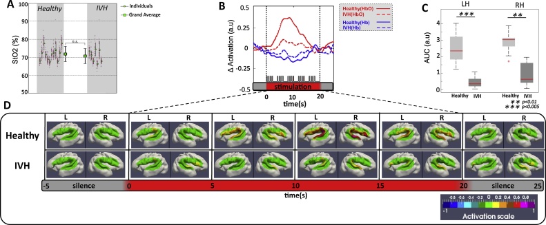
Intraventricular Hemorrhage (IVH) is the leading cause of neurological and cognitive impairment in preterm neonates with an incidence that increases with increasing prematurity. In the present study, we tested how preterm neonates with IVH react to external stimulation (i.e. speech syllables). We compared their neural responses measured by electroencephalography (EEG), and hemodynamic responses measured by functional near-infrared spectroscopy (fNIRS), with those of healthy preterms. A neural response to syllables was observed in these infants, but did not induce a vascular response in contrast with healthy neonates. These results clearly demonstrate that the cerebral vascular network in IVH preterm neonates was unable to compensate for the increased metabolism resulting from neuronal activation in response to external stimulation. Optical imaging is thus a sensitive tool to identify altered cerebral hemodynamic in critically ill preterms before behavioral changes are manifested or when only minor abnormalities on other functional monitoring techniques such as EEG are visible. We propose that a multi-modal approach provides unique opportunities for early monitoring of cognitive functions and opens up new possibilities for clinical care and recommended practices by studying the difficulties of the premature brain to adapt to its environment.

Keywords: Electroencephalogram (EEG); Functional Near Infrared Spectroscopy (fNIRS); Intraventricular hemorrhage; Neurovascular coupling; Preterm.


Figures

Fig. 1
Fig. 1
(A) Estimated projection of the optodes on the brain of a 30 w GA preterm infant. The geometric layout of the optical probe and its projection on the brain mesh of an individual 30 wGA preterm infant (courtesy of P. Hüppi and J. Dubois). Numbers 1–16 correspond to the light sources and letters A, B, C and D correspond to the detectors. (B) Auditory stimulation paradigm. Five series of four syllables were presented (SOA = 600 ms) in a block (duration 20 s), followed by 40 s of silence. Three types of blocks were randomly presented for a total duration of 108 min. In standard blocks, the same syllable (/ba/or/ga/) was presented, whereas, in deviant blocks, a change of voice (DV) or a change of phoneme (DP) was presented three times. (C) HB-ERP and HD-fOI system. Left: Preterm neonate with high-density EEG cap. The infants were tested while sleeping at night to avoid daylight and the intense daytime activity of a neonatal care unit. They were placed in the supine position on a comfortable pad in a dark and quiet incubator. The incubator was further protected from ambient light by dark sheets. Stimuli were binaurally presented at a comfortable hearing level (≈70 dB) via speakers placed at the infant’s feet, at a distance of 30 cm from the subject’s head. Right: A special probe made from soft, flexible foam (thickness = 5 mm) was designed to comfortably maintain the source and detector fibers on the infant’s head. A patch comprising two detectors and eight emitters (optodes) perpendicular to the head surface was placed on each side of the head. Each of the eight optodes contained two wavelength emitter glass fibers (690 and 830 nm). The probe was smoothly secured to the infant’s head to cover the perisylvian areas on each side with straps and foam padding. The probe was designed as a round-grid layout, maintaining a distance of 15 mm between the optodes (Fig. 1A). This layout allowed 10 measuring points (channels) that were simultaneously sampled on each hemisphere.
Fig. 2
Fig. 2
(A) High-resolution EEG. Recording of an individual IVH neonate showing pathological features (1) Abnormal frontal slow waves, (2) positive temporoparietal spikes and increased discontinuity. (B) Grand-average plot of the ERPs in IVH preterms; on the middle, response from each electrode and, on the left, three electrodes have been selected to show the decrease in amplitude with syllable repetition, especially observed between S1 and S2; on the right-up shows that IVH infants have smaller deflections than healthy infants, the right-down shows the difference in peak latencies (*** Three asterisks show significantly different peak latencies (pooled peaks S1-S4) between healthy and IVH infants (p < 0.001). * One asterisk indicates that the first peak latency (S1) is significantly (p < 0.05) different between healthy and IVH infants). (C) 2D maps of peaks 2, 3 and 4 in healthy and IVH preterm neonates. The maps are computed on a 40 ms time-window centered on 244 (P2), 378 (P3) and 576 ms (P4) after the first syllable of the trials. (D) Mean and standard deviation of the global field power (GFP) in healthy (blue) and IVH (red) neonates. The dotted lines indicate the onset of each of the 4 syllables of a trial. The amplitude of the response decreased with syllable repetition, but the peaks remained visible every 600 ms. Note the marked variability across subjects, especially in the IVH group due to the small number of subjects but also to the impact of the lesion. (E) Grand average (and standard deviation) of the projections of each pathological and healthy individual recording on spatial templates corresponding to the 2D maps of P2, P3 and P4 in healthy subjects. The repetition of the topographies after each syllable is revealed by the repetition of the same pattern of successive increase of the three coefficients after each syllable. Although the pattern is less obvious in IVH neonates, no significant difference was observed between the groups. (For interpretation of the references to colour in this figure legend, the reader is referred to the web version of this article.)
Fig. 3
Fig. 3
(A) Mean StO2 of Healthy (12 preterms) and IVH (7 preterms) infants during the experiment. Each thin black error bar indicates the mean and standard deviation across the 20 NIRS channels (red dots). Thick black error bars indicate mean and standard deviation of StO2 over Healthy and IVH preterm infants. (B) shows one sample fOI channel of grand-average of HbO (red lines), Hb (blue lines) changes for healthy (solid lines) and IVH (dotted lines) neonates during auditory stimulation. Horizontal red thick bars indicate the period of the stimulation (20 s). This response was pronounced for the healthy neonates, with a significant increase in HbO. (C) Comparison of the mean AUC changes of Oxy-Hb induced by auditory stimulation between healthy and IVH infants. For each hemisphere, activations were significantly reduced in the IVH group compared to the healthy group. (D) Topographies of the hemodynamic response in the two groups (Healthy, IVH). The HbO response, averaged each 5 s, was projected on a 3-D mesh of an individual 30 wGA preterm neonate. The red rectangle indicates the duration of auditory stimulation. The hemodynamic response in both hemispheres was weak in IVH neonates compared to healthy infants. (For interpretation of the references to colour in this figure legend, the reader is referred to the web version of this article.)

References

    1. Alderliesten T., Lemmers P.M., Smarius J.J., van de Vosse R.E., Baerts W., van Bel F. Cerebral oxygenation, extraction, and autoregulation in very preterm infants who develop peri-intraventricular hemorrhage. J. Pediatr. 2013;162:698–704. e692. - PubMed
    1. Ardila A., Ostrosky-Solis F. Gordon and Breach; 1984. The Right Hemisphere: Neurology and Neuropsychology.
    1. Azizollahi H., Aarabi A., Wallois F. Effects of uncertainty in head tissue conductivity and complexity on EEG forward modeling in neonates. Hum. Brain Mapp. 2016;37:3604–3622. - PMC - PubMed
    1. Bartocci M., Bergqvist L.L., Lagercrantz H., Anand K.J. Pain activates cortical areas in the preterm newborn brain. Pain. 2006;122:109–117. - PubMed
    1. Bisiacchi P.S., Mento G., Suppiej A. Cortical auditory processing in preterm newborns: an ERP study. Biol. Psychol. 2009;82:176–185. - PubMed

Publication types

MeSH terms