. 2018 Jan 12;19(1):227.
doi: 10.3390/ijms19010227.

Development of a Cytocompatible Scaffold from Pig Immature Testicular Tissue Allowing Human Sertoli Cell Attachment, Proliferation and Functionality

Affiliations

Development of a Cytocompatible Scaffold from Pig Immature Testicular Tissue Allowing Human Sertoli Cell Attachment, Proliferation and Functionality

Maxime Vermeulen et al. Int J Mol Sci. .

Abstract

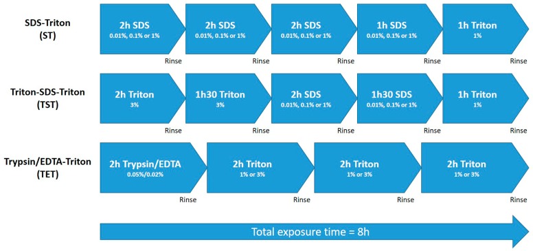
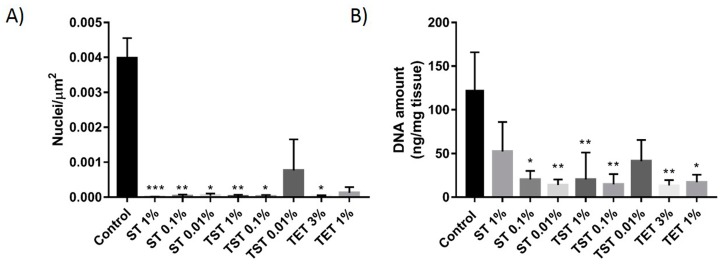
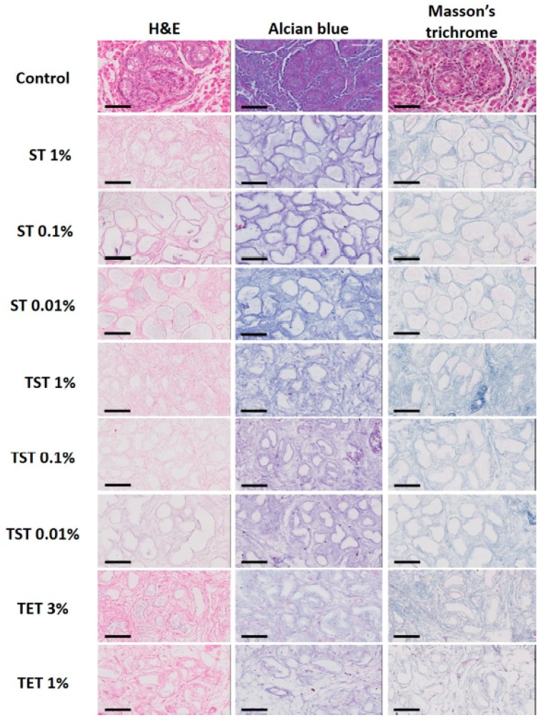
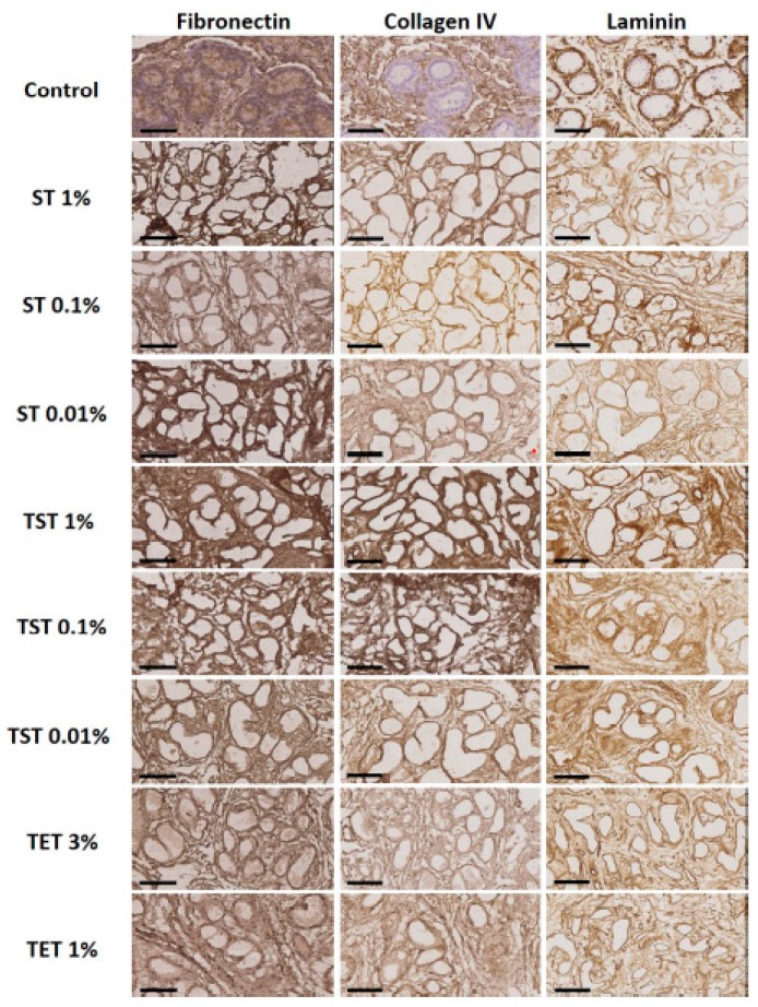
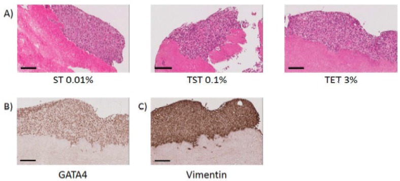
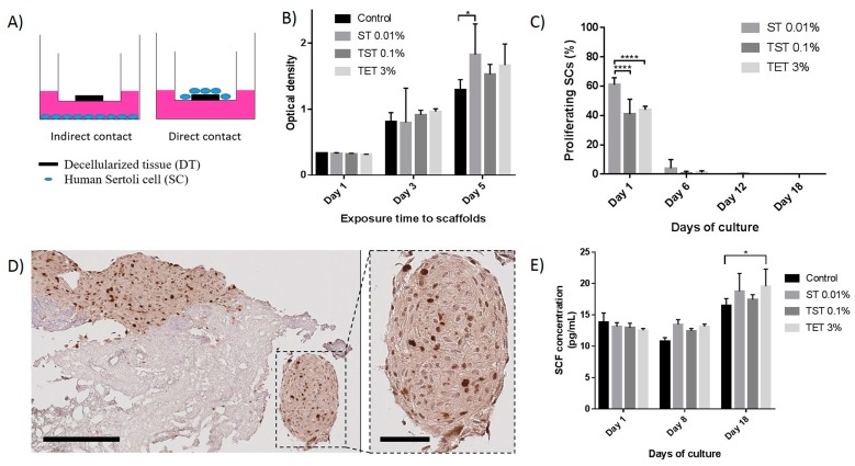
Cryopreservation of immature testicular tissue before chemo/radiotherapy is the only option to preserve fertility of cancer-affected prepubertal boys. To avoid reintroduction of malignant cells, development of a transplantable scaffold by decellularization of pig immature testicular tissue (ITT) able to support decontaminated testicular cells could be an option for fertility restoration in these patients. We, therefore, compared decellularization protocols to produce a cytocompatible scaffold. Fragments of ITT from 15 piglets were decellularized using three protocols: sodium dodecyl sulfate (SDS)-Triton (ST), Triton-SDS-Triton (TST) and trypsin 0.05%/ethylenediaminetetraacetic acid (EDTA) 0.02%-Triton (TET) with varying detergent concentrations. All protocols were able to lower DNA levels. Collagen retention was demonstrated in all groups except ST 1%, and a significant decrease in glycosaminoglycans was observed in the TST 1% and TET 1% groups. When Sertoli cells (SCs) were cultured with decellularized tissue, no signs of cytotoxicity were detected. A higher SC proliferation rate and greater stem cell factor secretion were observed than with SCs cultured without scaffold. ST 0.01% and TET 3% conditions offered the best compromise in terms of DNA elimination and extracellular matrix (ECM) preservation, while ensuring good attachment, proliferation and functionality of human SCs. This study demonstrates the potential of using decellularized pig ITT for human testicular tissue engineering purposes.

Keywords: decellularization; decellularized tissue; extracellular matrix; fertility preservation; immature testicular tissue; regenerative medicine; scaffold; testicular organoid; tissue engineering.


Conflict of interest statement

The authors declare no conflict of interest.

Figures

Figure 1
Figure 1
Schematic representation of decellularization processes. Total exposure time to decellularization solutions was eight hours in each case, punctuated by rinse steps in dH2O.
Figure 2
Figure 2
DNA quantification in DT. (A) Nuclei were counted on H and E-stained slides of native tissue and DT and represented as the number of nuclei per µm2; (B) Following tissue digestion, DNA was extracted and quantified using the Quant-iT™ PicoGreen® dsDNA assay. The results are presented as means ± SD. *** p < 0.001, ** p < 0.01, * p < 0.05.
Figure 3
Figure 3
Histological staining of native tissue and DT with different protocols in different conditions. H and E, Alcian blue and Masson’s trichrome staining evidenced preservation of structure, GAGs and collagen respectively. Scale bar = 70 μm.
Figure 4
Figure 4
Immunohistochemical analyses of native tissue and DT. Fibronectin, collagen IV and laminin were preserved in tissue decellularized with all protocols in all conditions. Scale bar = 70 µm.
Figure 5
Figure 5
Collagen and GAG quantification in native tissue and DTs. (A) Collagen in native tissue and DTs was quantified using the Sircol assay; (B) Quantification of GAGs in native tissue and DTs using the Blyscan assay. Results are presented as means ± SD. *** p < 0.001, ** p < 0.01, * p < 0.05.
Figure 6
Figure 6
Attachment of SCs onto DTs. (A) H and E staining of tissues decellularized with ST 0.01%, TST 0.1% and TET 3% onto which SCs can reattach. Scale bar = 100 µm; (B) SCs express GATA4 onto DTs. Scale bar = 100 µm; (C) SCs express vimentin onto DTs. Scale bar = 100 µm.
Figure 7
Figure 7
Effects of DTs on proliferation and functionality of SCs. (A) Representative drawing of direct and indirect contact culture systems. The indirect contact culture system was used for the WST-1 assay; (B) Proliferation of SCs seeded in the compartment below the transwell containing DTs evaluated using the WST-1 assay which is based on the reduction of a tetrazolium salt to a soluble violet formazan product by viable cells. Optical density, which is proportional to cell numbers/well, was recorded on days 1, 3 and 5 using a spectrophotometer; (C) Percentage of proliferative SCs seeded onto DTs on days 1, 6, 12 and 18 of culture. SCs seeded onto ST 0.01% DTs present a higher percentage of proliferation compared to TST 0.1% and TET 3% DTs on day 1; (D) Immunohistochemical staining of Ki67 in SCs seeded onto DT. The enlarged image shows a round shaped structure as observed in some cases. Scale bares = 200 and 60 µm; (E) SCs seeded onto DTs secreted at least the same amount of SCF than SCs seeded alone. Results are presented as mean ± SD. * p < 0.05, **** p < 0.0001.

References

    1. Gatta G., Botta L., Rossi S., Aareleid T., Bielska-Lasota M., Clavel J., Dimitrova N., Jakab Z., Kaatsch P., Lacour B., et al. Childhood cancer survival in Europe 1999–2007: Results of eurocare-5—A population-based study. Lancet Oncol. 2014;15:35–47. doi: 10.1016/S1470-2045(13)70548-5. - DOI - PubMed
    1. Wyns C., Curaba M., Petit S., Vanabelle B., Laurent P., Wese J.F., Donnez J. Management of fertility preservation in prepubertal patients: 5 years’ experience at the catholic university of louvain. Hum. Reprod. 2011;26:737–747. doi: 10.1093/humrep/deq387. - DOI - PubMed
    1. Wyns C., Collienne C., Shenfield F., Robert A., Laurent P., Roegiers L., Brichard B. Fertility preservation in the male pediatric population: Factors influencing the decision of parents and children. Hum. Reprod. 2015;30:2022–2030. doi: 10.1093/humrep/dev161. - DOI - PubMed
    1. Sato T., Katagiri K., Gohbara A., Inoue K., Ogonuki N., Ogura A., Kubota Y., Ogawa T. In vitro production of functional sperm in cultured neonatal mouse testes. Nature. 2011;471:504–507. doi: 10.1038/nature09850. - DOI - PubMed
    1. Liu Z., Nie Y.H., Zhang C.C., Cai Y.J., Wang Y., Lu H.P., Li Y.Z., Cheng C., Qiu Z.L., Sun Q. Generation of macaques with sperm derived from juvenile monkey testicular xenografts. Cell Res. 2016;26:139–142. doi: 10.1038/cr.2015.112. - DOI - PMC - PubMed

LinkOut - more resources