. 2018 Mar 28;217(8):1237-1246.
doi: 10.1093/infdis/jiy003.

1918 H1N1 Influenza Virus Replicates and Induces Proinflammatory Cytokine Responses in Extrarespiratory Tissues of Ferrets

Affiliations

1918 H1N1 Influenza Virus Replicates and Induces Proinflammatory Cytokine Responses in Extrarespiratory Tissues of Ferrets

Emmie de Wit et al. J Infect Dis. .

Abstract

Background: The 1918 Spanish H1N1 influenza pandemic was the most severe recorded influenza pandemic with an estimated 20-50 million deaths worldwide. Even though it is known that influenza viruses can cause extrarespiratory tract complications-which are often severe or even fatal-the potential contribution of extrarespiratory tissues to the pathogenesis of 1918 H1N1 virus infection has not been studied comprehensively.

Methods: Here, we performed a time-course study in ferrets inoculated intranasally with 1918 H1N1 influenza virus, with special emphasis on the involvement of extrarespiratory tissues. Respiratory and extrarespiratory tissues were collected after inoculation for virological, histological, and immunological analysis.

Results: Infectious virus was detected at high titers in respiratory tissues and, at lower titers in most extrarespiratory tissues. Evidence for active virus replication, as indicated by the detection of nucleoprotein by immunohistochemistry, was observed in the respiratory tract, peripheral and central nervous system, and liver. Proinflammatory cytokines were up-regulated in respiratory tissues, olfactory bulb, spinal cord, liver, heart, and pancreas.

Conclusions: 1918 H1N1 virus spread to and induced cytokine responses in tissues outside the respiratory tract, which likely contributed to the severity of infection. Moreover, our data support the suggested link between 1918 H1N1 infection and central nervous system disease.


Figures

Figure 1.
Figure 1.
Bodyweight loss and gross lung lesions in 1918 H1N1 virus–inoculated ferrets. Ferrets were inoculated intranasally with 106 50% tissue culture infectious dose (TCID50) of 1918 H1N1 influenza virus. A, Bodyweight of ferrets throughout the experiment was calculated as the percentage of start weight. Each line represents an individual ferret euthanized at 7 days postinoculation (dpi). B, Percentage of lung affected by gross lesions as observed at necropsy on 1, 3, 5, and 7 dpi. For each time point, average and standard deviation of 4 ferrets is shown.
Figure 2.
Figure 2.
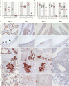
Virus shedding and replication in the respiratory tract of ferrets inoculated with 1918 influenza H1N1 virus. Ferrets were inoculated intranasally with 106 50% tissue culture infectious dose (TCID50) of 1918 H1N1 influenza virus. A, Virus titers in nose and throat swabs collected at 1, 3, 5, and 7 days postinoculation (dpi) from 4 ferrets euthanized at 7 dpi. Horizontal lines represent geometric mean titers. B, Virus titers in the nasal turbinates, trachea, and lung at 1, 3, 5, and 7 dpi. Horizontal lines represent geometric mean titers. C, Detection of influenza A virus nucleoprotein in the epithelium of the nasal respiratory mucosa, olfactory mucosa, bronchus, bronchioles, and alveoli at 1, 3, 5, and 7 dpi. An asterisk (*) in the respiratory mucosa indicates the detection of influenza virus antigen in sloughed-off epithelial cells. An arrowhead in the olfactory mucosa indicates influenza virus antigen in individual cells. An asterisk in the bronchus indicates influenza virus antigen in the submucosal glands.
Figure 3.
Figure 3.
Induction of tumor necrosis factor alpha (TNF-α), interleukin 6 (IL-6), and interleukin 8 (IL-8) in 1918 H1N1 virus–inoculated ferrets. Ferrets were inoculated intranasally with 106 50% tissue culture infectious dose (TCID50) of 1918 H1N1 influenza virus. Four ferrets were euthanized at each of the time points 1, 3, 5, and 7 days postinoculation (dpi), and tissue samples were collected for analysis of expression of proinflammatory cytokines. Log fold change expression (-ΔΔCt values) of TNF-α, IL-6, and IL-8 in the nasal turbinates and lungs of ferrets inoculated with 1918 H1N1 virus relative to the mean of the 4 Mock ferrets was calculated. Colors indicate up- or down-regulation of cytokines compared to the mean of Mock ferrets. Each square indicates expression levels in an individual ferret. Asterisks in the first square in a group indicate a statistically significant difference (t test) of the group of 4 ferrets as compared to the 4 Mock ferrets. *P < .05; **P < .01; ***P < .001; ****P < .0001.
Figure 4.
Figure 4.
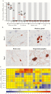
Virus titers, virus antigen, and cytokine expression in the nervous system of 1918 H1N1 virus–inoculated ferrets. Ferrets were inoculated intranasally with 106 50% tissue culture infectious dose (TCID50) of 1918 H1N1 influenza virus. Four ferrets were euthanized at each of the time points 1, 3, 5, and 7 days postinoculation (dpi) and tissue samples were collected for analysis. A, Virus titers in olfactory bulb, cerebrum, spinal cord, and cerebrospinal fluid (CSF) at 1, 3, 5, and 7 dpi. Horizontal lines represent geometric mean titers. B, Detection of influenza virus nucleoprotein in the brain stem at 1 and 5 dpi, the pituitary gland at 1 dpi, and the trigeminal ganglion at 5 dpi (×1000 magnification). C, Fold change expression of tumor necrosis factor alpha (TNF-α), interleukin 6 (IL-6), and interleukin 8 (IL-8) in the olfactory bulb, cerebrum, and spinal cord of ferrets inoculated with 1918 H1N1 virus relative to the mean of 4 Mock ferrets was calculated. Colors indicate up- or down-regulation of cytokines compared to the mean of the Mock ferrets. Each square indicates expression levels in an individual ferret. Asterisks in the first square in a group indicate a statistically significant difference (t test) of the group of 4 ferrets as compared to the 4 Mock ferrets. *P ≤ .05; **P ≤ .01; ***P ≤ .001; ****P ≤ .0001.
Figure 5.
Figure 5.
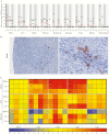
Virus titers, virus antigen, and cytokine expression in 1918 H1N1 influenza virus–inoculated ferrets. Ferrets were inoculated intranasally with 106 50% tissue culture infectious dose (TCID50) of 1918 H1N1 influenza virus. Four ferrets were euthanized at each of the time points 1, 3, 5, and 7 days postinoculation (dpi) and tissue samples were collected for analysis. A, Virus titers in heart, liver, spleen, pancreas, duodenum, jejunum, colon, kidney, and adrenal gland at 1, 3, 5, and 7 dpi. B, Detection of influenza virus nucleoprotein in hepatocytes of the liver of 2 ferrets euthanized at 5 dpi (left panel: ×400 magnification; right panel: ×1000 magnification). C, Fold change expression of tumor necrosis factor alpha (TNF-α), interleukin 6 (IL-6), and interleukin 8 (IL-8) in the heart, liver, and pancreas of ferrets inoculated with 1918 H1N1 virus relative to the mean of 4 Mock ferrets was calculated. Colors indicate up- or down-regulation of cytokines relative to the mean the Mock ferrets. Each square indicates expression levels in an individual ferret. Asterisks in the first square in a group indicate a statistically significant difference (t test) of the group of 4 ferrets as compared to the 4 Mock ferrets. *P ≤ .05; **P ≤ .01; ***P ≤ .001; ****P ≤ .0001.

References

    1. Korteweg C, Gu J. Pathology, molecular biology, and pathogenesis of avian influenza A (H5N1) infection in humans. Am J Pathol 2008; 172:1155–70. - PMC - PubMed
    1. Kuiken T, Taubenberger JK. Pathology of human influenza revisited. Vaccine 2008; 26(Suppl 4):D59–66. - PMC - PubMed
    1. Chi Y, Zhu Y, Wen T et al. Cytokine and chemokine levels in patients infected with the novel avian influenza A (H7N9) virus in China. J Infect Dis 2013; 208:1962–7. - PubMed
    1. de Jong MD, Simmons CP, Thanh TT et al. Fatal outcome of human influenza A (H5N1) is associated with high viral load and hypercytokinemia. Nat Med 2006; 12:1203–7. - PMC - PubMed
    1. Hayden FG, Fritz R, Lobo MC, Alvord W, Strober W, Straus SE. Local and systemic cytokine responses during experimental human influenza A virus infection. Relation to symptom formation and host defense. J Clin Invest 1998; 101:643–9. - PMC - PubMed

Publication types