. 2018 Mar 15;27(6):1002-1014.
doi: 10.1093/hmg/ddy017.

RAGE mediates Aβ accumulation in a mouse model of Alzheimer's disease via modulation of β- and γ-secretase activity

Affiliations

RAGE mediates Aβ accumulation in a mouse model of Alzheimer's disease via modulation of β- and γ-secretase activity

Fang Fang et al. Hum Mol Genet. .

Abstract

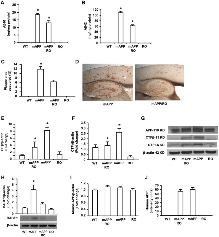
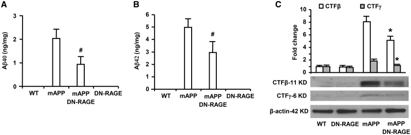
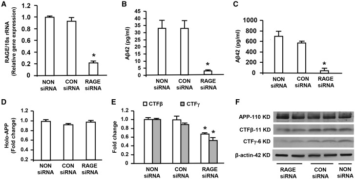
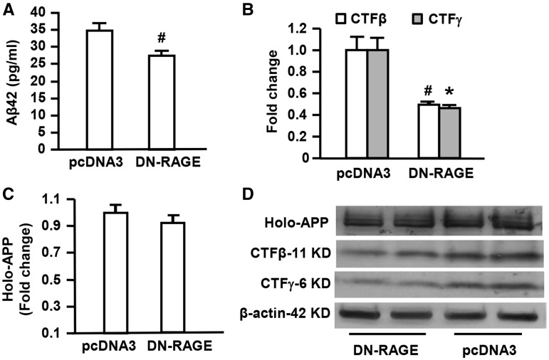
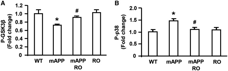
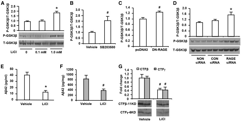
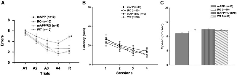
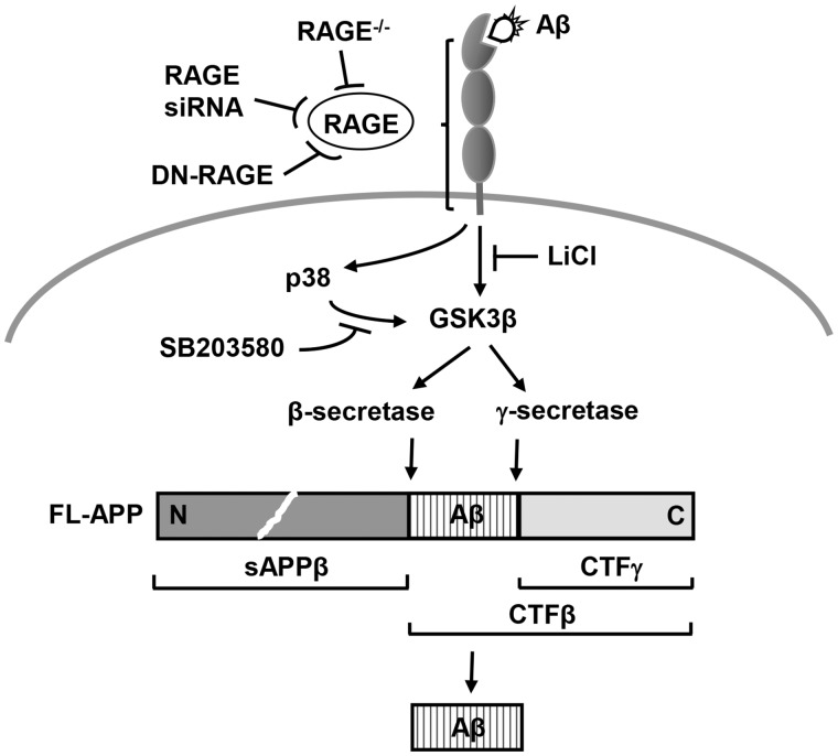
Receptor for Advanced Glycation End products (RAGE) has been implicated in amyloid β-peptide (Aβ)-induced perturbation relevant to the pathogenesis of Alzheimer's disease (AD). However, whether and how RAGE regulates Aβ metabolism remains largely unknown. Aβ formation arises from aberrant cleavage of amyloid pre-cursor protein (APP) by β- and γ-secretase. To investigate whether RAGE modulates β- and γ-secretase activity potentiating Aβ formation, we generated mAPP mice with genetic deletion of RAGE (mAPP/RO). These mice displayed reduced cerebral amyloid pathology, inhibited aberrant APP-Aβ metabolism by reducing β- and γ-secretases activity, and attenuated impairment of learning and memory compared with mAPP mice. Similarly, RAGE signal transduction deficient mAPP mice (mAPP/DN-RAGE) exhibited the reduction in Aβ40 and Aβ42 production and decreased β-and γ-secretase activity compared with mAPP mice. Furthermore, RAGE-deficient mAPP brain revealed suppression of activation of p38 MAP kinase and glycogen synthase kinase 3β (GSK3β). Finally, RAGE siRNA-mediated gene silencing or DN-RAGE-mediated signaling deficiency in the enriched human APP neuronal cells demonstrated suppression of activation of GSK3β, accompanied with reduction in Aβ levels and decrease in β- and γ-secretases activity. Our findings highlight that RAGE-dependent signaling pathway regulates β- and γ-secretase cleavage of APP to generate Aβ, at least in part through activation of GSK3β and p38 MAP kinase. RAGE is a potential therapeutic target to limit aberrant APP-Aβ metabolism in halting progression of AD.


Figures

Figure 1.
Figure 1.
Effect of RAGE deficiency on Aβ accumulation and β and γ secretases activity in mAPP mice. Aβ 40 (A) and Aβ 42 (B) levels were determined by ELISA in the cortex from 12 month-old mice of the indicated four genotypes (N = 7–10 mice/per genotype). Aβ deposits in the cortex (C) were quantified by histological image analysis in the brain sections stained with 3D6 antibody in the indicated groups (N = 5–10 mice/per genotype). Representative images show the sections stained with 3D6 from mAPP and mAPP/RO mice at 12 months of age (D). Scale bar = 30 µm. β- and γ-secretase activity was demonstrated by quantifying intensity of immunoreactive bands of CTFβ (E) or CTFγ (F) in the brain homogenates from indicated mice. N = 4–5 mice/per genotype. Representative immunoblots show immunoreactive bands for APP-110 KD, CTFβ-11 KD and CTFγ-6 KD as well as β-actin protein used as a loading control (G). Representative immunoblot and quantitative analysis of immunoreactive band for BACE1 were shown (H) and β-actin protein used as a loading control. Quantification of mouse APP (I) and human APP (J) was performed, respectively. *P < 0.01 compared with other groups of mice.
Figure 2.
Figure 2.
Effect of RAGE-deficient cytosolic domain in neurons on Aβ production and β- and γ-secretase activity in mAPP mice. Aβ40 (A) and Aβ42 (B) levels in the brain homogenate were determined by ELISA from mice of indicated four genotypes (N = 5–7 mice/per genotype). β- and γ-secretase activity was demonstrated by quantifying intensity of immunoreactive bands of CTFβ and CTFγ in brain homogenates from the indicated mice. (C) Representative immunoblots show the immunoreactive bands for CTFβ and CTFγ as well as β-actin protein used as a loading control (C). N = 4–5 mice/per genotype. #P < 0.05 or *P < 0.01 mAPP/DN-RAGE mice versus mAPP mice.
Figure 3.
Figure 3.
Effect of knockdown of RAGE on Aβ production, β- or γ-secretase activity in human APP/Aβ producing neuronal cells. RNA isolated from B103-wtAPP cells transfected with RAGE siRNA or negative control (CON) siRNA or Non-siRNA was subjected to Real Time PCR analysis of RAGE mRNA levels normalized to 18s rRNA (A). Aβ ELISA was used to measure Aβ42 levels in the media (B) and cell lysates (C) of B103-wtAPP cells transfected with RAGE siRNA or negative control (CON) siRNA or NON-siRNA. Quantification of the immunoreactive bands for APP, CTFβ and CTFγ in B103-wtAPP cells transfected with RAGE siRNA or CON siRNA or NON-siRNA was performed (D–F). Representative immunoblots show immunoreactive bands for APP, CTFβ and CTFγ as well as β-actin protein used as a loading control (F). Results come from at least three independent experiments. *P < 0.01 RAGE siRNA versus CON siRNA or NON siRNA transfected cells.
Figure 4.
Figure 4.
Effect of neuronal RAGE deficient cytosolic domain on Aβ production and β- or γ-secretase activity in human APP/Aβ producing neuronal cells. Aβ ELISA was used for measurement of Aβ42 levels in B103-wtAPP cells transfected with DN-RAGE or pcDNA3 (A). Quantification of intensity of immunoreactive bands for APP, CTFβ and CTFγ was performed in B103-wtAPP cells transfected with DN-RAGE or pcDNA3 vector (B–D). Representative immunoblots show immunoreactive bands for APP, CTFβ and APP-CTFγ in B103-wtAPP cells transfected with DN-RAGE or pcDNA3 as well as β-actin protein used as a loading control (D). Studies were repeated at least three times in each group. #P < 0.05 or *P < 0.01 DN-RAGE versus pcDNA3 vector-transfected cells.
Figure 5.
Figure 5.
Effect of RAGE deficiency on phosphorylation of GSK3β and p38 MAPK in mAPP mice. The levels of phosphorylated GSK3β at Ser-9 (P-GSK3β) (A) and phosphorylated p38 MAPK (B) in the mouse brain homogenates from indicated four genotypes were measured by ELISA. The bar graphs show fold increase in phosphorylated GSK3β (A) and phosphorylated p38 MAPK (B), respectively in the brain homogenates from indicated mice. N = 4–7 mice per group. *P < 0.01 mAPP versus WT mice or RO mice; #P < 0.05 mAPP/RO versus mAPP mice.
Figure 6.
Figure 6.
Effect of inhibition of GSK3β on Aβ production and β- and γ-secretase activity in human APP/Aβ producing neuronal cells. Immunoblotting with phosphor-GSK3β (Ser 9) (P-GSK3β) and total GSK3β (T-GSK3β) antibodies were performed in protein extracts from B103-wt APP cells with different treatments. The cells were treated with indicated doses of LiCl for 48 h (A,*P < 0.01 versus other groups of cells). The cells were treated with p38 inhibitor SB (203580, 1 µM) for 3 h (B, #P < 0.05 p38 inhibitor treated cells versus vehicle-treated cells). The cells were transfected with DN-RAGE 48 h (C, #P < 0.05 DN-RAGE versus pcDNA3 vector-transfected cells) and transfected with RAGE siRNA (D, *P < 0.01 RAGE siRNA versus CON siRNA or NON siRNA transfected cells). Studies were repeated at least three times in each group. After LiCl (1.0 mm) treatment for 48 h, Aβ42 levels was measured by ELISA in the media (E, *P < 0.01 LiCl-treated cells versus vehicle-treated cells) and cell lysates (F, #P < 0.05 LiCl treated cells versus vehicle-treated cells) of cultured neuronal cells (B103) stably expressed wild-type APP. Assessment of β- and γ-secretase activity was performed by quantification of intensity of immunoreactive bands for CTFβ and CTFγ (G, #P < 0.05 LiCl treated cells versus vehicle-treated cells) in B103-wtAPP cells treated with LiCl. Results come from at least three independent experiments.
Figure 7.
Figure 7.
Effect of RAGE deficiency on spatial learning/memory in mAPP mice. Analysis of spatial learning and memory in mAPP/RO mice at 12 months of age using radial arm water maze test, compared with mAPP, RO and WT littermates (A) (n = 9–10/group, #P < 0.05). Trials 1–4 acquisition trial and trail 5 denotes retention trail. The four genotypes showed no difference in their latency time (B) and speed (C) to reach the platform during the visible platform session.
Figure 8.
Figure 8.
Schematic depiction of RAGE-dependent signaling pathway that regulates β- and γ-secretase cleavage of APP to generate Aβ, at least in part through activation of GSK3β and p38 MAP kinase.

References

    1. Selkoe D.J. (1999) Translating cell biology into therapeutic advances in Alzheimer's disease. Nature, 399, A23–A31. - PubMed
    1. Vassar R., Citron M. (2000) Abeta-generating enzymes: recent advances in beta- and gamma-secretase research. Neuron, 27, 419–422. - PubMed
    1. Nunan J., Shearman M.S., Checler F., Cappai R., Evin G., Beyreuther K., Masters C.L., Small D.H. (2001) The C-terminal fragment of the Alzheimer's disease amyloid protein precursor is degraded by a proteasome-dependent mechanism distinct from gamma-secretase. Eur. J. Biochem., 268, 5329–5336. - PubMed
    1. Chang Y., Tesco G., Jeong W.J., Lindsley L., Eckman E.A., Eckman C.B., Tanzi R.E., Guenette S.Y. (2003) Generation of the beta-amyloid peptide and the amyloid precursor protein C-terminal fragment gamma are potentiated by FE65L1. J. Biol. Chem., 278, 51100–51107. - PubMed
    1. O'Brien R.J., Wong P.C. (2011) Amyloid precursor protein processing and Alzheimer's disease. Ann. Rev. Neurosci., 34, 185–204. - PMC - PubMed

Publication types

MeSH terms

Substances