

Bone formation in ankylosing spondylitis during anti-tumour necrosis factor therapy imaged by 18F-fluoride positron emission tomography
- PMID: 29329443
- PMCID: PMC5888961
- DOI: 10.1093/rheumatology/kex448
Bone formation in ankylosing spondylitis during anti-tumour necrosis factor therapy imaged by 18F-fluoride positron emission tomography
Erratum in
-
Bone formation in ankylosing spondylitis during anti-tumour necrosis factor therapy imaged by 18F-fluoride positron emission tomography.Rheumatology (Oxford). 2018 Apr 1;57(4):770. doi: 10.1093/rheumatology/key034. Rheumatology (Oxford). 2018. PMID: 29415219 Free PMC article. No abstract available.
Abstract
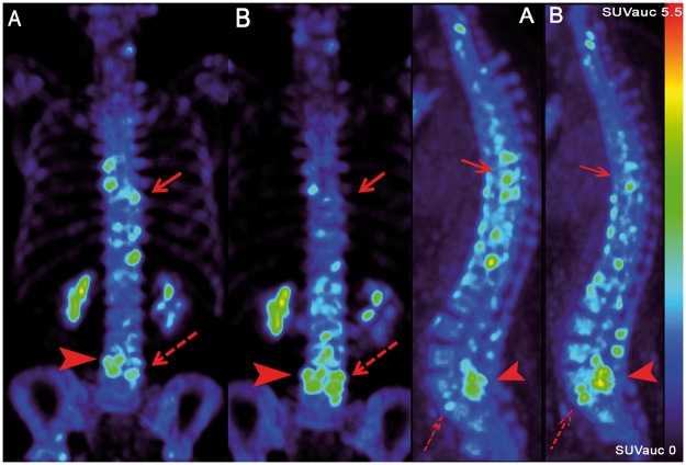
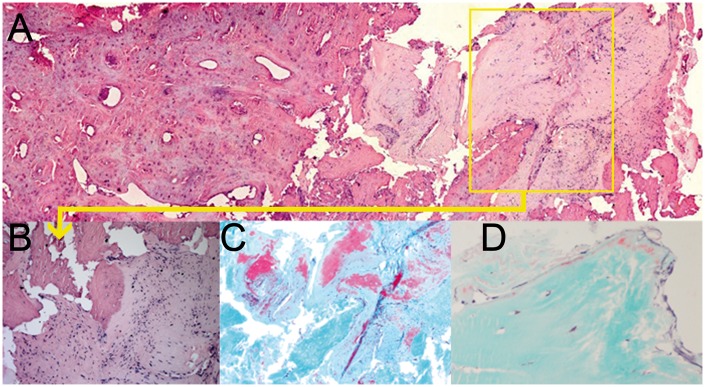
Objectives: Excessive bone formation is an important hallmark of AS. Recently it has been demonstrated that axial bony lesions in AS patients can be visualized using 18F-fluoride PET-CT. The aim of this study was to assess whether 18F-fluoride uptake in clinically active AS patients is related to focal bone formation in spine biopsies and is sensitive to change during anti-TNF treatment.
Methods: Twelve anti-TNF-naïve AS patients [female 7/12; age 39 years (SD 11); BASDAI 5.5 ± 1.1] were included. 18 F-fluoride PET-CT scans were performed at baseline and in two patients, biopsies were obtained from PET-positive and PET-negative spine lesions. The remaining 10 patients underwent a second 18F-fluoride PET-CT scan after 12 weeks of anti-TNF treatment. PET scans were scored visually by two blinded expert readers. In addition, 18F-fluoride uptake was quantified using the standardized uptake value corrected for individual integrated whole blood activity concentration (SUVAUC). Clinical response to anti-TNF was defined according to a ⩾ 20% improvement in Assessment of SpondyloArthritis international Society criteria at 24 weeks.
Results: At baseline, all patients showed at least one axial PET-positive lesion. Histological analysis of PET-positive lesions in the spine confirmed local osteoid formation. PET-positive lesions were found in the costovertebral joints (43%), facet joints (23%), bridging syndesmophytes (20%) and non-bridging vertebral lesions (14%) and in SI joints (75%). After 12 weeks of anti-TNF treatment, 18F-fluoride uptake in clinical responders decreased significantly in the costovertebral (mean SUVAUC -1.0; P < 0.001) and SI joints (mean SUVAUC -1.2; P = 0.03) in contrast to non-responders.
Conclusions: 18F-fluoride PET-CT identified bone formation, confirmed by histology, in the spine and SI joints of AS patients and demonstrated alterations in bone formation during anti-TNF treatment.
Figures
References
-
- Maksymowych WP, Mallon C, Morrow S. et al. Development and validation of the Spondyloarthritis Research Consortium of Canada (SPARCC) Enthesitis Index. Ann Rheum Dis 2009;68:948–53. - PubMed
-
- Rezvani A, Bodur H, Ataman S. et al. Correlations among enthesitis, clinical, radiographic and quality of life parameters in patients with ankylosing spondylitis. Mod Rheumatol 2014;24:651–6. - PubMed
-
- Baraliakos X, Haibel H, Listing J, Sieper J, Braun J.. Continuous long-term anti-TNF therapy does not lead to an increase in the rate of new bone formation over 8 years in patients with ankylosing spondylitis. Ann Rheum Dis 2014;73:710–5. - PubMed
Publication types
MeSH terms
Substances
LinkOut - more resources
Full Text Sources
Other Literature Sources
Medical
Research Materials
