. 2018 Jan 12;11(1):2.
doi: 10.1186/s13072-017-0172-y.

Systematic quantitative analysis of H2A and H2B variants by targeted proteomics

Affiliations

Systematic quantitative analysis of H2A and H2B variants by targeted proteomics

Sara El Kennani et al. Epigenetics Chromatin. .

Abstract

Background: Histones organize DNA into chromatin through a variety of processes. Among them, a vast diversity of histone variants can be incorporated into chromatin and finely modulate its organization and functionality. Classically, the study of histone variants has largely relied on antibody-based assays. However, antibodies have a limited efficiency to discriminate between highly similar histone variants.

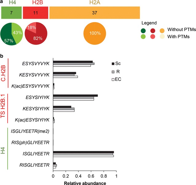
Results: In this study, we established a mass spectrometry-based analysis to address this challenge. We developed a targeted proteomics method, using selected reaction monitoring or parallel reaction monitoring, to quantify a maximum number of histone variants in a single multiplexed assay, even when histones are present in a crude extract. This strategy was developed on H2A and H2B variants, using 55 peptides corresponding to 25 different histone sequences, among which a few differ by a single amino acid. The methodology was then applied to mouse testis extracts in which almost all histone variants are expressed. It confirmed the abundance profiles of several testis-specific histones during successive stages of spermatogenesis and the existence of predicted H2A.L.1 isoforms. This methodology was also used to explore the over-expression pattern of H2A.L.1 isoforms in a mouse model of male infertility.

Conclusions: Our results demonstrate that targeted proteomics is a powerful method to quantify highly similar histone variants and isoforms. The developed method can be easily transposed to the study of human histone variants, whose abundance can be deregulated in various diseases.

Keywords: Chromatin; Histone variants; PRM; Proteomics; SRM; Spermatogenesis; Targeted proteomics.


Figures

Fig. 1
Fig. 1
Principles of discovery and targeted proteomics approaches. Major steps of discovery and targeted mass spectrometry analyses are schematized. Discovery proteomics (top panel) characterizes the global composition of a protein sample. With the quadrupole-orbitrap technology, peptide ions within a small window of mass-to-charge (m/z) ratio are isolated in the first quadrupole (Q1) and then fragmented in a collision cell; all ion fragments are finally monitored in the orbitrap analyzer. The processing of resulting MS/MS spectra allows identifying the proteins initially present in the samples (not shown). Targeted proteomics (bottom panel) precisely quantifies a predefined set of proteins. The SRM methodology first selects peptide ions representative of the proteins of interest in the first quadrupole (Q1); they are fragmented in the second quadrupole (Q2); finally, predefined representative ion fragments (F1, F2 and F3) are recorded in the last quadrupole (Q3). The reconstitution of each peptide elution profile, named SRM trace, allows for the integration and quantification of its abundance. The PRM methodology is similar to the SRM pipeline but the last quantification step is not restricted to a predefined set of fragment ions and can consider all of them, recorded in the Orbitrap analyzer
Fig. 2
Fig. 2
Sequence similarities between H2A, H2B and H3 histone variants. The similarity between H2A, H2B, and H3 variants is displayed in orange, red, and blue, respectively. Sequences were aligned with Clustal Omega tools available on the EMBL-EBI Website [39]. The size of plotted disks is proportional to the percentage of similarity between histone variants
Fig. 3
Fig. 3
Signature peptides used to quantify H2A and H2B variants by targeted proteomics. a Strategy used to select the signature peptides and validate their compatibility with targeted proteomic analysis. The sequences of 22 H2A and 3 H2B variants were obtained from our recently published list of mouse histone variants (MS_histone_DB, [8]). In silico digestion of these sequences produced a theoretical list of peptides, which were ranked according to their computed ESPP score, predictive of their compatibility with MS analysis [41]. Fifty-five peptides were selected and further analyzed to monitor the potential presence of post-translational modifications, which could interfere with their analysis by targeted proteomics. This analysis excluded seven of them (see Table 1). Then, heavy standard peptides, 13C,15N-labeled, were synthesized and analyzed on different MS instruments (LTQ-Orbitrap Velos, QTRAP 5500) to acquire full MS/MS spectra and create spectral libraries. They were used to select up to five more intense SRM transitions for each peptide. b Selected signature peptides presented on their corresponding histone variants. They are presented as black bars and numbered according to Table 1. Histone fold domains, also called globular domains, are presented as a rectangle for each histone, surrounded by N- and C-terminal tails. H2A (orange), H2B (red), H4 (green)
Fig. 4
Fig. 4
Signature peptides are mostly devoid of post-translational modifications. a Numbers of H2A, H2B and H4 peptides used in the targeted proteomic analysis that were identified to be fully non-modified or modified to some extent by discovery LC–MS/MS analyses. b Abundance of modified versus non-modified forms of the signature peptides of H2B, TS H2B.1 and H4. Analyses were performed on spermatocytes (Sc), round spermatids (R), elongating and condensing spermatids (EC)
Fig. 5
Fig. 5
Evaluation of the linearity of SRM quantification. a The SRM quantification is linear, using an increasing quantity of each isotopically labeled signature peptide spiked in a constant amount of protein matrix (acid-extracted histones from mouse testis). Please refer to the “Methods” section for experimental details. Data were normalized as described in Ref. [49]. b Similar results were obtained when a constant quantity of isotopically labeled signature peptide was spiked in an increasing amount of protein matrix (acid-extracted histones from mouse testis)
Fig. 6
Fig. 6
SRM-based quantification of H2A and H2B variants during mouse spermatogenesis a Experimental design. Spermatogenic cell fractions were analyzed [meiotic spermatocytes (Sc); round spermatids (R); elongating and condensing spermatids (EC)]. Histones were extracted with sulfuric acid, digested by trypsin and analyzed by LC-SRM. b LC-SRM quantification of the abundance of histone variants during spermatogenesis. Data were normalized to H4 levels as described in the “Methods” section and in Additional file 3. Two independent biological replicates are presented for each protein (replicate 1 and replicate 2) and were analyzed in technical triplicates. c Heat maps representing the abundance of H2A and H2B variants during spermatogenesis
Fig. 7
Fig. 7
Quantification of the abundance of H2A.L.1 isoforms in Sly-KD mice. The relative abundance of H2A and H2B variants was quantified by PRM in round spermatids from WT and Sly-KD mice. Two independent biological replicates are presented (top and bottom panels). H2A.L.2 and Y-ChrH2A.L.2 are expected to be expressed at similar levels between Sly-KD and WT spermatids [64] and peptide P29, shared by both variants, was used to normalize the abundance of peptides specific to H2A.L and subH2B (H2B.L.1) variants. The abundance of the other histone variants was normalized to H4 (P50–52). For more details, please refer to the “Methods” section

References

    1. Luger K, Mäder AW, Richmond RK, Sargent DF, Richmond TJ. Crystal structure of the nucleosome core particle at 2.8 A resolution. Nature. 1997;389:251–260. doi: 10.1038/38444. - DOI - PubMed
    1. Venkatesh S, Workman JL. Histone exchange, chromatin structure and the regulation of transcription. Nat Rev Mol Cell Biol. 2015;16:178–189. doi: 10.1038/nrm3941. - DOI - PubMed
    1. Seeber A, Gasser SM. Chromatin organization and dynamics in double-strand break repair. Curr Opin Genet Dev. 2017;43:9–16. doi: 10.1016/j.gde.2016.10.005. - DOI - PubMed
    1. Almouzni G, Cedar H. Maintenance of epigenetic information. Cold Spring Harb Perspect Biol. 2016;8:a019372. doi: 10.1101/cshperspect.a019372. - DOI - PMC - PubMed
    1. Böhmdorfer G, Wierzbicki AT. Control of chromatin structure by long noncoding RNA. Trends Cell Biol. 2015;25:623–632. doi: 10.1016/j.tcb.2015.07.002. - DOI - PMC - PubMed

Publication types

LinkOut - more resources