Review
. 2019 Mar:196:80-89.
doi: 10.1016/j.biomaterials.2017.12.025. Epub 2018 Jan 2.

Mesenchymal stem cell-macrophage crosstalk and bone healing

Affiliations
Review

Mesenchymal stem cell-macrophage crosstalk and bone healing

Jukka Pajarinen et al. Biomaterials. 2019 Mar.

Abstract

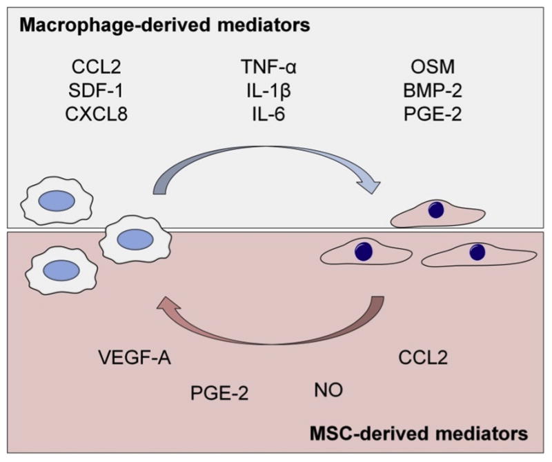
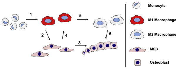
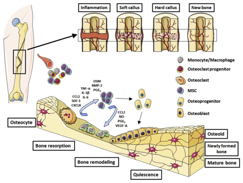
Recent research has brought about a clear understanding that successful fracture healing is based on carefully coordinated cross-talk between inflammatory and bone forming cells. In particular, the key role that macrophages play in the recruitment and regulation of the differentiation of mesenchymal stem cells (MSCs) during bone regeneration has been brought to focus. Indeed, animal studies have comprehensively demonstrated that fractures do not heal without the direct involvement of macrophages. Yet the exact mechanisms by which macrophages contribute to bone regeneration remain to be elucidated. Macrophage-derived paracrine signaling molecules such as Oncostatin M, Prostaglandin E2 (PGE2), and Bone Morphogenetic Protein-2 (BMP2) have been shown to play critical roles; however the relative importance of inflammatory (M1) and tissue regenerative (M2) macrophages in guiding MSC differentiation along the osteogenic pathway remains poorly understood. In this review, we summarize the current understanding of the interaction of macrophages and MSCs during bone regeneration, with the emphasis on the role of macrophages in regulating bone formation. The potential implications of aging to this cellular cross-talk are reviewed. Emerging treatment options to improve facture healing by utilizing or targeting MSC-macrophage crosstalk are also discussed.

Keywords: Bone Morphogenetic Protein-2; Fracture healing; Macrophage; Mesenchymal stem cell; Oncostatin M; Prostaglandin E2.


Figures

Fig. 1
Fig. 1. Contribution of macrophages and MSCs to first three stages of fracture healing
Contribution of monocyte-macrophages shown in blue and MSCs-osteoblasts in red background. Circulating monocytes arrive to the fracture site immediately following the injury. Monocyte-derived macrophages contribute to the clearance of the fracture site and amplification of the inflammatory reaction by secreting pro-inflammatory mediators and chemokines. As the fracture site is cleared of cell and extracellular matrix debris, the inflammation subsidizes. At the same time MSCs and other progenitor cells migrate to the area from periosteum, bone marrow and the circulation. MSCs proliferate and form cell-rich granulation tissue that ultimately differentiates into cartilage callus and woven bone. Macrophages are present throughout the fracture repair likely contributing to MSC differentiation by secreting growth factors but their numbers decrease as the repair progresses. The role of macrophages and MSCs in the final, remodeling phase, of the fracture healing is poorly understood but is likely dominated by signals derived from other monocyte-lineage cells, the osteoclasts.
Fig. 2
Fig. 2. Paracrine signaling molecules involved in the cross-talk between macrophages and MSCs
Macrophages regulate the recruitment and differentiation of the MSCs. On the upper part of the image are shown the best known macrophage derived chemokines, pro-inflammatory cytokines, and osteoinductive factors involved in the regulation of MSC functions. MSCs reciprocally regulate macrophage recruitment and function, generally having PGE2 and iNOS mediated immunosuppressive impact on macrophages.
Fig. 3
Fig. 3. Interactions of macrophages and MSCs during fracture healing
(1) Local damage associated molecular patterns (DAMPs) and other danger signals released from necrotic cells and damaged extracellular matrix as well as complement and coagulation system components activate recruited monocytes into inflammatory M1 macrophages. (2) Inflammatory macrophages secrete chemokines that recruit MSCs from periosteum, bone marrow, and circulation. Once recruited to the fracture site MSCs are exposed to macrophage derived inflammatory cytokines and osteoinductive factors. (3) Together with other microenviromental signals such as oxygen tension and mechanical cues these cytokines and growth factors guide the MSC differentiation along osteogenic and chondrogenic pathways. (4) MSCs reciprocally modulate the function of macrophages, possibly contributing to further monocyte/macrophage recruitment, induction of osteoinductive molecules, and eventual suppression of the inflammatory reaction. (5) As the fracture site is cleared of tissue debris and other signals maintaining the inflammatory macrophage phenotype, macrophages change their phenotype to tissue regenerative M2. (6) These tissue regenerative macrophages possibly contribute to bone formation by MSCs and osteoblasts via osteoinductive signaling molecules that might be different from M1-derived osteoinductive signals.
Fig. 4
Fig. 4. Therapeutic potential of utilizing macrophage-MSC crosstalk for bone regeneration
(a) The beneficial impact of macrophages on MSC-mediated bone formation can potentially be enhanced by increasing the number of macrophages at the fracture site, either delivering monocyte/macrophages directly to the fracture site or by introducing macrophage growth factors. (b) Macrophage polarization can be modulated towards an anti-inflammatory and tissue regenerative M2 phenotype either pharmacologically or by utilizing bioactive materials. This approach is likely particularly beneficial in cases of excessive or chronic M1-mediated inflammation, that is detrimental to bone healing. (c) An attractive strategy to optimize bone heling appears to be facilitating a short period (several days) of inflammation prior to modulation of macrophages to an anti-inflammatory phenotype. This approach might optimally utilize the bone regenerative properties of both M1 and M2 macrophages in a succession that mimics the physiological fracture healing.
Fig. 5
Fig. 5. Summary of the MSC-macrophage interactions during bone regeneration
Shown the key cells and signaling molecules involved to the cross-talk between inflammatory and bone forming cells.

References

    1. C. Control. National Hospital Ambulatory Medical Care Survey: 2013 Emergency Department Summary Tables. 2013 Available from: https://www.cdc.gov/nchs/data/ahcd/nhamcs_emergency/2013_ed_web_tables.pdf.
    1. Miranda M, Moon M. Treatment strategy for nonunions and malunions. In: Stannard J, Schmidt P, editors. Surgical Treatment of Orthopaedic Trauma. Thieme; New York: 2007. pp. 77–100.
    1. Hak DJ, Fitzpatrick D, Bishop JA, Marsh JL, Tilp S, Schnettler R, Simpson H, Alt V. Delayed union and nonunions: epidemiology, clinical issues, and financial aspects. Injury. 2014 Jun;45(Suppl 2):S3–S7. - PubMed
    1. Thompson Z, Miclau T, Hu D, Helms JA. A model for intramembranous ossification during fracture healing. J Orthop Res. 2002 Sep;20(5):1091–1098. - PubMed
    1. Uhthoff HK, Rahn BA. Healing patterns of metaphyseal fractures. Clin Orthop Relat Res. 1981 Oct;(160):295–303. - PubMed

Publication types

LinkOut - more resources