. 2018 Apr:517:108-121.
doi: 10.1016/j.virol.2017.12.027. Epub 2018 Jan 10.

Feline coronavirus: Insights into viral pathogenesis based on the spike protein structure and function

Affiliations

Feline coronavirus: Insights into viral pathogenesis based on the spike protein structure and function

Javier A Jaimes et al. Virology. 2018 Apr.

Abstract

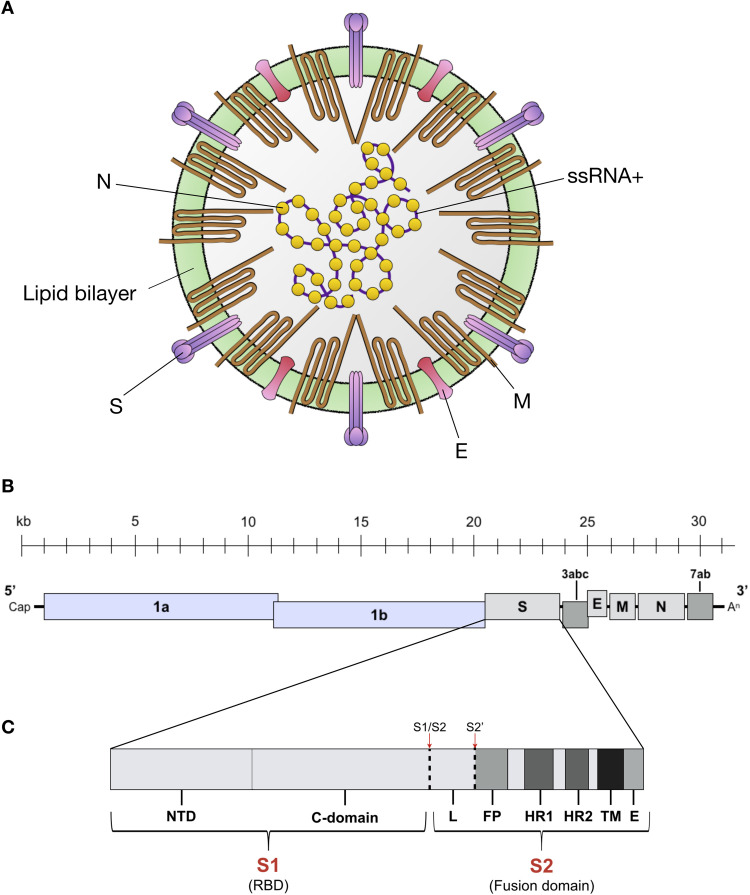
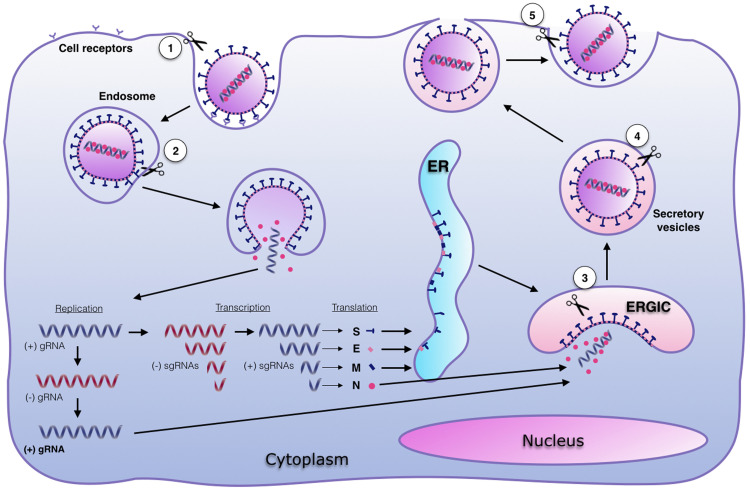
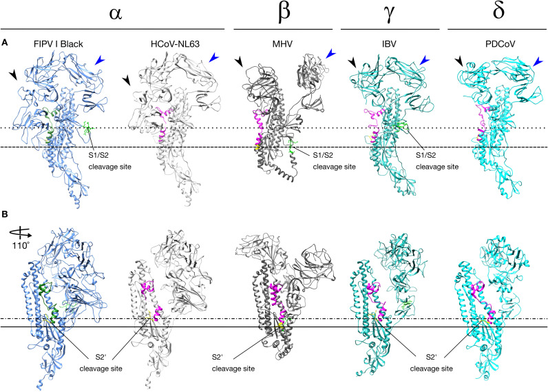
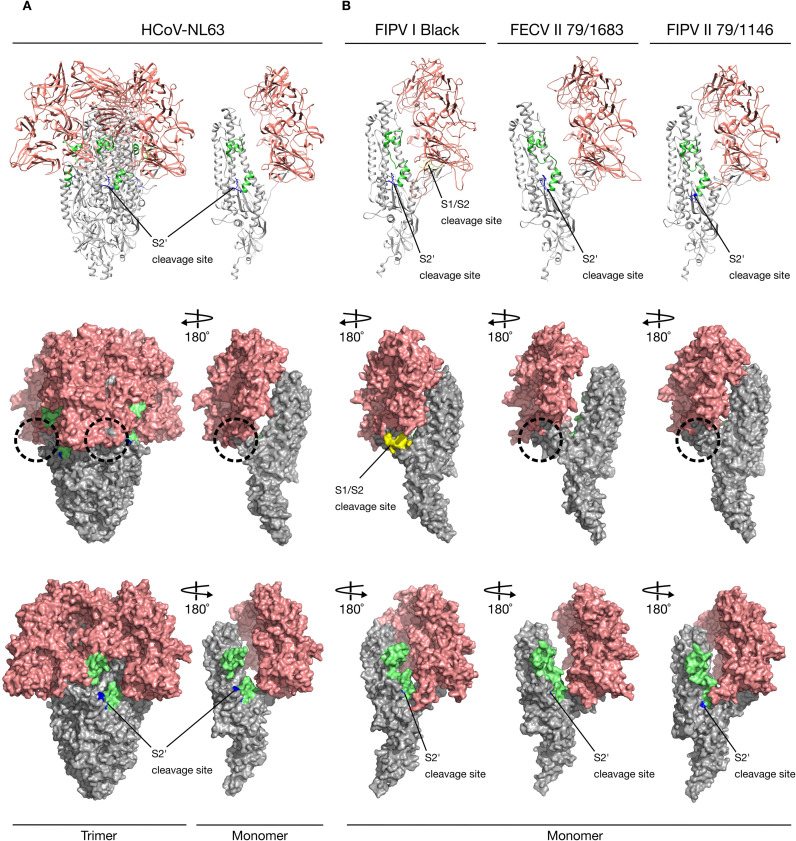
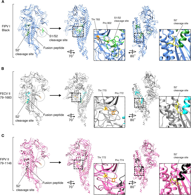
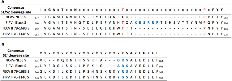
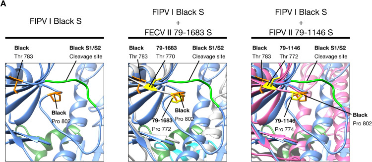
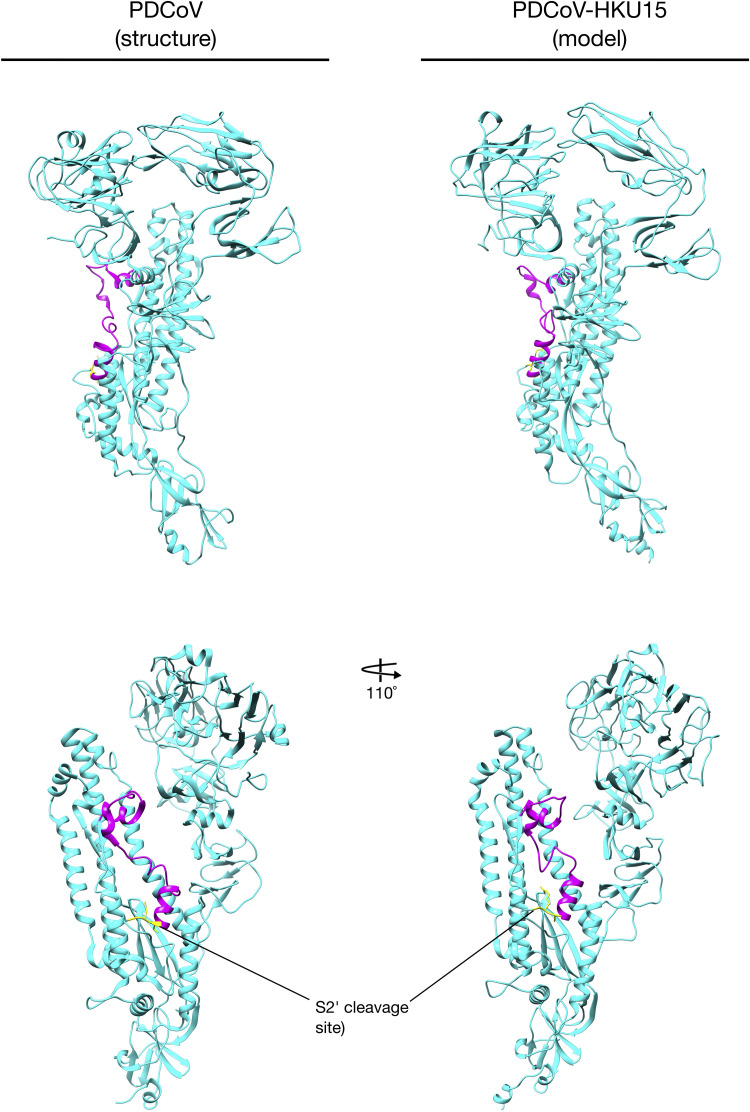
Feline coronavirus (FCoV) is an etiological agent that causes a benign enteric illness and the fatal systemic disease feline infectious peritonitis (FIP). The FCoV spike (S) protein is considered the viral regulator for binding and entry to the cell. This protein is also involved in FCoV tropism and virulence, as well as in the switch from enteric disease to FIP. This regulation is carried out by spike's major functions: receptor binding and virus-cell membrane fusion. In this review, we address important aspects in FCoV genetics, replication and pathogenesis, focusing on the role of S. To better understand this, FCoV S protein models were constructed, based on the human coronavirus NL63 (HCoV-NL63) S structure. We describe the specific structural characteristics of the FCoV S, in comparison with other coronavirus spikes. We also revise the biochemical events needed for FCoV S activation and its relation to the structural features of the protein.

Keywords: Cleavage activation; Coronavirus; Feline coronavirus; Feline infectious peritonitis; Pathogenesis; Serotype; Spike protein; Spike structure; Tropism.


Figures

Fig. 1
Fig. 1
FCoV structure and genome. (A) FCoV structure and proteins. Spike (S), matrix (M), envelope (E) and nucleocapsid (N). Adapted from: Kipar and Meli, 2013. (B) FCoV ssRNA+ is about ~ 29 kilobases (kb) long and has 11 ORF encoding 7 non-structural proteins (Replicase proteins 1a and 1b, and accessory proteins 3a, 3b, 3c, 7a and 7b); and 4 structural proteins (S, M, E, and N). Adapted from: Masters and Pearlman, 2013. (C) S gene diagram. FCoV S is composed by two subunits: S1 (receptor binding domain – RBD) and S2 (fusion domain). The S1 subunit is divide in two functional domains: N-terminal domain (NTD) and C-terminal domain (C-domain or CTD). The S2 subunit is composed by the fusion peptide (FP), two heptad repeats (HR1 and HR2), a transmembrane domain (TM) and an endodomain (E). The two S activation sites S1/S2 and S2’ are indicated (red arrows and dashed lines), as well as the linker (L) region between S1/S2 and S2’. Adapted from: Millet and Whittaker (2015).
Fig. 2
Fig. 2
FCoV replication cycle and sites for S activation. Replication cycle starts with viral binding to the cellular receptor. The virus is endocyted and viral-cell fusion allows the delivery of the (+) genomic RNA (gRNA) to the cytoplasm, to initiate the genome replication and the protein synthesis. Sub-genomic RNAs (sgRNAs) are transcribed and translated. Structural proteins S, E and M are folded and post-translationally modified at the ER. Viral assembly takes place in the ERGIC and viruses are released through exocytosis. Activation of S proteins (scissors) can take place at: 1. The cell membrane during viral attachment (S1/S2 site); 2. The endosome to induce viral-cell fusion (S2’ site); 3. The trans-Golgi network during viral assembly (S1/S2 site); 4. The secretory vesicle during viral egress (S1/S2 site); and 5. The cell membrane during viral release (S1/S2 site). Adapted from: Millet and Whittaker (2015).
Fig. 3
Fig. 3
Alphacoronavirus (α), Betacoronavirus (β), Gammacoronavirus (γ) and Deltacoronavirus (δ) spike protein. Comparison between S protein monomer structures from HCoV-NL63 (RCSB PDB # 5SZS), MHV (RCSB PDB # 3JCL) and PDCoV (RCSV PDB # 6B7N), and the S protein monomer models from FIPV I Black and IBV strain Beaudette. The NTD (black arrow heads) and C-domain (blue arrow heads) are signaled. (A) S1/S2 (bright green) cleavage site is present in FCoV, MHV and IBV. FCoV and IBV S1/S2 cleavage site are located at the same relative position (dotted line), while MHV S1/S2 cleavage site is located lower compared with FCoV and IBV dashed line). (B) S2’ (yellow) cleavage site is present in all CoVs spike. All CoV S2’ cleavage sites are located at the same relative position (dashed and dotted line). MHV S2’ is located lower compared with other CoV (solid line). The fusion peptide located at the same relative position in all spikes (FCoV = green, other CoV = magenta).
Fig. 4
Fig. 4
Alphacoronavirus S proteins. Ribbon and surface images of the structure (HCoV-NL63) and models (FCoV) of S proteins and their cleavage sites. S1 domain (salmon), S2 domain (gray), fusion peptide (lime green). (A) HCoV-NL63 S protein structures (RCSB PDB # 5SZS) with its unique S2’ cleavage site (blue) and the missing S1/S2 cleavage site expected location (dashed circle). (B) FIPV I Black, FECV II 78/1683 and FIPV II 79-1146S proteins models based on HCoV-NL63 structure. Notice that only FIPV I Black possesses two cleavage sites: S1/S2 (yellow) and S2’ (blue) while FCoV II viruses possess the S2’ cleavage site (blue) but no S1/S2 (dashed circles).
Fig. 5
Fig. 5
FCoV I and II spike protein. Comparison between FCoV I and II S protein monomer models. (A) FIPV I Black S protein model and cleavage sites. S1/S2 cleavage site is protruding from the protein forming a 19 amino acids loop (dotted line circle). Location and magnification of FIPV I Black S1/S2 cleavage site (bright green) and flanking amino acids (orange): Thr 783 and Pro 802. Location and magnification of FIPV I Black S2’ cleavage site (yellow). (B) FECV II 79-1683S protein model and cleavage sites. Notice the absence of the S1/S2 cleavage site and loop (dotted line circle). Expected location of FECV II 79–1683 S1/S2 loop and flanking amino acids (yellow): Thr 770 and Pro 772. Location and magnification of FECV II 79–1683 S2’ cleavage site (yellow). (C) FIPV II 79-1146S protein model and cleavage sites. Notice the absence of the S1/S2 cleavage site and loop (dotted line circle). Expected location of FIPV II 79-1146 S1/S2 loop and flanking amino acids (yellow): Thr 772 and Pro 774. Location and magnification of FIPV II 79-1146 S2’ cleavage site (yellow).
Fig. 6
Fig. 6
HCoV-NL63, FCoV I and II S1/S2 and S2’ amino acid sequence alignment. Amino acid alignment of HCoV-NL63, FIPV I Black, FECV II 1683 and FIPV II 1146 S1/S2 and S2’ region. (A) A 16 amino acids gap corresponding to the S1/S2 cleavage site and loop is missing in HCoV-NL63 and in both FCoV II S sequences. FIPV I Black S1/S2 cleavage site is highlighted in blue. Corresponding S1/S2 flanking amino acids (Thr and Pro) for all FCoVs are highlighted in red. (B) A three amino acids gap immediately before the S2’ cleavage site is observed in HCoV-NL63 and FIPV I Black S. The S2’ cleavage sites are highlighted in blue.
Fig. 7
Fig. 7
FCoV S1/S2 cleavage site. (A) FIPV I Black S1/S2 cleavage site (bright green) and flanking amino acids (orange): Thr 783 and Pro 802. (B) Superposition of FECV II 79-1683 (light gray) and FIPV I Black (light blue) S1/S2 regions. FECV II 79-1683 amino acids Thr 770 and Pro 772 (yellow), are located in the same position than FIPV I Black S1/S2 flanking amino acids (orange). (C) Superposition of FIPV II 79-1146 (pink) and FIPV I Black (light blue) S1/S2 regions. FIPV II 79-1146 amino acids Thr 772 and Pro 774 (yellow), are located in the same position than FIPV I Black S1/S2 flanking amino acids (orange). Notice that in both FCoV II S proteins the complete S1/S2 loop (including the S1/S2 cleavage site) is missing.
Fig. S1
Fig. S1
PDCoV spike structure and predicted model. PDCoV spike structure (RCSV PDB # 6B7N) and predicted PDCoV-HKU15 strain MI6148 spike model (GenBank # AIA99518.1). Supplementary material 1: HCoV-NL63 S and FIPV I Black S amino acid alignment. HCoV-NL63 (RCSV PDB # 5SZS) and FIPV I Black S (Susan Baker, Loyola University Chicago, personal communication). Supplementary material 2: HCoV-NL63 S and FECV II 79-1683 S amino acid alignment. HCoV-NL63 S (RCSV PDB # 5SZS) and FECV II 79-1683 S (GenBank # AFH58021.1). Supplementary material 3: HCoV-NL63 S and FIPV II 79-1146 S amino acid alignment. HCoV-NL63 S (RCSV PDB # 5SZS) and FIPV II 79-1146 S (GenBank # AAY32596.1). Supplementary material 4: HCoV-NL63 S and IBV strain Beaudette S amino acid alignment. HCoV-NL63 S (RCSV PDB # 5SZS) and IBV strain Beaudette S (GenBank # AJ311362.1).

References

    1. An D.J., Jeoung H.Y., Jeong W., Park J.Y., Lee M.H., Park B.K. Prevalence of Korean cats with natural feline coronavirus infections. Virol. J. 2011;8:455. - PMC - PubMed
    1. Armstrong J., Niemann H., Smeekens S., Rottier P., Warren G. Sequence and topology of a model intracellular membrane protein, E1 glycoprotein, from a coronavirus. Nature. 1984;308:751–752. - PMC - PubMed
    1. Balint A., Farsang A., Zadori Z., Hornyak A., Dencso L., Almazan F., Enjuanes L., Belak S. Molecular characterization of feline infectious peritonitis virus strain DF-2 and studies of the role of ORF3abc in viral cell tropism. J. Virol. 2012;86:6258–6267. - PMC - PubMed
    1. Barcena M., Oostergetel G.T., Bartelink W., Faas F.G., Verkleij A., Rottier P.J., Koster A.J., Bosch B.J. Cryo-electron tomography of mouse hepatitis virus: insights into the structure of the coronavirion. Proc. Natl. Acad. Sci. USA. 2009;106:582–587. - PMC - PubMed
    1. Barker E.N., Stranieri A., Helps C.R., Porter E.L., Davidson A.D., Day M.J., Knowles T., Kipar A., Tasker S. Limitations of using feline coronavirus spike protein gene mutations to diagnose feline infectious peritonitis. Vet. Res. 2017;48:60. - PMC - PubMed

Publication types

MeSH terms

Substances

LinkOut - more resources