. 2018 Mar 2;430(5):695-709.
doi: 10.1016/j.jmb.2018.01.002. Epub 2018 Jan 10.

Conformation and Trimer Association of the Transmembrane Domain of the Parainfluenza Virus Fusion Protein in Lipid Bilayers from Solid-State NMR: Insights into the Sequence Determinants of Trimer Structure and Fusion Activity

Affiliations

Conformation and Trimer Association of the Transmembrane Domain of the Parainfluenza Virus Fusion Protein in Lipid Bilayers from Solid-State NMR: Insights into the Sequence Determinants of Trimer Structure and Fusion Activity

Myungwoon Lee et al. J Mol Biol. .

Abstract

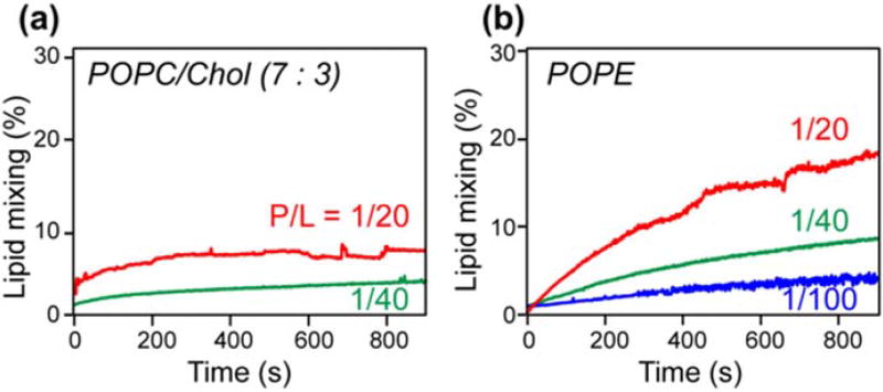
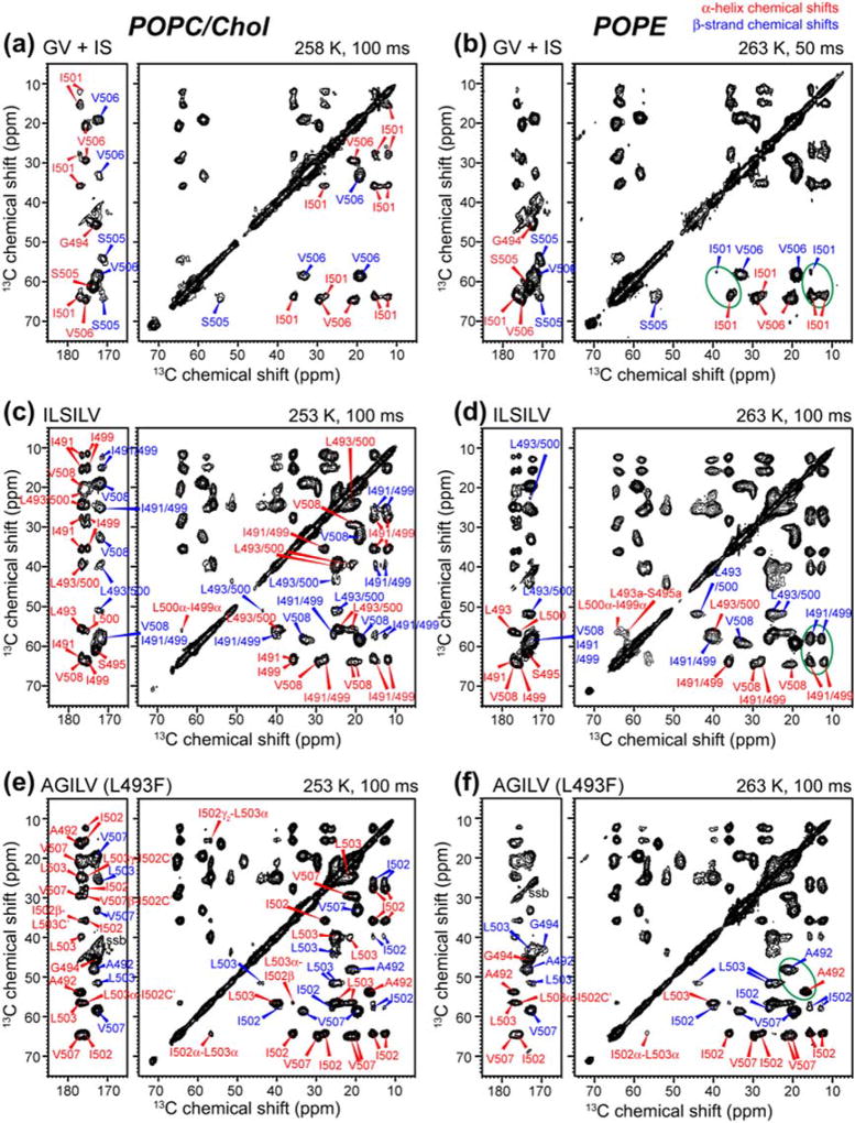
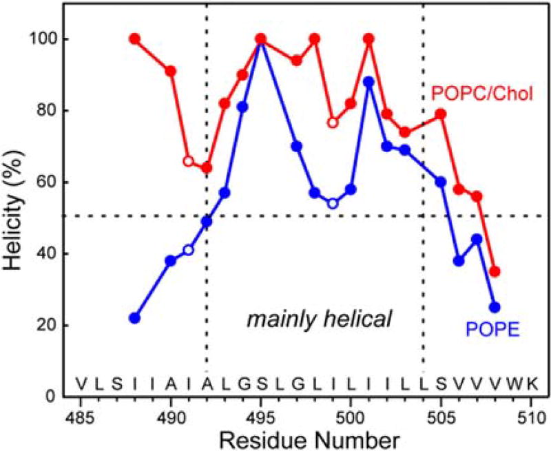
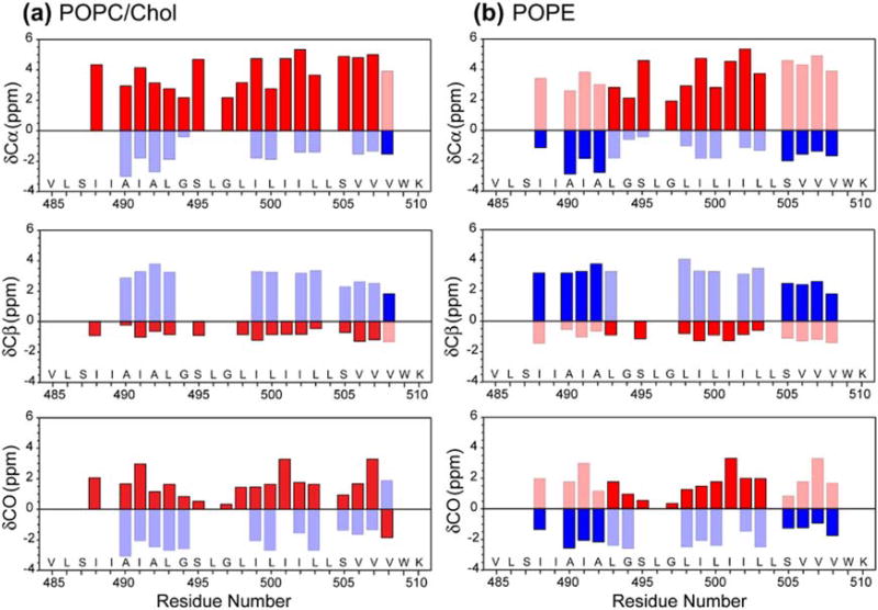
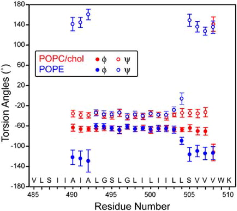
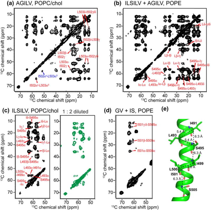
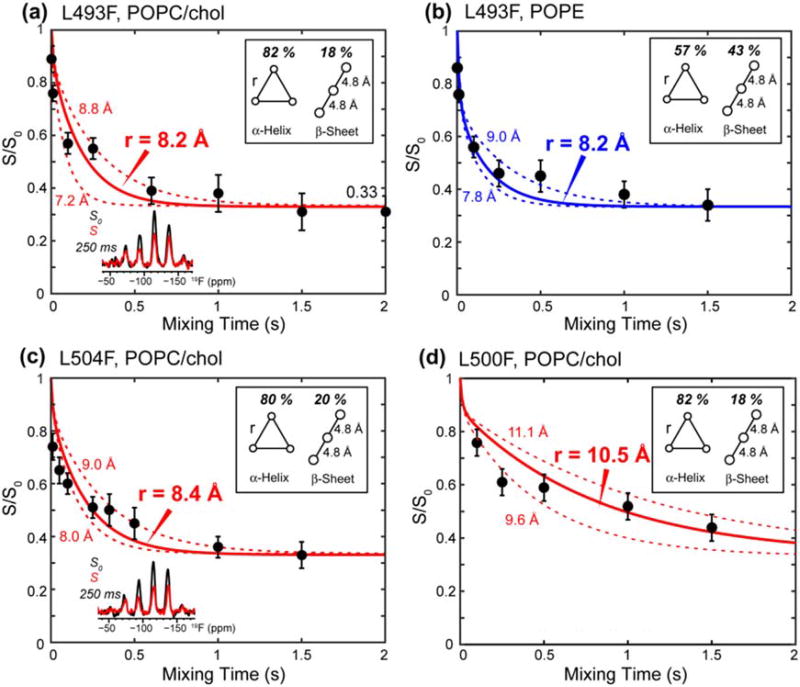
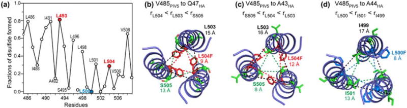
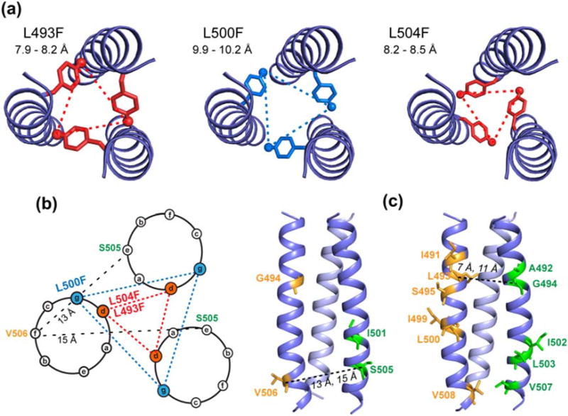
Enveloped viruses enter cells by using their fusion proteins to merge the virus lipid envelope and the cell membrane. While crystal structures of the water-soluble ectodomains of many viral fusion proteins have been determined, the structure and assembly of the C-terminal transmembrane domain (TMD) remains poorly understood. Here we use solid-state NMR to determine the backbone conformation and oligomeric structure of the TMD of the parainfluenza virus 5 fusion protein. 13C chemical shifts indicate that the central leucine-rich segment of the TMD is α-helical in POPC/cholesterol membranes and POPE membranes, while the Ile- and Val-rich termini shift to the β-strand conformation in the POPE membrane. Importantly, lipid mixing assays indicate that the TMD is more fusogenic in the POPE membrane than in the POPC/cholesterol membrane, indicating that the β-strand conformation is important for fusion by inducing membrane curvature. Incorporation of para-fluorinated Phe at three positions of the α-helical core allowed us to measure interhelical distances using 19F spin diffusion NMR. The data indicate that, at peptide:lipid molar ratios of ~1:15, the TMD forms a trimeric helical bundle with inter-helical distances of 8.2-8.4Å for L493F and L504F and 10.5Å for L500F. These data provide high-resolution evidence of trimer formation of a viral fusion protein TMD in phospholipid bilayers, and indicate that the parainfluenza virus 5 fusion protein TMD harbors two functions: the central α-helical core is the trimerization unit of the protein, while the two termini are responsible for inducing membrane curvature by transitioning to a β-sheet conformation.

Keywords: conformational plasticity; magic-angle-spinning NMR; spin diffusion; trimer formation.


Figures

Fig. 1
Fig. 1
PIV5 TMD induces mixing of POPC/cholesterol vesicles (a) and POPE vesicles (b). The peptide causes stronger lipid mixing in the POPE membrane than in the POPC/cholesterol membrane. Moreover, the extent of lipid mixing increases with the peptide/lipid molar ratio.
Fig. 2
Fig. 2
2D 13C–13C DARR correlation spectra of the TMD in the POPC/cholesterol (left) and POPE (right) membranes. (a, b) Mixed GV and IS-labeled TMD. (c, d) ILSILV-labeled TMD. (e, f) AGILV-labeled TMD. Red and blue assignments denote α-helical and β-strand chemical shifts. The peptide shows a mixture of α-helix and β-strand chemical shifts in the membrane.
Fig. 3
Fig. 3
Residue-specific α-helicity of the TMD in POPC/cholesterol and POPE membranes, obtained from the relative intensities of the cross peaks in 2D 13C–13C correlation spectra. The peptide has much lower helicity at the N- and C-termini than at the central segment in the POPE membrane. In the POPC/cholesterol membrane, the peptide is mostly helical except for the C-terminal end.
Fig. 4
Fig. 4
13C secondary chemical shifts of the TMD in the POPC/cholesterol (a) and POPE membrane (b). α-Helical secondary shifts are shown in red, while β-strand secondary shifts are shown in blue. Shaded bars denote the minor conformation. The TMD exhibits mainly α-helical structure in the POPC/cholesterol membrane but predominant β-strand conformation at the N- and C-terminal regions in the POPE membrane.
Fig. 5
Fig. 5
Backbone (φ, ψ) torsion angles of the TMD in the POPC/cholesterol (red) and POPE (blue) membrane, predicted by TALOS-N from the measured 13C and 15N chemical shifts. Only the torsion angles of the major conformer are shown.
Fig. 6
Fig. 6
2D 13C–13C PDSD spectra of the TMD with long mixing times of 0.5 and 1.0 s, measured at 253–263 K. Only inter-residue cross peaks are assigned. (a) AGILV (L493F)-labeled TMD in the POPC/cholesterol membrane. (b) 1:1 mixture of AGILV (L493F) and ILSILV-labeled TMD in the POPE membrane. All inter-residue cross peaks are intramolecular ones. (c) Undiluted and 1:2 diluted (green) ILSILV-labeled TMD in the POPC/cholesterol membrane. The same inter-residue cross peaks were observed, indicating that all cross peaks are intramolecular. (d) Mixed GV and IS-labeled TMD in the POPE membrane, showing I501–S505 cross peaks. (e) Intramolecular Cα–Cα distances of the TMD generated using the measured (φ, ψ) torsion angles.
Fig. 7
Fig. 7
19F CODEX data of membrane-bound TMD. (a) L493F-TMD in the POPC/cholesterol membrane. Representative S0 and S spectra are shown. (b) L493F-TMD in the POPE membrane. (c) L504F-TMD in the POPC/cholesterol membrane. (d) L500F-TMD in the POPC/cholesterol membrane. The data were acquired at 230 K under 8 kHz MAS. The percentages of α-helix and β-strand are taken from Table S1, and intermolecular 19F–19F distances in the β-sheet are fixed to 4.8 Å in the simulation. The CODEX intensities of L493F and L504F equilibrate to 0.33, indicating that the TMD is trimerized. The L500F CODEX decay is slower, indicating longer interhelical distances.
Fig. 8
Fig. 8
Interhelical 19F–19F distances and disulfide crosslinking data rule out several trimer structural models. (a) Fractions of disulfide bond formation reproduced from Ref. [21]. The three Leu residues that were replaced by 4-19F-Phe in this study are colored. (b) Proposed trimer structure model, obtained by aligning PIV5 V485 with hemagglutinin Q47. The interhelical Cα–Cα distances for L504 are shorter than the distances for the neighboring L503 and S505, consistent with the crosslinking data. (c) Alternative trimer model obtained by aligning PIV5 V485 with hemagglutinin A43. The interhelical Cα–Cα distance of S505 is shorter than that of L504, which is inconsistent with the disulfide crosslinking data. (d) Alternative trimer model obtained by aligning V485 of PIV5 with A44 of hemagglutinin. L500 gives shorter interhelical Cα–Cα distances than the neighboring I499 and I501, which is inconsistent with the crosslinking data.
Fig. 9
Fig. 9
Proposed trimer structure model of the PIV5 TMD. (a) Top view of the trimer structure, showing L493F, L500F, and L504F interhelical 19F–19F distances. These agree with the experimentally measured 19F–19F distances. (b) Helical wheel diagram for the trimeric TMD, with L493F and L504F at the d position of the heptad repeat, forming close interhelical contacts, while L500F lies at the g position, giving longer interhelical distances. (c) Side views of the trimer structure, showing the locations of 13C, 15N-labeled residues in the two mixed labeled samples (Table 1). Representative interhelical distances between S505 and V506, and between G494 and L495, are much longer than can be measured using 13C spin diffusion NMR. This is further illustrated in the helical wheel diagram for the S505–V506 pair.

Similar articles

Cited by

References

    1. Lamb RA, Jardetzky TS. Structural basis of viral invasion: lessons from paramyxovirus F. Curr. Opin. Struct. Biol. 2007:427–436. - PMC - PubMed
    1. Eckert DM, Kim PS. Mechanisms of viral membrane fusion and its inhibition. Annu. Rev. Biochem. 2001;70:777–810. - PubMed
    1. Kim YH, Donald JE, Grigoryan G, Leser GP, Fadeev A, Lamb RA, et al. Capture and imaging of prehairpin fusion intermediate of the paramyxovirus PIV5. Proc. Natl. Acad. Sci. U. S. A. 2011;108:20992–20997. - PMC - PubMed
    1. Yin HS, Wen XL, Paterson RG, Lamb RA, Jardetzky TS. Structure of the parainfluenza virus 5F protein in its metastable, prefusion conformation. Nature. 2006;439:38–44. - PMC - PubMed
    1. White JM, Delos SE, Brecher M, Schornberg K. Structures and mechanisms of viral membrane fusion proteins: multiple variations on a common theme. Crit. Rev. Biochem. Mol. Biol. 2008;43:287–288. - PMC - PubMed

Publication types

MeSH terms

LinkOut - more resources