. 2018 Mar 15;78(6):1484-1496.
doi: 10.1158/0008-5472.CAN-17-1518. Epub 2018 Jan 12.

Kindlin-1 Promotes Pulmonary Breast Cancer Metastasis

Affiliations

Kindlin-1 Promotes Pulmonary Breast Cancer Metastasis

Sana Sarvi et al. Cancer Res. .

Abstract

In breast cancer, increased expression of the cytoskeletal adaptor protein Kindlin-1 has been linked to increased risks of lung metastasis, but the functional basis is unknown. Here, we show that in a mouse model of polyomavirus middle T antigen-induced mammary tumorigenesis, loss of Kindlin-1 reduced early pulmonary arrest and later development of lung metastasis. This phenotype relied on the ability of Kindlin-1 to bind and activate β integrin heterodimers. Kindlin-1 loss reduced α4 integrin-mediated adhesion of mammary tumor cells to the adhesion molecule VCAM-1 on endothelial cells. Treating mice with an anti-VCAM-1 blocking antibody prevented early pulmonary arrest. Kindlin-1 loss also resulted in reduced secretion of several factors linked to metastatic spread, including the lung metastasis regulator tenascin-C, showing that Kindlin-1 regulated metastatic dissemination by an additional mechanism in the tumor microenvironment. Overall, our results show that Kindlin-1 contributes functionally to early pulmonary metastasis of breast cancer.Significance: These findings provide a mechanistic proof in mice that Kindin-1, an integrin-binding adaptor protein, is a critical mediator of early lung metastasis of breast cancer. Cancer Res; 78(6); 1484-96. ©2018 AACR.


Conflict of interest statement

The authors declare no conflicts of interest.

Figures
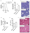
Figure 1
Figure 1. Kindlin-1 loss delays tumor onset but not growth of established tumors.
(A) Kaplan–Meier analysis of tumor onset in MT-Kin-1fl/fl (n = 16) and MT-Kin-1wt/wt (n = 10) mice (P < 0.0001, log-rank test). (B) Quantification of area covered by focal lesions in mammary glands from MT-Kin-1fl/fl and MT-Kin-1wt/wt mice. Values are means ± SEM (n = 4 mice; **P < 0.01). (C) Representative H&E staining of fat pads from 28-day-old MT-Kin-1fl/fl and MT-Kin-1wt/wt mice. Scale bar, 50 μm. (D) Box-and-whisker plots of tumor doubling times in MT-Kin-1fl/fl and MT-Kin-1wt/wt mice (n = 6 mice; non-significant, P = 0.1797). (E) Quantification of percentage of Ki67-positive (Ki67+) cells within size-matched tumors from MT-Kin-1fl/fl and MT-Kin-1wt/wt mice. Values are means ± SEM (n = 4 mice; non-significant, P = 0.0812). (F) Representative H&E staining of tumors from MT-Kin-1fl/fl and MT-Kin-1wt/wt mice. Scale bar, 50 μm.
Figure 2
Figure 2. Kindlin-1 loss reduces pulmonary metastasis.
(A) Incidence of metastatic lesions in the lungs of MT-Kin-1fl/fl (n = 16) or MT-Kin-1wt/wt (n = 10) mice. (B) Representative H&E staining of lung metastases in MT-Kin-1fl/fl and MT-Kin-1wt/wt mice. Scale bar, 50 μm. (C) PCR analysis of PyV MT in lungs from MT-Kin-1fl/fl or MT-Kin-1wt/wt mice normalized to the total primary tumor burden in each mouse. Values are means ± SEM (n = 7 mice; *P < 0.05, Student’s t-test). (D) Box-and-whisker plots of lung weights collected 8 weeks after injection of dissociated tumors from MT-Kin-1fl/fl or MT-Kin-1wt/wt mice (n = 20 mice; **P < 0.01, Student’s t-test). (E) Box-and-whisker plots of total area covered by metastatic lesions in lungs after injection of dissociated tumors from MT-Kin-1fl/fl or MT-Kin-1wt/wt mice (n = 20 mice; **P < 0.01, Student’s t-test). (F) Quantification of number of metastatic nodules per lung. Each point represents an individual mouse (n = 17 mice; non-significant, P = 0.1329). (G) Quantification of the size of individual metastatic nodules per lung. Each point represents an individual metastatic nodule (n = 7 mice; ****P < 0.0001, Student’s t-test). (H) Quantification of percentage of Ki67-positive cells in lung metastases (n = 7 mice; non-significant, P = 0.1192).
Figure 3
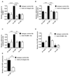
Figure 3. Kindlin-1 regulates integrin activation and metastatic colonization.
(A, B) Expression of active β1 integrin (A) and total β1 integrin (B) expression assessed by flow cytometry in mammary tumors from MT-Kin-1fl/fl and MT-Kin-1wt/wt mice. Values are means ± SEM (n = 3 mice; *P < 0.05, Student’s t-test). (C) Western blot analysis demonstrating the re-expression of His-tagged Kindlin-1-WT or Kindlin-1-AA into Kindlin-1-Null A1 cells. Actin was used as a loading control. (D) Ratio of active β1 integrin to total β1 integrin expression. Values are normalized to WT cells and are means + SEM (n = 4; **P < 0.01, ***P < 0.001, one-way ANOVA with Tukey’s post hoc correction). (E) Adhesion to fibronectin expressed as number of adherent cells relative to WT cells. Values are means ± SEM (n = 3; **P < 0.01, one-way ANOVA with Tukey’s post hoc correction). (F) PCR analysis of PyV MT in lungs 21 days post-injection of A1 Null, WT or AA cells. Values are normalized to WT cells and are means ± SEM (n = 6 mice; ****P < 0.0001, one-way ANOVA with Tukey’s post hoc correction). (G) PCR analysis of PyV MT in lungs 30 minutes post-injection of A1 Null, WT or AA cells. Values are normalized to WT cells and are means ± SEM (n = 6 mice; **P < 0.01, ***P < 0.001, one-way ANOVA with Tukey’s post hoc correction).
Figure 4
Figure 4. Kindlin-1 regulates cellular adhesion to endothelial cells and pulmonary arrest in an integrin-dependent manner.
(A) Adhesion to VCAM-1 in the presence of function-blocking anti-α4 integrin antibody or isotype IgG control expressed as number of adherent cells relative to WT. Values are means ± SEM (n = 3; ****P < 0.0001). (B) Adhesion to endothelial cells expressed as number of adherent cells relative to WT cells. Values are means ± SEM (n = 3; ***P < 0.001). (C) Adhesion to endothelial cells in the presence of function-blocking anti-VCAM-1 antibody or isotype IgG control expressed as number of adherent cells relative to WT. Values are means ± SEM (n = 3; **P < 0.01, ***P < 0.001). (D) PCR analysis of PyV MT in lungs 30 minutes post-injection of A1 Null, WT or AA cells in the presence of function-blocking anti-VCAM-1 antibody or isotype IgG control. Values are means ± SEM (n = 4 mice; **P < 0.01, two-way ANOVA with Bonferroni’s post hoc correction). (E) PCR analysis of PyV MT in lungs 30 minutes post-injection of A1 WT in the presence of function-blocking anti-α4 integrin antibody or isotype IgG control. Values are means ± SEM (n = 12 mice; **P < 0.01, Student’s t-test).
Figure 5
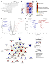
Figure 5. Kindlin-1 regulates the mammary tumor cell secretome.
(A) Gene ontology enrichment analysis of top-level cellular component terms significantly overrepresented in the proteomic dataset of the A1 Null and WT secretomes (P < 0.001, hypergeometric test with Benjamini–Hochberg post hoc correction). (B) Hierarchically clustered heatmap of median-centered, normalized protein intensities quantified by label-free MS. Significantly differentially regulated extracellular proteins are shown (FDR = 5%, Student’s t-test). Gray bars indicate reported associations with tumor metastasis; adjacent colored bars (Met. reguln) indicate reported up-regulation (red), down-regulation (blue) or both (gray) in metastasis signatures. (C) Functional enrichment analysis of the significantly differentially regulated extracellular proteins in (B). Parent or sibling overrepresented functions are grouped together and displayed as a heatmap (log10 color scale) (P < 0.05, hypergeometric test with Benjamini–Hochberg post hoc correction). (D) Volcano plot of extracellular proteins quantified in the A1 Null and WT secretomes. Black curves show the threshold for significant differential regulation (FDR = 5%, Student’s t-test). Proteins differentially regulated by more than 16-fold (P < 0.001) are labeled. (E) Topological analysis of the Kindlin-1-dependent Met-1 secretome network (see Supplementary Fig. S6). Clustering coefficient represents the capacity for groups of proteins to be connected. Proteins with a clustering coefficient greater than 0.35 are labeled. (F) Integrative network analysis of the Kindlin-1-dependent secretome directly associated with PAI-1 and tenascin-C. Protein node (circle) size is proportional to FDR-adjusted P of differential regulation (Q); node color represents log2-transformed fold change. Connecting edge (line) color represents relationship type; edge thickness is proportional to a linear regression–derived network weighting. The graph was clustered using a force-directed algorithm.
Figure 6
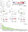
Figure 6. Kindlin-1 dependent secretome is associated with lung metastasis and is elevated in cells metastatic to lung.
(A) PCR analysis of Tnc and Serpine1 in A1 Null, WT or AA cells. Expression values are normalized to WT cells and are means ± SEM (n = 4; **P < 0.01, one-way ANOVA with Tukey’s post hoc correction). (B) MDA-MB-231 subpopulations with enhanced lung metastatic activity have elevated expression of FERMT1 and TNC. (C) Comparison of expression of FERMT1, TNC and SERPINE1 in MDA-MB-231 subpopulations predisposed or not predisposed to lung metastasis (*P = 0.05, **P = 0.008, ***P = 0.001, Wilcoxon test). (D) Comparison of expression of FERMT1, TNC and SERPINE1 in 2999 primary breast tumors split into those where FERMT1 expression is detected (***P < 0.001, Wilcoxon test). (E) Cox proportional hazards survival analysis of the Kindlin-1-dependent secretome genes represented in a primary breast cancer dataset (MSK82, GSE2603; n = 82). Patients were stratified at all possible cut-points for all secretome genes, and the proportions of cut-points significantly associated with lung (green bars) and non-lung (gray diamonds) metastasis (P < 0.05) are shown for each gene. For clarity, genes not significantly associated with lung metastasis at any cut-point (P ≥ 0.05) are not displayed. The secretome genes were significantly more associated with worse outcomes in terms of lung metastasis as compared to non-lung metastasis (****P = 3.96 × 10⁻⁵, Wilcoxon test).

References

    1. Driouch K, Bonin F, Sin S, et al. Confounding effects in "a six-gene signature predicting breast cancer lung metastasis": reply. Cancer Res. 2009;69:9507–9511. - PubMed
    1. Landemaine T, Jackson A, Bellahcene A, et al. A six-gene signature predicting breast cancer lung metastasis. Cancer Res. 2008;68:6092–6099. - PubMed
    1. Sin S, Bonin F, Petit V, et al. Role of the focal adhesion protein kindlin-1 in breast cancer growth and lung metastasis. J Natl Cancer Inst. 2011;103:1323–1337. - PubMed
    1. Rognoni E, Ruppert R, Fassler R. The kindlin family: functions, signaling properties and implications for human disease. J Cell Sci. 2016;129:17–27. - PubMed
    1. Has C, Castiglia D, del Rio M, et al. Kindler syndrome: extension of FERMT1 mutational spectrum and natural history. Hum Mutat. 2011;32:1204–1212. - PubMed

Publication types

MeSH terms