. 2018 Jan 12;3(19):eaal2736.
doi: 10.1126/sciimmunol.aal2736.

Production of BMP4 by endothelial cells is crucial for endogenous thymic regeneration

Affiliations

Production of BMP4 by endothelial cells is crucial for endogenous thymic regeneration

Tobias Wertheimer et al. Sci Immunol. .

Abstract

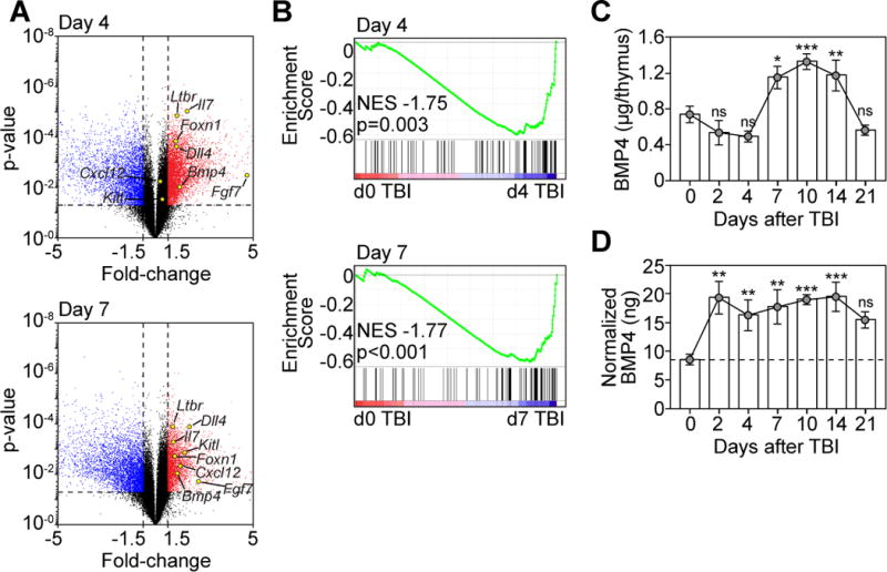
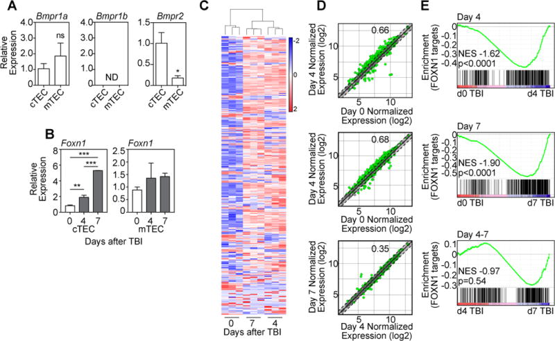
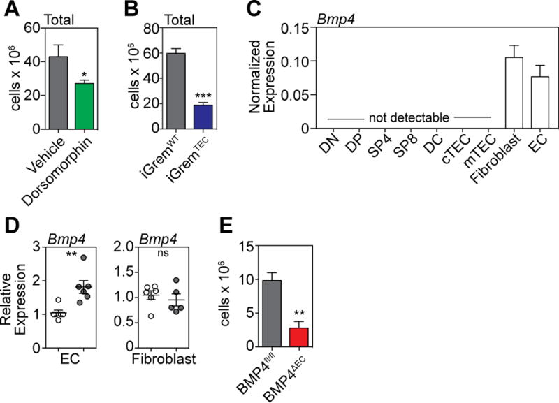
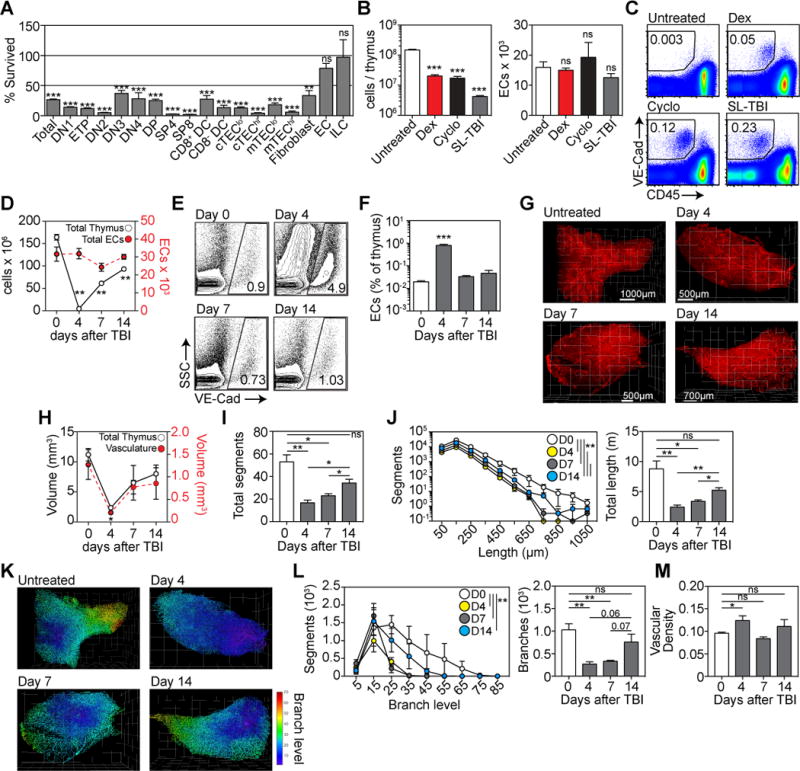
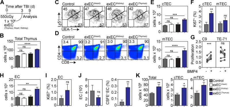
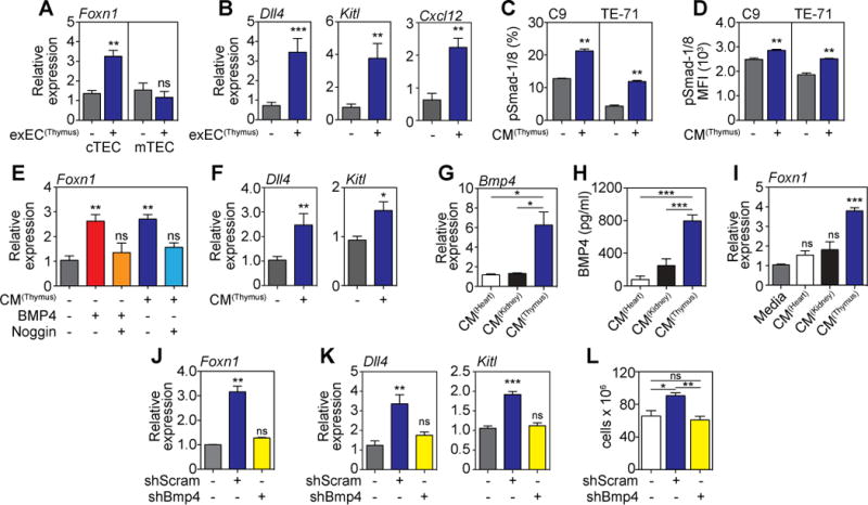
The thymus is not only extremely sensitive to damage but also has a remarkable ability to repair itself. However, the mechanisms underlying this endogenous regeneration remain poorly understood, and this capacity diminishes considerably with age. We show that thymic endothelial cells (ECs) comprise a critical pathway of regeneration via their production of bone morphogenetic protein 4 (BMP4) ECs increased their production of BMP4 after thymic damage, and abrogating BMP4 signaling or production by either pharmacologic or genetic inhibition impaired thymic repair. EC-derived BMP4 acted on thymic epithelial cells (TECs) to increase their expression of Foxn1, a key transcription factor involved in TEC development, maintenance, and regeneration, and its downstream targets such as Dll4, a key mediator of thymocyte development and regeneration. These studies demonstrate the importance of the BMP4 pathway in endogenous tissue regeneration and offer a potential clinical approach to enhance T cell immunity.


Figures

Figure 1
Figure 1. BMP signaling pathways are upregulated in the thymus after thymic damage
(A–B) Thymuses were pooled 6-week-old C57BL/6 mice and microarray analysis was performed on CD45 cells enriched from either untreated mice (d0) or 4, and 7 days after TBI (550 cGy, n=3/timepoint with each n pooled from 3–5 mice). (A) Volcano plot outlining genes that changed >1.5 fold, p<0.05 with some key thymus-related genes highlighted. (B) GSEA analysis was performed on the transcriptome derived from CD45 cells after TBI (Fig. 1A) with BMP target genes (GO: 0030510). (C–D) Thymuses were harvested at days 0, 2, 4, 7, 10, 14, and 21 after TBI (n=5–14/timepoint) and BMP4 levels were measured by ELISA. (C) Absolute amount of BMP4 in the thymus. (D) Amount of BMP4 normalized to the weight of the thymus (ng BMP4/μg thymus). Data combined from 2–3 independent experiments. *, p<0.05; **, p<0.01, ***, p<0.001.
Figure 2
Figure 2. BMP4 targets thymic epithelial cells and induces expression of Foxn1 and its downstream targets after damage
(A) TEC subsets were FACS purified from untreated 6 week-old C57BL/6 mice and expression of the BMPR subunits Bmpr1a, Bmpr1b, and Bmpr2 were measured by qPCR (n=3/subset). (B) cTECs or mTECs were FACS purified from the thymus at days 0, 4 and 7 after TBI and expression of Foxn1 was assessed by qPCR (n=4–6/timepoint from 2–3 independent experiments). (C–E) Thymuses were pooled from 6 week-old C57BL/6 mice and microarray analysis was performed on CD45 cells enriched from either untreated mice (d0) or 4, and 7 days after TBI (550 cGy, n=3/timepoint with each n pooled from 3–5 mice). (C) Heat map of high confidence FOXN1 target genes in cTECs (21) showing clustering between samples and between days 0, 4 and 7. (D) Dot plots showing normalized expression values comparing days 0, 4 and 7 after TBI. Green dots represent individual genes significantly different (p<0.05), and dotted lines (and shaded section) represent genes with a fold change <1.5 (+/−). The proportion of significantly changed genes is denoted within each plot. (E) GSEA analysis was performed comparing gene changes with a FOXN1 target gene signature comprising the list of high confidence FOXN1 gene targets (Table S1). Bar graphs represent mean ± SEM. *, p<0.05; **, p<0.01, ***, p<0.001.
Figure 3
Figure 3. BMP4 production by ECs represents a non-redundant pathway of regeneration after thymic damage
(A) Six week old C57BL/6 mice were administered with the BMP type I receptor inhibitor Dorsomorphin dihydrochloride (12.5mg/kg) i.p. at day −1 before TBI and twice daily after TBI. Thymus was harvested and total cellularity assessed at day 7 after TBI (n=10 mice/treatment). (B) Tamoxifen was administered to iGremlin∷K5-CreER+(iGremTEC) and iGremlin∷K5-CreER (iGremWT) mice on days −1, 0 and +1 surrounding TBI (550cGy). Thymus was harvested and total cellularity assessed on day 9. (C) Cell subsets (n=3/population) comprising approximately 99.5% of the known cellular subsets in the thymus were FACS purified and assessed for their expression of Bmp4 at steady-state by qPCR: double negative (DN, CD4-CD8−), double positive (DP, CD4+CD8+), single positive (SP)-4 (CD4+CD8-CD3+), SP8 (CD4CD8+CD3+), dendritic cells (DC, CD11c+MHCII+), cTEC (CD45EpCAM+MHCII+Ly51+UEA1lo), mTEC (CD45EpCAM+MHCII+Ly51loUEA1+), Fibroblasts (CD45EpCAMPDGFRa+), ECs (CD45EpCAMVE-Cad+). (D) ECs and Fibroblasts were FACS sorted at days 0 and 4 after TBI and expression of Bmp4 was assessed by qPCR (n=6/population/timepoint). (E) Tamoxifen was administered to BMP4fl/fl∷Cdh5-CreERT2+ (BMP4∆EC, n=18) and BMP4fl/fl∷Cdh5-CreERT2 (BMP4fl/fl, n=17) mice on days −2, −1, 0, 1 and 2 surrounding TBI (550cGy). Thymus was harvested and total cellularity measured on day 7. Bar graphs represent mean ± SEM of at least 2 independent experiments. *, p<0.05; **, p<0.01, ***, p<0.001.
Figure 4
Figure 4. Endothelial cells form a damage-resistant regenerative niche in the thymus
(A) Cell subsets in the thymus were assessed at day 7 after TBI and the depletion calculated compared to an untreated age-matched control cohort (n=10–25/subset). Subsets analyzed include DN1 (CD44+CD25), ETP ((CD44+CD25ckit+), DN2 ((CD44+CD25), DN3 (CD44+CD25), DN4 (CD44+CD25), DP, SP4, SP8, CD8+ or CD8 DCs, MHCIIhi or MHCIIlo cTEChi/lo (CD45EpCAM+MHCII+Ly51+UEA1lo), MHCIIhi or MHCIIlo mTEChi/lo (CD45EpCAM+MHCII+Ly51loUEA1+), fibroblast, ECs, and innate lymphoid cells (CD45+CD3CD8-IL7Rα+CD4+RORγt+CCR6+). (B–C) 6 week-old female C57BL/6 mice were treated with PBS (n=10), Dexamethasone (Dex, 50mg/kg ip on day 0, n=10), cyclophosphamide (Cyclo, 100mg/kg/day ip on days −1 and 0, n=10) or TBI (550 cGy on day 0, n=10). On day 4, mice were perfused with 25μg anti-VE-cadherin antibody (BV13) conjugated to Alexa 647 sacrificed and total thymic cellularity and endothelial cell number assessed. (B) Total thymic cellularity and absolute number of ECs. (C) Concatenated flow cytometry plots detailing the proportion of VE-Cadherin+CD45− cells in the thymus. (D–E), C57BL/6 mice were given TBI (n=10–15/group) and assessed on days 4, 7 and 14. On the day of harvest mice were perfused with 25μg anti-VE-cadherin antibody (BV13) conjugated to Alexa 647. (D) Total cellularity (open circles) and absolute number of endothelial cells (closed circles) in the thymus calculated using flow cytometry. (E) Proportion of VE-cadherin+ ECs as a function of CD45-stromal cells. Flow cytometry plots represent concatenated data from one experiment. (F) Proportion of ECs as a function of total thymic cellularity (n=14–20 from 4 independent experiments). (GM) 3D reconstruction of thymus vasculature at days 0, 4, 7 and 14 after TBI using light sheet field microscopy (n=3/timepoint). (G) Visualization of VE-Cadherin staining in the thymus. (H) Calculation of the volume of whole thymus and vasculature. (I) Total number of vessel segments in the thymus vasculature after damage where segments were defined as the length of vessel between two branching points. (J) Vessel segments were binned according to their length. Total vasculature length was calculated. (K) Vascular segments were color-coded based on branch level. (L) Number of segments/branch level and the total number of vessel branches. (M) Vascular density was calculated as a ratio of vascular network volume as a function of total thymus volume (from Fig. 4H). Bar graphs represent mean ± SEM. *, p<0.05; **, p<0.01, ***, p<0.001.
Figure 5
Figure 5. Thymic ECs can be cultivated ex vivo and mediate enhanced thymic regeneration upon exogenous administration after damage
ECs were FACS sorted from the thymus (n=25), heart (n=10) or kidney (n=10) based on expression of VE-cadherin and transduced with the viral gene E4ORF1. These cells are referred to as exEC. In order to model immune injury we exposed 6–8 week-old C57BL/6 mice to TBI and 1 × 10⁶ exEC were administered iv at day 3 after TBI (n=25 in control group). (A) Experiment schematic. (B) Total thymic cellularity at day 9 after TBI. (C–D) Concatenated flow cytometric plots detailing the proportion of (C) TECs and (D) thymocyte subsets. (E) Absolute number of cortical and medullary thymic epithelial cells (cTEC and mTEC respectively). (F) TEC proliferation was measured on day 3 by Ki-67 staining. (G) C9 or TE-71 cells were stimulated with BMP4 (100ng/ml) for 24 hours after which proliferation was assessed (n=6–7 independent experiments). (H) Absolute number of ECs. (I) EC proliferation on day 3 after exEC administration. (J) exECs were generated and labeled with CFSE and 10 × 106 cells were transferred on day 3 after TBI. 4 hours after transfer CFSE expression was assessed by VE-Cad+ cells in the thymus. Displayed are total EC number and proportion of CFSE+ ECs (n=13–15 from three independent experiments). (K) Total thymic cellularity and absolute number of cTEC and mTEC 28 days after TBI and administration of 1 × 10⁶ thymus-derived exEC on day 3 (n=10/group). Graphs represent mean ± SEM of at least 2 independent experiments. *, p<0.05; **, p<0.01, ***, p<0.001.
Figure 6
Figure 6. exEC-produced BMP4 mediates thymic regeneration via activation of Foxn1 in TECs
(A–B) 6–8 week-old C57BL/6 mice were given TBI and 1 × 10⁶ exEC were administered iv at day 3 after TBI. Thymus was harvested 4 days later and cortical and medullary TECs were FACS purified and expression of Foxn1 (A) and its downstream target genes Dll4, Kitl, and Cxcl12 in cTECs (B) was measured by qPCR (n=8–9/group). (C–D) Conditioned media (CM) from in vitro cultures of exEC derived from thymus was incubated with C9 or TE-71 cells for 20 minutes after which phosphorylation of Smad1/8 was measured by flow cytometry (n=3/group). (E–F) CM from in vitro cultures of exEC derived from thymus was incubated with the cTEC cell line C9 for 24 hours (n=7–10). Recombinant BMP4 (30ng/ml) or and/or Noggin (100ng/ml) were added to marked wells as controls. (E) Foxn1 measured by qPCR. (F) Expression of Dll4 and Kitl measured by qPCR. (G) Bmp4 expression in heart, kidney and thymus exEC. (H) BMP4 protein was measured by ELISA in exEC CM derived from thymus, heart or kidney (n=4/group). (I) CM from exEC generated from heart, kidney or thymus was incubated with C9 cells for 24 hours after which expression of Foxn1 was measured by qPCR. (J–L) exEC were generated from thymus-derived ECs and transduced to express either a Bmp4 shRNA (shBMP4) or scrambled (shScram) control. (J–K) CM derived from thymus shBMP4 or shScram exEC cultures was incubated with C9 cells for 24 hours when Foxn1 (J) and or the Foxn1 downstream genes Dll4 and Kitl (K) expression was assessed by qPCR. (L) Transduced exEC were transplanted into mice previously given TBI on day 3. Total thymus cellularity at day 9 after shBmp4 or shScram was administered 3 days after TBI (n=10/group). Bar graphs represent mean ± SEM of at least 2 independent experiments. *, p<0.05; **, p<0.01, ***, p<0.001.

References

    1. Abramson J, Anderson G. Thymic Epithelial Cells. Annu Rev Immunol. 2017 - PubMed
    1. Chaudhry MS, Velardi E, Dudakov JA, van den Brink MRM. Thymus: the next (re)generation. Immunological Reviews. 2016;271:56–71. - PMC - PubMed
    1. Jaffe HL. The Influence of the Suprarenal Gland on the Thymus: I. Regeneration of the Thymus Following Double Suprarenalectomy in the Rat. J Exp Med. 1924;40:325–342. - PMC - PubMed
    1. Miller JF. Immunological function of the thymus. Lancet. 1961;2:748–749. - PubMed
    1. Dudakov JA, Perales MA, van den Brink MRM. In: Thomas’ Hematopoietic Cell Transplantation. Forman S, Negrin RS, Antin JH, Appelbaum FA, editors. Vol. 1. John Wiley & Sons, Ltd; West Sussex, UK: 2016. pp. 160–165. chap. 2B.

Publication types

MeSH terms

Substances