. 2018 Apr;16(4):599-609.
doi: 10.1158/1541-7786.MCR-17-0492. Epub 2018 Jan 12.

Foxo-dependent Par-4 Upregulation Prevents Long-term Survival of Residual Cells Following PI3K-Akt Inhibition

Affiliations

Foxo-dependent Par-4 Upregulation Prevents Long-term Survival of Residual Cells Following PI3K-Akt Inhibition

Jeffrey S Damrauer et al. Mol Cancer Res. 2018 Apr.

Abstract

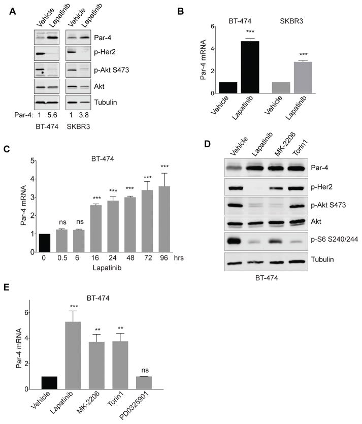
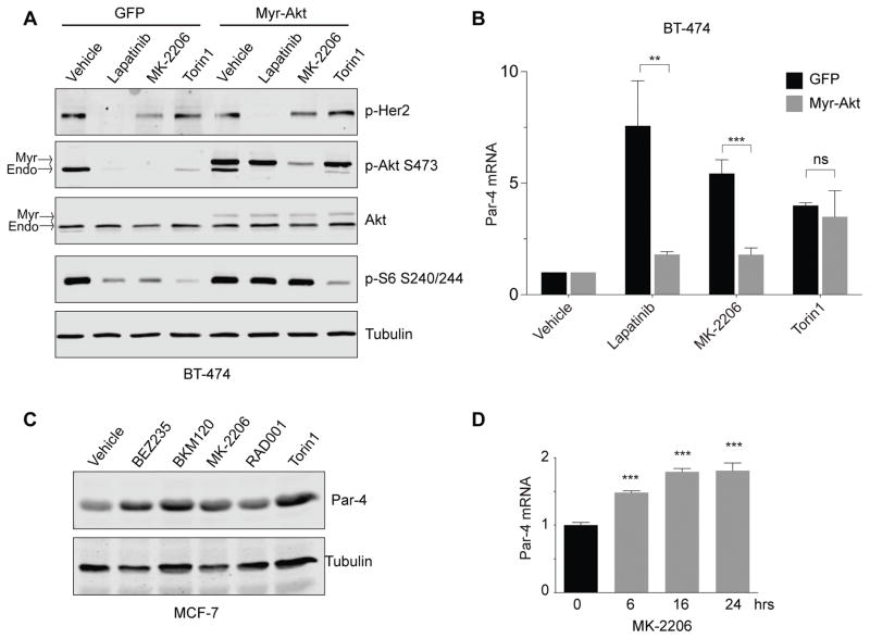
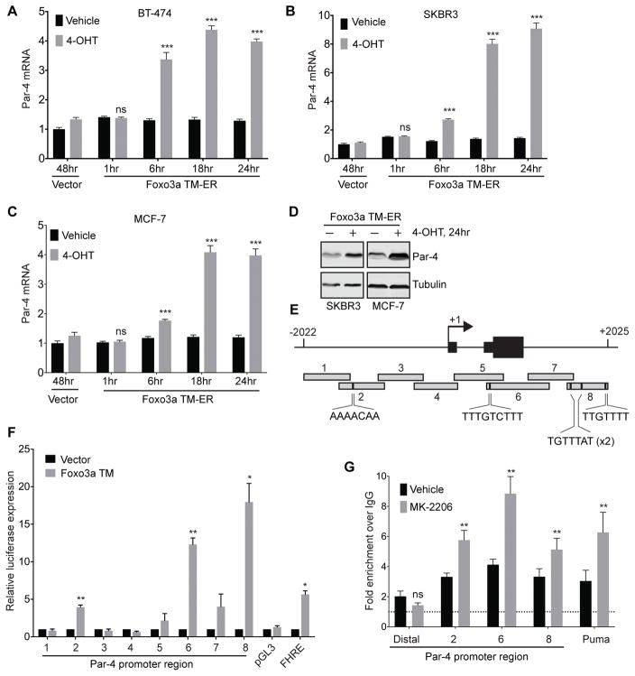
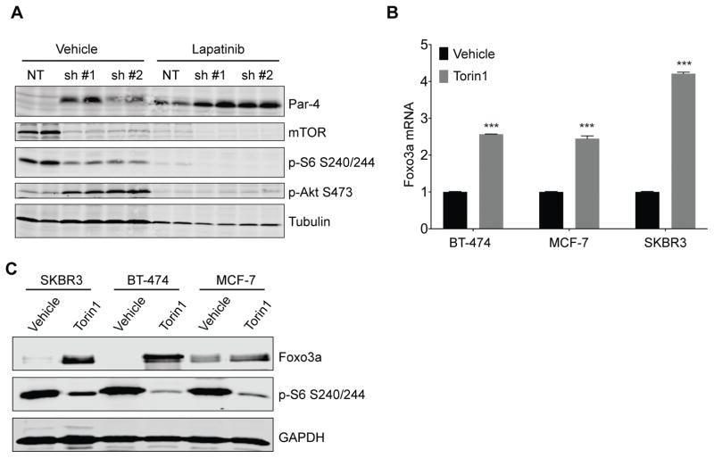
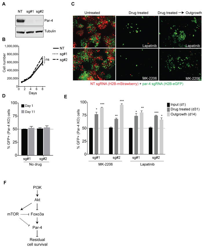
Tumor recurrence is a leading cause of death and is thought to arise from a population of residual cells that survive treatment. These residual cancer cells can persist, locally or at distant sites, for years or decades. Therefore, understanding the pathways that regulate residual cancer cell survival may suggest opportunities for targeting these cells to prevent recurrence. Previously, it was observed that the proapoptotic protein (PAWR/Par-4) negatively regulates residual cell survival and recurrence in mice and humans. However, the mechanistic underpinnings on how Par-4 expression is regulated are unclear. Here, it is demonstrated that Par-4 is transcriptionally upregulated following treatment with multiple drugs targeting the PI3K-Akt-mTOR signaling pathway, and identify the Forkhead family of transcription factors as mediators of this upregulation. Mechanistically, Foxo3a directly binds to the Par-4 promoter and activates its transcription following inhibition of the PI3K-Akt pathway. This Foxo-dependent Par-4 upregulation limits the long-term survival of residual cells following treatment with therapeutics that target the PI3K-Akt pathway. Taken together, these results indicate that residual breast cancer tumor cell survival and recurrence requires circumventing Foxo-driven Par-4 upregulation and suggest that approaches to enforce Par-4 expression may prevent residual cell survival and recurrence. Mol Cancer Res; 16(4); 599-609. ©2018 AACR.


Conflict of interest statement

The authors declare that they have no conflicts of interest relevant to this work.

Figures

Figure 1
Figure 1
Drugs targeting the Her2-Akt-mTOR pathway induce Par-4 upregulation in Her2-amplified breast cancer cells. A and B, Treatment of the Her2-amplified cell lines BT-474 and SKBR3 with Lapatinib for 2 days induces Par-4 protein (A) and mRNA (B) upregulation. C, Time-course of Par-4 mRNA upregulation following Lapatinib treatment in BT-474 cells. D and E, Western blot (D) and qPCR (E) analysis showing Par-4 upregulation in BT-474 cells treated with Lapatinib, the Akt inhibitor MK-2206, or the mTOR inhibitor Torin1 for 2 days. Significance was determined by Student’s t-test and data are presented as mean plus standard deviation (SD). **, p<0.01; ***, p<0.001.
Figure 2
Figure 2
Inhibition of the Akt pathway is required for Par-4 upregulation following drug treatment. A, BT-474 cells expressing a control vector or myristoylated Akt were treated with the indicated drugs, and the effects on downstream signaling were measured by Western blotting. B, qPCR analysis showing that constitutively active Akt prevents Par-4 upregulation following inhibition of Her2 and Akt but not mTOR. C, The PIK3CA-mutant breast cancer cell line MCF-7 was treated with drugs targeting PI3K (BEZ235 and BKM120), Akt (MK-2206), or mTOR (RAD001 and Torin) for 2 days and Par-4 levels were measured by Western blot. D, Time-course of Par-4 mRNA upregulation following MK-2206 treatment in MCF-7 cells. Data are presented as mean plus SD. **, p<0.01; ***, p<0.001.
Figure 3
Figure 3
Par-4 is a direct target of Foxo3a. A – C, Time-course of Par-4 mRNA upregulation following 4-OH Tamoxifen treatment in BT-474 (A), SKBR3 (B), or MCF-7 (C) cells stably expressing Foxo3a TM-ER fusion. D, Western analysis showing Par-4 upregulation 24 hours after 4-OHT treatment. E, Schematic of the Par-4 promoter surrounding the transcriptional start site. Eight regions (~500bp each) were cloned upstream of luciferase for use in reporter gene experiments. Putative Foxo3a binding sites in region 2, 6, and 8 are shown. F, 293T cells were transfected with luciferase constructs containing each promoter region together with empty vector or constitutively active Foxo3a TM, and luciferase expression was measured 24 hours later. pGL3: empty vector (negative control); FHRE: forkhead response element (positive control) G, Chromatin immunoprecipitation (ChIP) analysis of Foxo3a occupancy at indicated regions of the Par-4 promoter in BT-474 cells treated with vehicle or MK-2206 for 24 hours. A distal region (-10 kb) of the Par-4 promoter was used as a negative control, and the Puma promoter was used as a positive control. Data are expressed as fold-enrichment over IgG IP. Significance was determined by Student’s t-test and data are presented as mean plus SD. **, p<0.01; ***, p<0.001.
Figure 4
Figure 4
Inhibition of the mTOR pathway induces upregulation of Par-4 and Foxo3a. A, BT-474 cells were transduced with lentivirus expressing a control shRNA or one of two shRNAs targeting mTOR. Four days later, cells were treated with vehicle or Lapatinib for 3 days and levels of Par-4 and components of the mTOR pathway were measured by Western blotting. B and C, BT-474, MCF-7, and SKBR3 cells were treated with Torin1 for 2 days and levels of Foxo3a were measured by qRT-PCR (B) or Western blot (C). Significance was determined by Student’s t-test and data are presented as mean plus SD. **, p<0.01; ***, p<0.001.
Figure 5
Figure 5
Par-4 expression prevents the long-term survival of cells following inhibition of the PI3K-Akt pathway. A, Western blot showing Par-4 expression in BT-474 cells expressing a non-targeting sgRNA or one of two sgRNAs targeting Par-4. B, Growth curves for control and Par-4 knockout BT-474 cells. C, Fluorescent micrographs from a cellular competition assay. Control cells labeled with H2B-mStrawberry were mixed in a 1:1 ratio with Par-4 knockout cells labeled with H2B-eGFP. Cells were left untreated or treated with Lapatinib or MK-2206 for 31 days, at which point the drug was removed and surviving cells were allowed to grow out for 2 weeks. D, Quantification of the percentage of Par-4 knockout (GFP+) cells in untreated cells at day 1 and after 5 population doublings (d11). E, Quantification of the percentage of Par-4 knockout (GFP+) cells in the residual population that survives following treatment with Lapatinib or MK-2206 (d31), and after removal of drug to allow the outgrowth of residual cells (d14). F, Model for Par-4 regulation following therapies targeting the PI3K-Akt-mTOR pathway. Data are presented as the mean of three biological replicates plus SD. *, p<0.05; **, p<0.01; ***, p<0.001.

References

    1. Siegel RL, Miller KD, Jemal A. Cancer statistics, 2016. CA Cancer J Clin. 2016;66:7–30. - PubMed
    1. Symmans WF, Peintinger F, Hatzis C, Rajan R, Kuerer H, Valero V, et al. Measurement of residual breast cancer burden to predict survival after neoadjuvant chemotherapy. J Clin Oncol. 2007;25:4414–22. - PubMed
    1. Bednarz-Knoll N, Alix-Panabieres C, Pantel K. Clinical relevance and biology of circulating tumor cells. Breast Cancer Res. 2011;13:228. - PMC - PubMed
    1. Pantel K, Muller V, Auer M, Nusser N, Harbeck N, Braun S. Detection and clinical implications of early systemic tumor cell dissemination in breast cancer. Clin Cancer Res. 2003;9:6326–34. - PubMed
    1. Janni W, Vogl FD, Wiedswang G, Synnestvedt M, Fehm T, Juckstock J, et al. Persistence of disseminated tumor cells in the bone marrow of breast cancer patients predicts increased risk for relapse--a European pooled analysis. Clin Cancer Res. 2011;17:2967–76. - PubMed

Publication types

MeSH terms

Substances