. 2018 Mar 2;293(9):3056-3072.
doi: 10.1074/jbc.M117.812784. Epub 2018 Jan 12.

Effects of allelic variations in the human myxovirus resistance protein A on its antiviral activity

Affiliations

Effects of allelic variations in the human myxovirus resistance protein A on its antiviral activity

Laura Graf et al. J Biol Chem. .

Abstract

Only a minority of patients infected with seasonal influenza A viruses exhibit a severe or fatal outcome of infection, but the reasons for this inter-individual variability in influenza susceptibility are unclear. To gain further insights into the molecular mechanisms underlying this variability, we investigated naturally occurring allelic variations of the myxovirus resistance 1 (MX1) gene coding for the influenza restriction factor MxA. The interferon-induced dynamin-like GTPase consists of an N-terminal GTPase domain, a bundle signaling element, and a C-terminal stalk responsible for oligomerization and viral target recognition. We used online databases to search for variations in the MX1 gene. Deploying in vitro approaches, we found that non-synonymous variations in the GTPase domain cause the loss of antiviral and enzymatic activities. Furthermore, we showed that these amino acid substitutions disrupt the interface for GTPase domain dimerization required for the stimulation of GTP hydrolysis. Variations in the stalk were neutral or slightly enhanced or abolished MxA antiviral function. Remarkably, two other stalk variants altered MxA's antiviral specificity. Variations causing the loss of antiviral activity were found only in heterozygous carriers. Interestingly, the inactive stalk variants blocked the antiviral activity of WT MxA in a dominant-negative way, suggesting that heterozygotes are phenotypically MxA-negative. In contrast, the GTPase-deficient variants showed no dominant-negative effect, indicating that heterozygous carriers should remain unaffected. Our results demonstrate that naturally occurring mutations in the human MX1 gene can influence MxA function, which may explain individual variations in influenza virus susceptibility in the human population.

Keywords: Mx proteins; allelic variations; antiviral response; dynamin; genetic polymorphism; influenza virus; innate immunity; interferon.


Conflict of interest statement

The authors declare that they have no conflicts of interest with the contents of this article

Figures

Figure 1.
Figure 1.
Structure of human MxA and positions of the variations. A, schematic presentation of the MxA primary structure. B, bundle signaling element. B, structure of the MxA monomer from the crystal structure (Protein Data Bank code 3SZR) (3). Domains are color-coded, and secondary structure elements are labeled. Orange, G domain; red, BSE; green, stalk; blue dotted line, unstructured loop L4. Positions of the G domain and stalk variations are highlighted in blue. Amino acid residues of WT MxA are shown in stick representations. *, positions of previously characterized artificial mutations; T103A in the G domain is GTPase-deficient, and M527D in the stalk is a monomeric mutant. Both mutants are antivirally inactive (2, 9, 32).
Figure 2.
Figure 2.
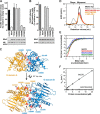
Characterization of MxA G domain variants. A, antiviral activity of the G domain variants in a FLUAV minireplicon system. 293T cells were co-transfected with expression plasmids for the MxA variants (300 ng) and the minireplicon system of VN/04, including a reporter construct encoding firefly luciferase under the control of the viral promoter. After 24 h, firefly luciferase activity was determined and normalized to the activity of constitutively co-expressed Renilla luciferase. Results are presented relative to the activity in the absence of MxA, the vector control (see “Experimental procedures” for calculation), and as arithmetic means ± S.D. of three independent experiments. Protein expression of FLAG-tagged MxA, viral NP, and actin was determined by Western blot analysis. B, restriction of FLUAV replication by G domain variants in tissue culture. A549 cells were transfected with MxA expression plasmids (500 ng) and 24 h later infected with SC35MNS1_2A_GFP-NEP (H7N7) at an MOI of 0.5. After fixation of the cells at 10 h post-infection, MxA was stained, and cells were analyzed by FACS. MxA-positive cells were selected, and the percentage of infected (GFP-positive) cells was determined. The percentage of GFP-positive cells expressing the inactive mutant T103A was set to 100%. Arithmetic means ± S.D. (error bars) of four independent experiments are shown. C, G domain dimer of GMPPCP-bound (gray) stalkless MxA (4P4S, G domain A in yellow (residues 70–340), G domain B in blue (residues 68–340)) (8). Positions of MxA G domain variations are highlighted in red. Amino acid residues of WT MxA are shown in stick representations. D, analytical gel-filtration analysis of the indicated mutants in the presence and absence (apo) of GDP-AlFx. E, nucleotide binding analysis of monomeric MxA G domain variants by ITC at 8 °C. GTPγS was titrated stepwise into the protein solution. The resulting heat changes were integrated, and the obtained values were fitted to a quadratic binding equation (one-site binding model). The following KD values were derived from the fittings: M527D (black), KD = 13 ± 5 μm, n = 0.83 ± 0.08; M527D/N220D (red), KD = 13 ± 2 μm, n = 0.50 ± 0.04; M527D/G255E (blue), KD = 5 ± 2 μm, n = 0.48 ± 0.18; M527D/V268M (green), KD = 17 ± 3 μm, n = 0.54 ± 0.04. The MxA constructs showed a varying degree of precipitation in these assays, which may explain the reduced binding numbers. F, protein concentration-dependent GTPase activities of monomeric M527D (□) and M527D/N220D (○) were determined at 37 °C by an HPLC-based assay. The mean kobs was calculated from two independent experiments for each concentration. The error bars show the range of the two data points. mAU, milliabsorbance units.
Figure 3.
Figure 3.
MxA G-interface variants have no dominant-negative effect on WT MxA. A, co-immunoprecipitation of WT MxA with G domain variants. 293T cells were co-transfected with HA-tagged WT MxA and FLAG-tagged MxA mutants. At 24 h post-transfection, cell lysates were subjected to FLAG-specific immunoprecipitations (IP). Precipitates and whole-cell lysates (WCL) were analyzed by Western blotting. B, effect of MxA G domain variants on the antiviral activity of WT MxA in the FLUAV minireplicon system of VN/04, as described in the legend to Fig. 2A. HA-tagged WT MxA (300 ng) was co-transfected with the components of the minireplicon and increasing amounts (50, 100, and 200 ng) of the indicated FLAG-tagged MxA variants. Protein expression was monitored by Western blot analysis. Data are presented as described in the legend to Fig. 2A. C, GTPase activity of M527D can be stimulated by the monomeric G domain mutant N220D. M527D (2.5 μm) was incubated with increasing concentrations of M527D/N220D, and GTPase activity was measured as described in the legend to Fig. 2F. Vertical lines in the Western blots indicate cuts combining two blots of one experiment run in parallel. Error bars, S.D.
Figure 4.
Figure 4.
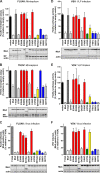
Antiviral activity of MxA stalk variants. A, antiviral activity against FLUAV in the VN/04 minireplicon system, as described in the legend to Fig. 2A. The vector control and the antiviral activity of WT MxA and T103A have already been shown in Fig. 2A. B, antiviral activity against THOV in the SiAr126 minireplicon system. MxA variants (100 ng of expression plasmids) and the components of the minireplicon system, including a reporter construct encoding firefly luciferase under the control of the THOV promoter and Renilla luciferase to monitor transfection efficiency, were co-expressed in 293T cells. Firefly luciferase activity was measured in the cell lysates at 24 h post-transfection and normalized to the activity of the Renilla luciferase. Data are presented as described in the legend to Fig. 2A. C, restriction of FLUAV replication by stalk variants in A549 cells analyzed by FACS, as described in the legend to Fig. 2B. The antiviral activities of WT MxA and T103A have already been shown in Fig. 2B. D and E, antiviral activity against VSV. D, 293T cells were co-transfected with FLAG-tagged MxA variants (300 ng) and VSV-G (300 ng). At 24 h post-transfection, cells were infected with VSV*ΔG(Luc) at an MOI of 1. Another 24 h later, supernatants were collected, the cells were harvested, and firefly luciferase activity was measured (VLP infection). E, the supernatants containing newly produced VLPs were used to infect naive 293T cells, which were lysed 24 h later to determine firefly luciferase activity (VLP titration). The values are presented relative to the activity in the absence of MxA, the vector control (see “Experimental procedures” for calculation). Arithmetic means ± S.D. (error bars) of three biological replicates are shown. F, restriction of VSV replication by stalk variants in tissue culture. 24 h after transfection with MxA expression plasmids (500 ng), A549 cells were infected with VSV-GFP at an MOI of 0.5 for 6.5 h. Fixed cells stained with an MxA-specific antibody were analyzed by FACS. MxA-positive cells were selected, and the percentage of infected (GFP-positive) cells was determined. The percentage of GFP-positive cells expressing the inactive mutant T103A was set to 100%. Results are displayed as arithmetic means ± S.D. of three independent experiments. Protein expression of FLAG-tagged MxA, actin, FLUAV, and THOV NP was verified by Western blot analyses. Vertical lines in the Western blots indicate cuts combining two blots of one experiment run in parallel. The color code of the bars is explained under “Results.”
Figure 5.
Figure 5.
Characterization of the MxA stalk variants E394K, R408Q, E419ter, and F454C. A, protein concentration–dependent GTPase activities of WT MxA (gray), R408Q (red), and F454C (orange) were determined as in Fig. 2F. B, analytical gel-filtration analysis, as in Fig. 2D, for WT MxA (gray), M527D (black), R408Q (red), and F454C (orange) in the absence of nucleotide. C, co-immunoprecipitation of WT MxA with the indicated stalk variants. 293T cells were transfected either with HA-tagged WT MxA (500 ng) or FLAG-tagged MxA variants (500 ng). At 24 h post-transfection, the lysates of cells expressing WT MxA and the indicated mutant were mixed and subjected to FLAG-specific immunoprecipitation (IP). Precipitates and whole-cell lysates (WCL) were analyzed by Western blotting. *, immunoglobulin heavy chain; **, nonspecific bands. D, effect of the stalk variants on the antiviral activity of WT MxA in the VN/04 minireplicon system, as described in the legend to Fig. 2A. HA-tagged WT MxA (300 ng) was co-transfected with the components of the minireplicon and increasing amounts (50, 100, and 200 ng) of the indicated FLAG-tagged variants. Protein expression was monitored by Western blot analysis. Data are presented as described in the legend to Fig. 2A. Vertical lines in the Western blots indicate cuts combining two blots of one experiment run in parallel. Error bars, S.D.
Figure 6.
Figure 6.
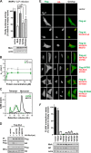
Characterization of the MxA stalk variants V470G and E516del. A, antiviral activity against RVFV. MxA (100 ng) and RVFV-N and -L (50 ng each) were co-expressed in 293T cells. 24 h post-transfection, cells were infected with Rift Valley fever VLPs encoding firefly luciferase as a reporter. Luciferase activities were measured 24 h later and are presented relative to the activity in the absence of MxA, the vector control (see “Experimental procedures” for calculation). Arithmetic means ± S.D. (error bars) of four biological replicates are shown. Western blot analysis was performed to control protein expression of FLAG-tagged MxA and actin. Significance was calculated using one-way analysis of variance with Dunnett's post hoc test. ns, not significant; **, p ≤ 0.01; ****, p ≤ 0.0001. B, protein concentration–dependent GTPase activities of WT MxA (gray; same control as in Fig. 5A) and V470G (green), as in Fig. 5A. C, analytical gel filtration of WT MxA, M527D (gray and black; same controls as in Fig. 5B), and V470G (green) in the absence of nucleotide, as described in the legend to Fig. 2D. D, co-immunoprecipitation of HA-tagged WT MxA (500 ng) with the FLAG-tagged variants (IP) V470G and E516del (500 ng) as described in the legend to Fig. 5C. Precipitates and whole-cell lysates (WCL) were analyzed by Western blotting. E, nuclear co-translocation of the MxA stalk variants V470G and E516del with WT MxA. Artificial nuclear forms of WT and MxA variants carrying an HA tag and the NLS of the SV40 large T antigen (HA-NLS-MxA) were co-expressed with FLAG-tagged WT MxA or MxA variants in HeLa cells. At 24 h post-transfection, cells were fixed and stained with antibodies directed against the HA tag (red) and the FLAG tag (green). The right column displays the overlay of the two signals. F, effect of different amino acid substitutions at position 470 on the antiviral activity of MxA. The antiviral activity of the mutants against FLUAV was determined in the VN/04 minireplicon system, as described in the legend to Fig. 2A.
Figure 7.
Figure 7.
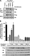
The loop L4 variant F561L has no negative effect on WT MxA. A, co-immunoprecipitation of HA-tagged WT MxA (500 ng) with the FLAG-tagged variants (IP) F561L and S566Y (500 ng) as described in Fig. 5C. Precipitates and whole-cell lysates (WCL) were analyzed by Western blotting. Vertical lines in the Western blots indicate cuts combining two blots of one experiment run in parallel. B, effect of MxA loop L4 variants on the antiviral activity of WT MxA in the THOV minireplicon system, as described in the legend to Fig. 4B. HA-tagged WT MxA (50 ng) was co-transfected with the components of the THOV minireplicon system and increasing amounts (25, 50, and 100 ng) of the indicated FLAG-tagged MxA variants. Protein expression was monitored by Western blot analysis. Error bars, S.D.

References

    1. Haller O., Staeheli P., Schwemmle M., and Kochs G. (2015) Mx GTPases: dynamin-like antiviral machines of innate immunity. Trends Microbiol. 23, 154–163 10.1016/j.tim.2014.12.003 - DOI - PubMed
    1. Gao S., von der Malsburg A., Paeschke S., Behlke J., Haller O., Kochs G., and Daumke O. (2010) Structural basis of oligomerization in the stalk region of dynamin-like MxA. Nature 465, 502–506 10.1038/nature08972 - DOI - PubMed
    1. Gao S., von der Malsburg A., Dick A., Faelber K., Schröder G. F., Haller O., Kochs G., and Daumke O. (2011) Structure of myxovirus resistance protein a reveals intra- and intermolecular domain interactions required for the antiviral function. Immunity 35, 514–525 10.1016/j.immuni.2011.07.012 - DOI - PubMed
    1. Mitchell P. S., Patzina C., Emerman M., Haller O., Malik H. S., and Kochs G. (2012) Evolution-guided identification of antiviral specificity determinants in the broadly acting interferon-induced innate immunity factor MxA. Cell Host Microbe 12, 598–604 10.1016/j.chom.2012.09.005 - DOI - PMC - PubMed
    1. Patzina C., Haller O., and Kochs G. (2014) Structural requirements for the antiviral activity of the human MxA protein against Thogoto and influenza A virus. J. Biol. Chem. 289, 6020–6027 10.1074/jbc.M113.543892 - DOI - PMC - PubMed

Publication types

Substances

Associated data

LinkOut - more resources