. 2017 Dec 1;31(23-24):2416-2429.
doi: 10.1101/gad.308163.117. Epub 2018 Jan 12.

The RES complex is required for efficient transformation of the precatalytic B spliceosome into an activated Bact complex

Affiliations

The RES complex is required for efficient transformation of the precatalytic B spliceosome into an activated Bact complex

Penghui Bao et al. Genes Dev. .

Abstract

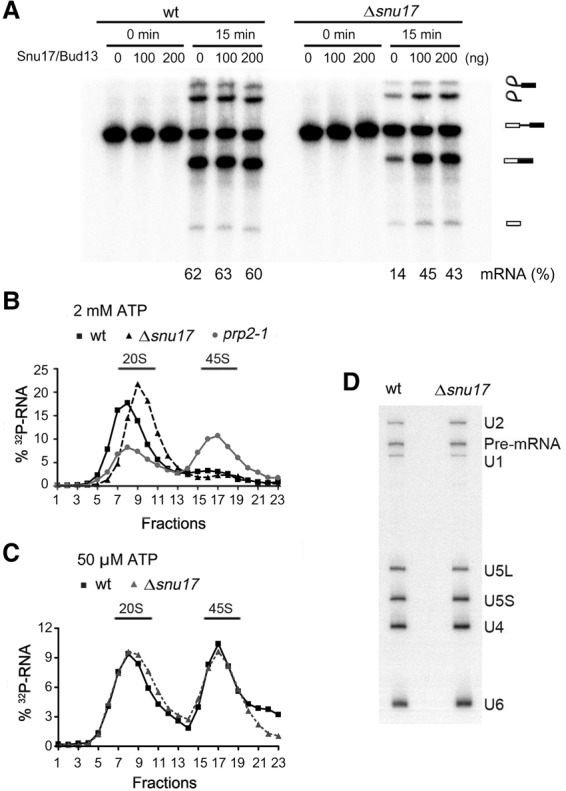
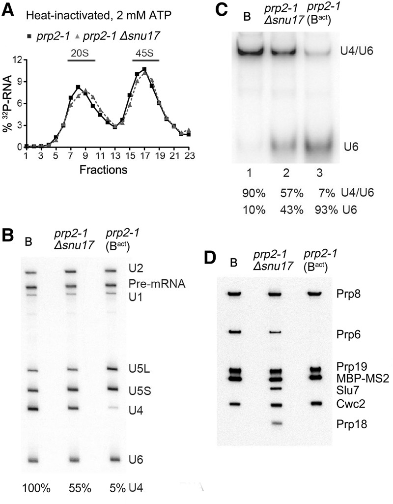
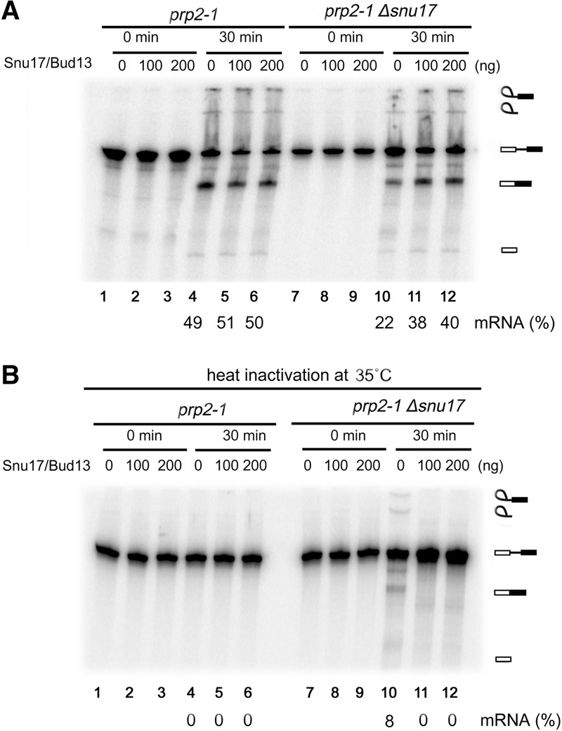
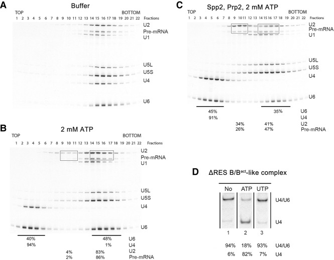
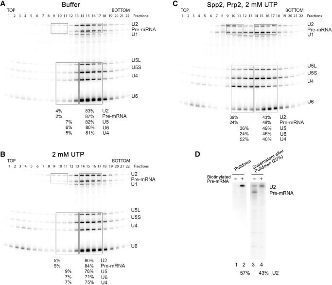
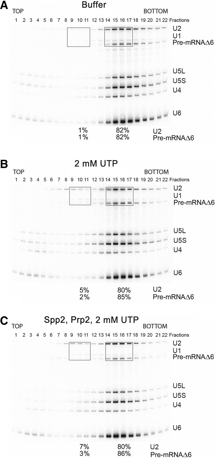
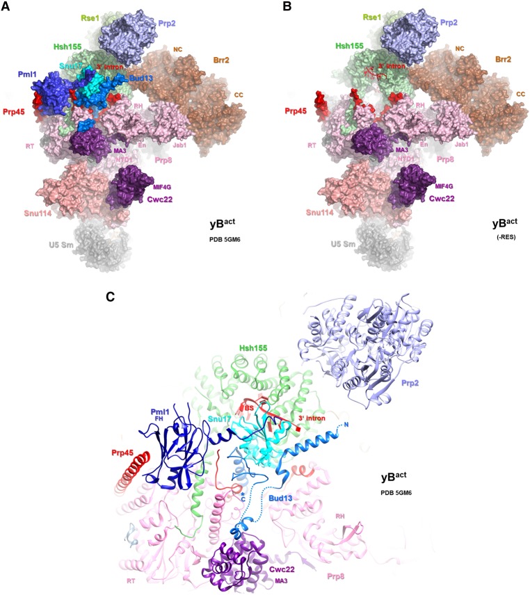
The precise function of the trimeric retention and splicing (RES) complex in pre-mRNA splicing remains unclear. Here we dissected the role of RES during the assembly and activation of yeast spliceosomes. The efficiency of pre-mRNA splicing was significantly lower in the absence of the RES protein Snu17, and the recruitment of its binding partners, Pml1 (pre-mRNA leakage protein 1) and Bud13 (bud site selection protein 13), to the spliceosome was either abolished or substantially reduced. RES was not required for the assembly of spliceosomal B complexes, but its absence hindered efficient Bact complex formation. ΔRES spliceosomes were no longer strictly dependent on Prp2 activity for their catalytic activation, suggesting that they are structurally compromised. Addition of Prp2, Spp2, and UTP to affinity-purified ΔRES B or a mixture of B/Bact complexes formed on wild-type pre-mRNA led to their disassembly. However, no substantial disassembly was observed with ΔRES spliceosomes formed on a truncated pre-mRNA that allows Prp2 binding but blocks its activity. Thus, in the absence of RES, Prp2 appears to bind prematurely, leading to the disassembly of the ΔRES B complexes to which it binds. Our data suggest that Prp2 can dismantle B complexes with an aberrant protein composition, suggesting that it may proofread the spliceosome's RNP structure prior to activation.

Keywords: Prp2 ATPase/RNA helicase; RES complex; pre-mRNA splicing; spliceosome activation; spliceosome disassembly.


Figures

Figure 1.
Figure 1.
Pre-mRNA splicing, but not spliceosomal B complex formation, is less efficient in extracts from an S. cerevisiae Δsnu17 strain. (A) Splicing of 32P-labeled Actin pre-mRNA in whole-cell extracts from wild-type or Δsnu17 S. cerevisiae cells. A mixture of affinity-purified Snu17 and Snu17–Bud13 dimer (100 or 200 ng as indicated) or the corresponding buffer alone was added prior to incubation of the splicing reaction. The identities of the 32P-labeled RNA species are indicated at the right. The percentage of mRNA formed is indicated at the bottom. (B) Glycerol gradient sedimentation profile of affinity-purified spliceosomes formed on 32P-labeled Actin pre-mRNA under splicing conditions at 2 mM ATP for 60 min in extracts from wild-type, Δsnu17, and prp2-1 S. cerevisiae cells. The prp2-1 extract was preincubated at 35°C to inactivate Prp2. (C) Glycerol gradient sedimentation of affinity-purified spliceosomes formed at 50 µM ATP for 60 min in extracts from wild-type or Δsnu17 yeast cells. (D) RNA compositions of affinity-purified wild-type or Δsnu17 splicing complexes (formed at 50 µM ATP) migrating in the 45S peak fractions of the glycerol gradient. RNA was detected by Northern blotting, and the snRNAs are indicated at the right. The 32P-labeled Actin pre-mRNA was detected by autoradiography.
Figure 2.
Figure 2.
The transformation of the spliceosomal B complex into Bact is less efficient in the absence of RES complex proteins. (A) Glycerol gradient sedimentation profile of MS2-MBP affinity-purified spliceosomal complexes formed after initial heat inactivation of Prp2 on 32P-labeled Actin pre-mRNA in extracts from prp2-1 or prp2-1 Δsnu17 yeast cells after 60 min in the presence of 2 mM ATP. (B) RNA compositions of affinity-purified B complexes (formed at 50 µM ATP) or Bact or splicing complexes formed in extracts from prp2-1 Δsnu17 cells (at 2 mM ATP) sedimenting in the 45S peak fractions of the glycerol gradient. The pre-mRNA and snRNAs were separated by denaturing polyacrylamide gel electrophoresis (PAGE) and detected as described in the legend for Figure 1. The amount of U4 snRNA in each lane (shown below) was quantitated as a percentage of the pre-mRNA band (which acts as a spliceosome loading control) and then normalized to the amount of U4 in the B complex, which was set to 100%. (C) Quantitation of the amount of U4/U6 duplex and U6 snRNA in affinity-purified B, Bact, or spliceosomal complexes formed in extracts from prp2-1 Δsnu17 cells. RNAs were analyzed by native PAGE, and U6 snRNA was visualized by Northern blotting. The percent of free U6 versus U6 base-paired with U4 was quantitated with a PhosphorImager and is shown below each lane. The identity of the slower-migrating band as U4/U6 was confirmed by Northern blotting with a probe against U4 snRNA. (D) Proteins from affinity-purified B complexes (formed at 50 µM ATP) or spliceosomal complexes formed at 2 mM ATP in extracts from prp2-1 (predominantly Bact complexes) or prp2-1 Δsnu17 cells (as indicated) after heat inactivation of Prp2 were analyzed by Western blotting using antibodies against the indicated proteins. Antibodies against Prp8 were used to ensure equal loading.
Figure 3.
Figure 3.
The requirement for Prp2 activity can be bypassed in the absence of Snu17. Pre-mRNA splicing in extracts from prp2-1 or prp2-1 Δsnu17 yeast cells without heat inactivation of Prp2 (A) or after heat inactivation of Prp2 at 35°C (B). Splicing was performed without or after addition of affinity-purified Snu17/Bud13 (100 or 200 ng as indicated). RNA was separated by denaturing PAGE and visualized using a PhosphorImager. The identities of the 32P-labeled RNA species are indicated at the right. The percentage of mRNA formed after 30 min is indicated below.
Figure 4.
Figure 4.
Affinity-purified ΔRES spliceosomes are partially disassembled upon addition of Prp2, Spp2, and ATP. Spliceosomal complexes formed in extracts from prp2-1 Δsnu17 yeast cells after initial heat inactivation of Prp2 (designated ΔRES B/Bact-like complexes) were affinity-purified, and 45S complexes were subjected to a second glycerol gradient after incubation with buffer (A), 2 mM ATP (B), or ATP, Prp2, and Spp2 (C). RNA was isolated from each gradient fraction, separated by denaturing PAGE, and detected by Northern blotting with 32P-labeled probes against the snRNAs indicated at the right. The 32P-labeled Actin pre-mRNA was detected by autoradiography. The percentage of the pre-mRNA or the indicated snRNAs in the boxed or underlined fractions is shown below the gel in B and C. (D) RNA was isolated from ΔRES B/Bact-like complexes and analyzed by native PAGE. Free U4 snRNA or U4 base-paired with U6 was subsequently visualized by Northern blotting with a 32P probe complementary to U4 snRNA. The percentage of free U4 versus U4 base-paired with U6 was quantitated with a PhosphorImager and is shown below each lane.
Figure 5.
Figure 5.
The Prp2 RNA helicase facilitates the disassembly of ΔRES spliceosomes. ΔRES B/Bact-like complexes were affinity-purified, and 45S complexes were subjected to a second glycerol gradient after incubation with buffer (A), 2 mM UTP (B), or UTP, Prp2, and Spp2 (C). RNA was analyzed as described in the legend for Figure 4. The percentage of the pre-mRNA or the indicated snRNAs in the boxed or underlined fractions is shown below each panel. (D) Affinity-purified 45S spliceosomes formed on nonbiotinylated or biotinylated Actin pre-mRNA in prp2-1 Δsnu17 extract (after heat inactivation of Prp2) were incubated with UTP, Prp2, and Spp2 and then analyzed on a second glycerol gradient. RNP complexes in gradient fractions 8–10 were subjected to streptavidin agarose pull-downs. RNA precipitated together with the biotinylated pre-mRNA (which was not released from the streptavidin beads) as well as RNA remaining in the supernatant were analyzed by denaturing PAGE followed by Northern blotting (to visualize the U2 snRNA) or autoradiography (to detect the radiolabeled pre-mRNA). The percentage of U2 in the pulled-down fraction versus the supernatant is shown below the gel.
Figure 6.
Figure 6.
ΔRES spliceosomes formed on truncated ActΔ6 pre-mRNA are not disassembled upon addition of UTP, Prp2, and Spp2. Affinity-purified ΔRES B/Bact-like spliceosomes formed on truncated ActΔ6 pre-mRNA at 2 mM ATP were subjected to a second glycerol gradient after incubation with buffer (A), 2 mM UTP (B), or UTP, Prp2, and Spp2 (C). RNA was analyzed as described in the legend for Figure 4. The percentage of U2 and the pre-mRNA in the boxed fractions is shown below the gel in each panel.
Figure 7.
Figure 7.
The location of the RES complex proteins in the yeast Bact complex. (A,B) Overview of the location of selected proteins, shown with space-filling models, in the yeast Bact structure (Rauhut et al. 2016). In B, Snu17, Pml1, and Bud13 have been removed. (C) Close-up of the RES proteins and other spliceosomal proteins that they contact or that are in close proximity in the yeast Bact complex. Proteins are depicted as ribbon diagrams.

References

    1. Arenas JE, Abelson JN. 1997. Prp43: an RNA helicase-like factor involved in spliceosome disassembly. Proc Natl Acad Sci 94: 11798–11802. - PMC - PubMed
    1. Bao P, Höbartner C, Hartmuth K, Lührmann R. 2017. Yeast Prp2 liberates the 5′ splice site and the branch site adenosine for catalysis of pre-mRNA splicing. RNA 23: 1770–1779. - PMC - PubMed
    1. Bertram K, Agafonov DE, Dybkov O, Haselbach D, Leelaram MN, Will CL, Urlaub H, Kastner B, Lührmann R, Stark H. 2017. Cryo-EM structure of a pre-catalytic human spliceosome primed for activation. Cell 170: 701–713. - PubMed
    1. Boon KL, Auchynnikava T, Edwalds-Gilbert G, Barrass JD, Droop AP, Dez C, Beggs JD. 2006. Yeast ntr1/spp382 mediates prp43 function in postspliceosomes. Mol Cell Biol 26: 6016–6023. - PMC - PubMed
    1. Brooks MA, Dziembowski A, Quevillon-Cheruel S, Henriot V, Faux C, van Tilbeurgh H, Séraphin B. 2009. Structure of the yeast Pml1 splicing factor and its integration into the RES complex. Nucleic Acids Res. 37: 129–143. - PMC - PubMed

MeSH terms

Substances

LinkOut - more resources