. 2018 Jan 12;8(1):679.
doi: 10.1038/s41598-017-19068-1.

Induction of protein citrullination and auto-antibodies production in murine exposed to nickel nanomaterials

Affiliations

Induction of protein citrullination and auto-antibodies production in murine exposed to nickel nanomaterials

Bashir M Mohamed et al. Sci Rep. .

Abstract

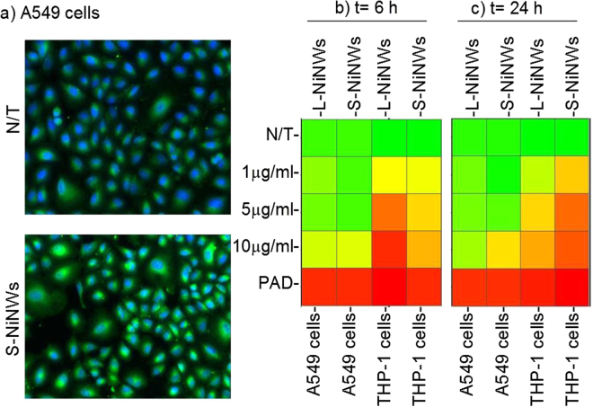
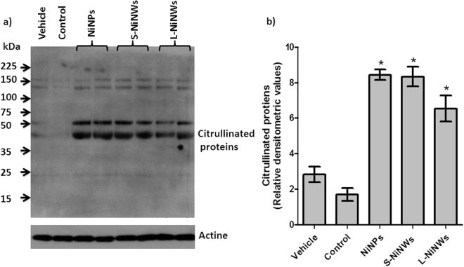
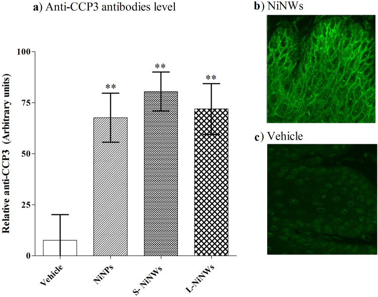
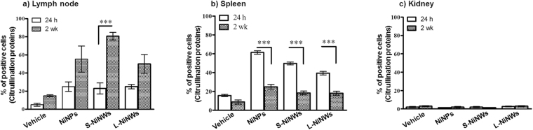
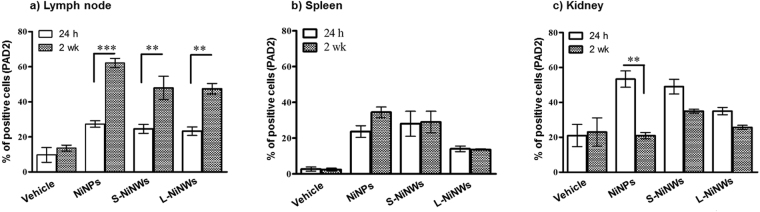
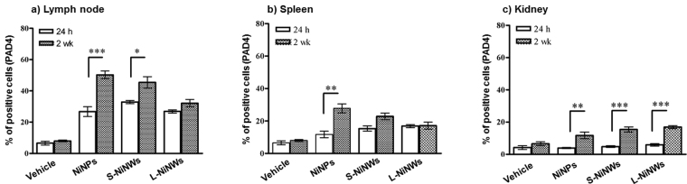
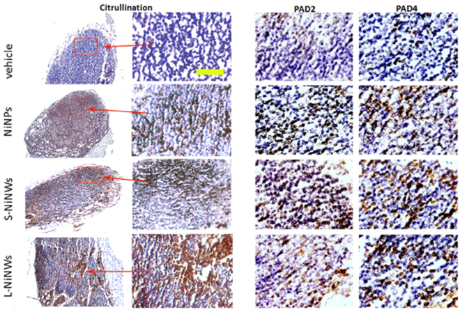
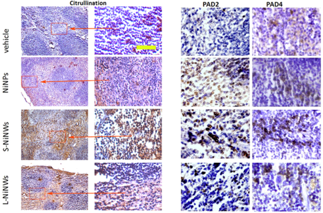
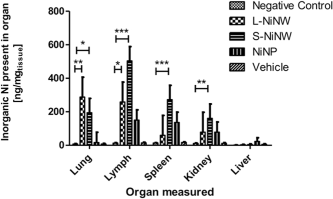
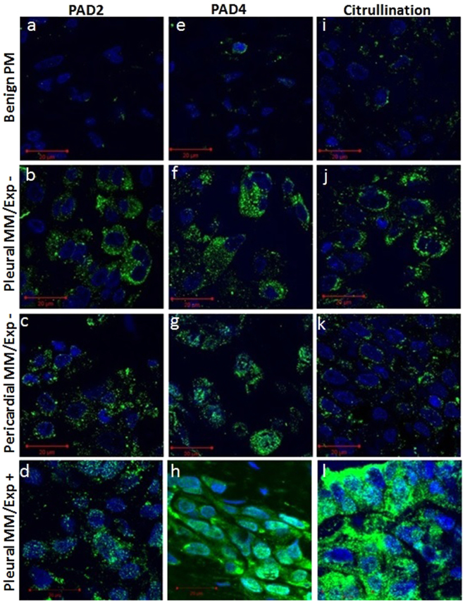
Citrullination, or the post-translational deimination of polypeptide-bound arginine, is involved in several pathological processes in the body, including autoimmunity and tumorigenesis. Recent studies have shown that nanomaterials can trigger protein citrullination, which might constitute a common pathogenic link to disease development. Here we demonstrated auto-antibody production in serum of nanomaterials-treated mice. Citrullination-associated phenomena and PAD levels were found to be elevated in nanomaterials -treated cell lines as well as in the spleen, kidneys and lymph nodes of mice, suggesting a systemic response to nanomaterials injection, and validated in human pleural and pericardial malignant mesothelioma (MM) samples. The observed systemic responses in mice exposed to nanomaterials support the evidence linking exposure to environmental factors with the development of autoimmunity responses and reinforces the need for comprehensive safety screening of nanomaterials. Furthermore, these nanomaterials induce pathological processes that mimic those observed in Pleural MM, and therefore require further investigations into their carcinogenicity.


Conflict of interest statement

The authors declare that they have no competing interests.

Figures

Figure 1
Figure 1
Citrullination levels in THP-1 and A549 cells exposed to nickel nanomaterials. (a) Representative fluorescent images of untreated controls (N/T), short (S)-NiNWs-treated A549 cells after 24 h exposure. Images show cytoplasmic citrullinated protein (green) and nuclei (blue). (b and c) Heatmaps reflect the citrullination levels indicated by colours ranging from red (≥75%) to yellow (50%) to green (≤25%) compared to negative control (N/T). PAD was used as positive control.
Figure 2
Figure 2
Induction of citrullinated proteins in mice sera following nickel nanomaterials exposure. (a) Serum samples from nickel nanomaterial-injected mice, vehicle control and untreated mice (N/T) were probed by immunoblotting technique for citrullinated proteins. (b) Densitometric quantitation of citrullinated protein bands indicated a significant increase in protein citrullination due to nickel nanomaterials exposure compared to vehicle and untreated controls. Data are representative of three independent experiments (mean ± S.E.). *p < 0.05 with respect to corresponding controls. Note: western blot gels were adapted to fit within composite image, full gel images are provided in Supplemental Information (Figure S2).
Figure 3
Figure 3
Detection of anti-CCP3 antibodies in murine sera. (a) Serum samples from nickel nanomaterial-injected and vehicle-injected mice were examined for anti-CCP3 antibodies by ELISA. (b,c) Reactivity of anti-citrulline antibody was confirmed by indirect immunofluorescence after incubating sera from mice exposed to S-NiNW and vehicle controls on oesophageal tissue sections.
Figure 4
Figure 4
Immunohistochemical analysis of citrullinated proteins in murine lymphoid organs and kidneys. Mice were injected with PBS (vehicle) or NiNPs, S-NiNWs and L-NiNWs and the organs were analysed after 24 h and 2 weeks. LN and kidney tissue sections were stained with anti-citrulline antibody. Protein citrullination in LN (a) spleen (b) and kidney tissue sections (c) sections were compared to their group controls. p-values of <0.0001 are indicated by ***. Statistical analysis between the different experimental groups is reported in Table S1.
Figure 5
Figure 5
Immunohistochemical analysis of PAD2 expression in murine lymphoid organs and kidneys. Mice were injected with PBS (vehicle) or NiNPs, S-NiNWs and L-NiNWs for 24 h and 2 weeks. LN and kidney tissue sections were stained with anti-PAD2 antibody. The histograms shown the percentage of positively stained cells for PAD2 expression in LN (a), spleen (b), and kidney tissue sections (c) p-values of <0.0001 are indicated by ***. Statistical analysis between the different experimental groups is reported in Table S2.
Figure 6
Figure 6
Immunohistochemical analysis of PAD4 expression in murine lymphoid organs and kidneys. Mice were injected with PBS (vehicle) or NiNPs, S-NiNWs and L-NiNWs for 24 h and 2 weeks. LN, spleen and kidney tissue sections were stained with anti-PAD4 antibody. PAD4 in LN (a) spleen (b) and kidney tissue (c) sections were compared to their group controls. p-values of <0.0001 are indicated by ***.
Figure 7
Figure 7
Immunohistochemistry of anti-citrulline, anti-PAD2 and PAD4 antibodies on murine lymph node (LN) tissue sections. Immunodetection of citrullinated proteins, PAD2 and PAD4 enzymes in murine LN tissue sections 2-week post treatment with nickel nanomaterials and vehicle controls. Brown staining demonstrates positive immunohistochemical reaction for citrulline, PAD2 and PAD4. First two panels from left (Citrulline) represent the corresponding microscopic fields at lower (10×) and higher (20×) magnification stained for citrullinated proteins, to illustrate the topography of positive staining within the LN. Red rectangles indicated by arrows outline the regions of interest magnified in the adjacent panel. Two panels on the right show representative topographically similar, but not identical microscopic fields stained for PAD2 and PAD4 enzymes, respectively. Scale bar (yellow): 100 μm.
Figure 8
Figure 8
Immunohistochemistry of anti-citrulline, anti-PAD2 and PAD4 antibodies on murine spleen tissue sections. Immunodetection of citrullinated proteins, PAD2 and PAD4 enzymes in murine spleen tissue sections 2-week post exposure to nickel nanomaterials and vehicle controls. Brown staining demonstrates positive immunohistochemical reaction for citrulline, PAD2 and PAD4. First two panels from left (Citrulline) represent the corresponding microscopic fields at lower (10×) and higher (20×) magnification stained for citrullinated proteins, to illustrate the topography of positive staining within the spleen. Red rectangles indicated by arrows outline the regions of interest magnified in the adjacent panel. Two panels on the right show representative topographically similar, but not identical microscopic fields stained for PAD2 and PAD4 enzymes, respectively. Scale bar (yellow): 100 μm.
Figure 9
Figure 9
Quantification of inorganic nickel magnetic material in murine organs after 2-week exposure to nanomaterials (n = 3 animals). Comparison of total mass of nickel nanomaterial measured accumulated in each organ versus organ mass. Inorganic nickel content was measured by SQUID magnetometry as total amount of magnetic signal compared to vehicle. Data are shown as average ± standard error of the mean (nrepeats = 3). The symbols (*), (**) and (***) indicate statistical significant changes as compared to the values measured against negative control samples (p < 0.05, 0.01 and 0.001, respectively) (two-way ANOVA and Bonferroni post-test).
Figure 10
Figure 10
Immunodetection of PAD2, PAD4 expression and citrullinated proteins in human MM. abbreviations: MM/Exp+: Malignant mesothelioma from the patients with a known asbestos exposure, and MM/Exp −: Malignant mesothelioma from the patients without a history of asbestos exposure. PAD2 expression in tissue sections of (a) benign PM (b) and (c) pleural and pericardial MM/Exp− and (d) pleural MM/Exp+. PAD4 expression in tissue sections of (e) benign PM, (f) and (g) pleural, and pericardial MM/Exp− and (h) pleural MM/Exp+. Induction of protein citrullination in tissue sections of (i) benign PM, (j) and (k) pleural and pericardial MM/Exp− and (l) pleural MM/Exp+.

References

    1. Chavanas S, et al. Comparative analysis of the mouse and human peptidylarginine deiminase gene clusters reveals highly conserved non-coding segments and a new human gene, PADI6. Gene. 2004;330:19–27. doi: 10.1016/j.gene.2003.12.038. - DOI - PubMed
    1. Foulquier C, et al. Peptidyl arginine deiminase type 2 (PAD-2) and PAD-4 but not PAD-1, PAD-3, and PAD-6 are expressed in rheumatoid arthritis synovium in close association with tissue inflammation. Arthritis and rheumatism. 2007;56:3541–3553. doi: 10.1002/art.22983. - DOI - PubMed
    1. Wegner N, et al. Autoimmunity to specific citrullinated proteins gives the first clues to the etiology of rheumatoid arthritis. Immunological reviews. 2010;233:34–54. doi: 10.1111/j.0105-2896.2009.00850.x. - DOI - PubMed
    1. Wang S, Wang Y. Peptidylarginine deiminases in citrullination, gene regulation, health and pathogenesis. Biochimica et biophysica acta. 2013;1829:1126–1135. doi: 10.1016/j.bbagrm.2013.07.003. - DOI - PMC - PubMed
    1. Darrah E, Rosen A, Giles JT, Andrade F. Peptidylarginine deiminase 2, 3 and 4 have distinct specificities against cellular substrates: novel insights into autoantigen selection in rheumatoid arthritis. Annals of the rheumatic diseases. 2012;71:92–98. doi: 10.1136/ard.2011.151712. - DOI - PMC - PubMed

Publication types

MeSH terms

LinkOut - more resources