. 2018 Jan 12;8(1):670.
doi: 10.1038/s41598-017-18428-1.

Human Placental-Derived Adherent Stromal Cells Co-Induced with TNF-α and IFN-γ Inhibit Triple-Negative Breast Cancer in Nude Mouse Xenograft Models

Affiliations

Human Placental-Derived Adherent Stromal Cells Co-Induced with TNF-α and IFN-γ Inhibit Triple-Negative Breast Cancer in Nude Mouse Xenograft Models

Hoshea Allen et al. Sci Rep. .

Abstract

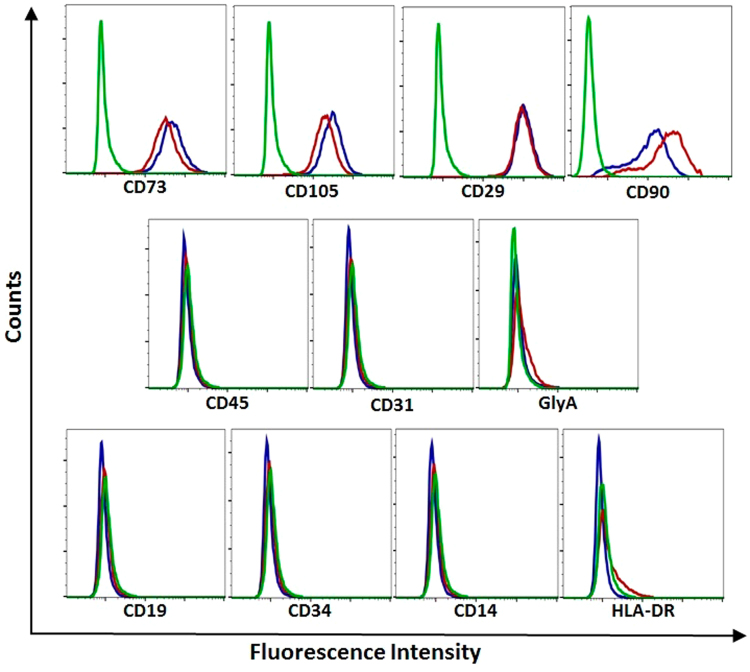
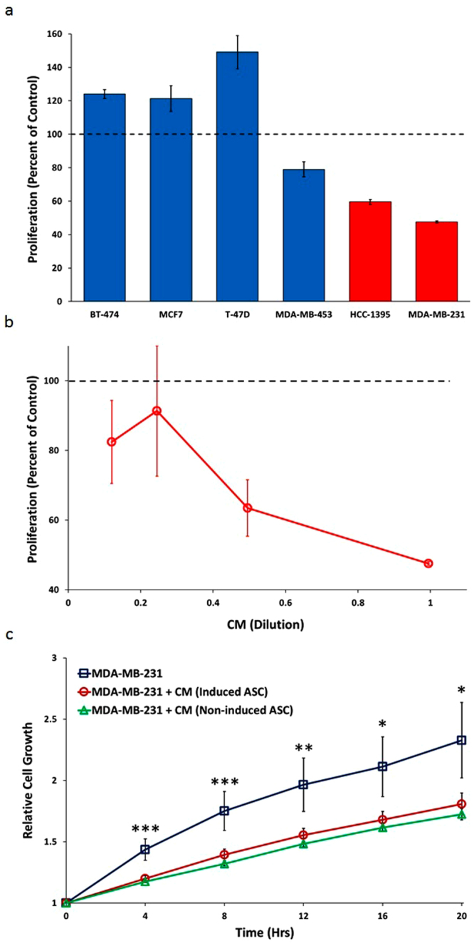
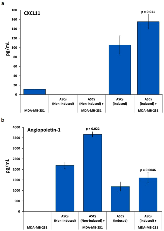
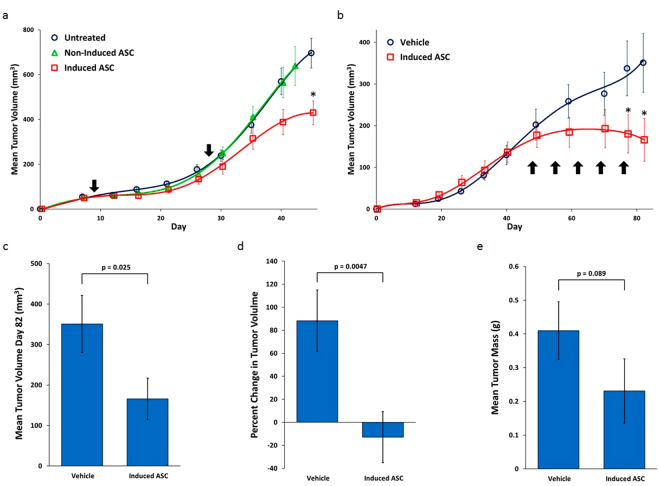
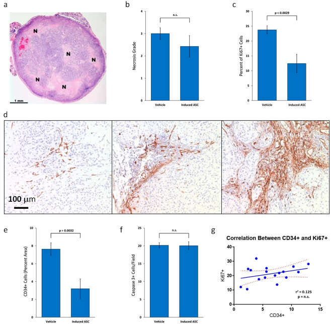
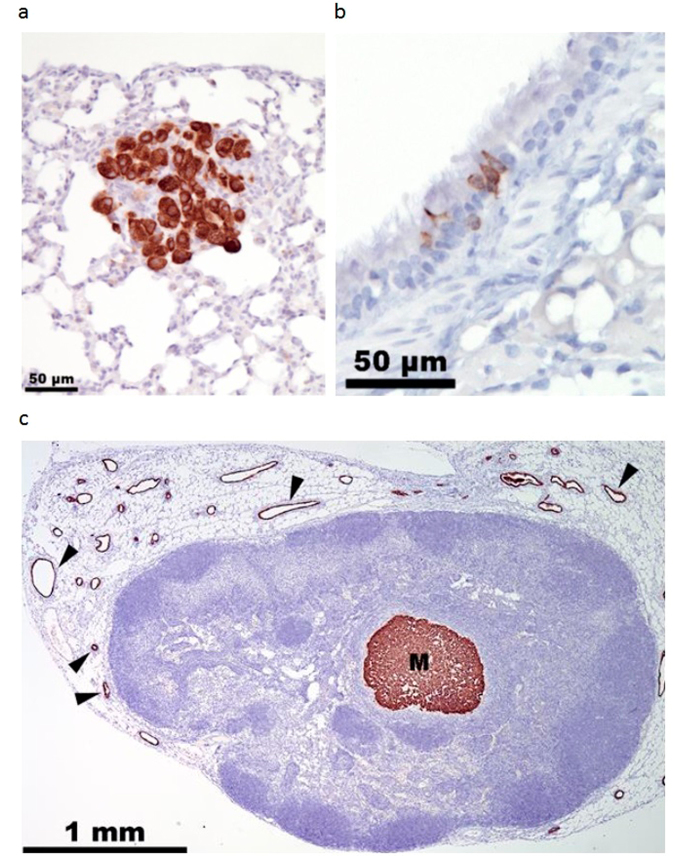
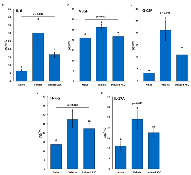
Culturing 3D-expanded human placental-derived adherent stromal cells (ASCs) in the presence of tumor necrosis factor-alpha (TNF-α) and interferon-gamma (IFN-γ) transiently upregulated the secretion of numerous anti-proliferative, anti-angiogenic and pro-inflammatory cytokines. In a 3D-spheroid screening assay, conditioned medium from these induced-ASCs inhibited proliferation of cancer cell lines, including triple-negative breast cancer (TNBC) lines. In vitro co-culture studies of induced-ASCs with MDA-MB-231 human breast carcinoma cells, a model representing TNBC, supports a mechanism involving immunomodulation and angiogenesis inhibition. In vivo studies in nude mice showed that intramuscular administration of induced-ASCs halted MDA-MB-231 cell proliferation, and inhibited tumor progression and vascularization. Thirty percent of treated mice experienced complete tumor remission. Murine serum concentrations of the tumor-supporting cytokines Interleukin-6 (IL-6), Vascular endothelial growth factor (VEGF) and Granulocyte-colony stimulating factor (G-CSF) were lowered to naïve levels. A somatic mutation analysis identified numerous genes which could be screened in patients to increase a positive therapeutic outcome. Taken together, these results show that targeted changes in the secretion profile of ASCs may improve their therapeutic potential.


Conflict of interest statement

The authors are current and former employees and shareholders of Pluristem Ltd.

Figures

Figure 1
Figure 1
Surface Marker Expression Profiles of TNF-α/IFN-γ-Induced and Non-Induced Placental-Derived ASCs. CD marker expression in human placental-derived ASCs. Cells were stained with PE-conjugated antibodies against CD90, CD73, CD105, CD29, CD45, CD31, GlyA, CD19, CD34, CD14 and HLA-DR. CD marker expression was analyzed by flow cytometry. Isotype control IgG1 (green), TNF-α/IFN-γ-induced-ASCs (red), and non-induced-ASCs (blue).
Figure 2
Figure 2
Proliferative Response of Breast Cancer Cell Lines to CM from TNF-α/IFN-γ- Induced and Non-Induced Placental-Derived ASCs. (a) Proliferative response of the six breast cancer cell lines to undiluted CM from TNF-α/IFN-γ-induced-ASC in the 3D-spheroid assay. Red bars represent TNBC cell lines. Error bars represent SD (n = 3). (b) POC response curves for MDA-MB-231 in the 3D-spheroid assay upon serial dilution. Error bars represent SD (n = 3). (c) Inhibition of MDA-MB-231 2D growth in real time with CM from TNF-α/IFN-γ-induced and non-induced-ASCs. Error bars represent SEM (n = 4 for MDA-MB-231 with regular growth medium, n = 11 for MDA-MB-231 + induced-ASC CM, n = 8 for MDA-MB-231 + non-induced-ASC CM). P-values are based on ANOVA: ***p < 0.01, **p < 0.01, and *p < 0.05.
Figure 3
Figure 3
Co-Culturing of MDA-MB-231 with Human Placental-Derived ASCs Affects Cytokine Secretion Profiles. ASCs (either non-induced or induced with TNF-α/IFN-γ) were co-cultured with MDA-MB-231 and CM was collected for analysis. Error bars represent SEM (n = 3 for induced-ASCs, n = 2 for non-induced-ASCs). (a) CXCL11. (b) Angiopoietin-1. CM was collected after 48 hours. P-values are based on paired sample t-test.
Figure 4
Figure 4
TNF-α/IFN-γ-Induced Human Placental-Derived ASCs Inhibit MDA-MB-231 Tumor Growth in Nude Mice. (a) Growth curves of tumor volume for MDA-MB-231 SubQ tumors in nude mice. Arrows mark days when TNF-α/IFN-γ-induced placental-derived ASCs were injected. (b) Growth curves of tumor volume for MDA-MB-231 mammary fat pad tumors in nude mice. The arrows mark days of the weekly IM injections of induced-ASCs. (c) Mean tumor volume at day 82 in the mammary fat pad model. (d) Percent change in mean tumor volume in the mammary fat pad model from the start of weekly IM injections of induced-ASCs. (e) Mean tumor mass at day 84 in the mammary fat pad model. n = 10 per group. Error bars are SEM. P-values are based on a one-tailed student’s t-test. Asterisks indicate p < 0.05.
Figure 5
Figure 5
TNF-α/IFN-γ-Induced Human Placental-Derived ASCs Inhibit Tumor Cell Proliferation and Vascularization in MDA-MB-231 Tumors in Nude Mice. (a) Representative section of H&E-stained MDA-MB-231 mammary fat pad tumor with grade 2 necrosis from mouse treated with TNF-α/IFN-γ-induced placental-derived ASCs. “N” marks areas of necrosis. (b) Mean necrosis grade in MDA-MB-231 mammary fat pad tumors from nude mice treated with either induced-ASCs or vehicle. (c) Mean percentage of proliferating cells in MDA-MB-231 mammary fat pad tumors from nude mice treated with either induced-ASCs or vehicle. (d) Representative sections of CD34 immunostained MDA-MB-231 mammary fat pad tumors showing different levels of vascularization all from different areas of tumors from mice treated with induced-ASCs. (e) Mean area occupied by blood vessels in MDA-MB-231 mammary fat pad tumors from nude mice treated with either induced-ASCs or vehicle. (f) Mean active caspase 3-expressing cells per field in MDA-MB-231 mammary fat pad tumors from nude mice treated with either induced-ASCs or vehicle. (g) Correlation between percent CD34+ stained area and percent Ki67+ cells in MDA-MB-231 mammary fat pad tumors in nude mice. n = 10. Error bars are SEM. P-values are based on a one-tailed student’s t-test.
Figure 6
Figure 6
Lymph Node and Lung Micro-Metastases. (a) Representative MDA-MB-231 metastatic cells in lung section immunostained for CK18. (b) A group of normal respiratory epithelial cells showing weak immunostaining for CD18. (c) Representative metastasis (labeled “M”) in lymph node section immunostained for CD18. Black wedges mark endothelial cells in mammary gland ducts surrounding lymph nodes.
Figure 7
Figure 7
TNF-α/IFN-γ-Induced Placental-Derived ASCs Reverse the Increased Concentration of Pro-Tumorigenic Murine Cytokines in the Serum of Nude Mice Bearing MDA-MB-231 Tumors. Concentration of murine cytokines in the serum of nude mice. (a) IL-6. (b) VEGF. (c) G-CSF. (d) TNF-α. (e) II-17A. Data are based on n = 10 with outliers removed prior to performing statistical analysis. One way ANOVA was performed with the Duncan post hoc test. “A” and “B” indicate groups resulting from the post hoc test (α < 0.05). Error bars are SEM.

References

    1. Cleator S, Heller W, Coombes RC. Triple-negative breast cancer: therapeutic options. Lancet. Oncol. 2007;8:235–44. doi: 10.1016/S1470-2045(07)70074-8. - DOI - PubMed
    1. Karnoub AE, et al. Mesenchymal stem cells within tumour stroma promote breast cancer metastasis. Nature. 2007;449:557–63. doi: 10.1038/nature06188. - DOI - PubMed
    1. Rowan BG, et al. Human adipose tissue-derived stromal/stem cells promote migration and early metastasis of triple negative breast cancer xenografts. PLoS One. 2014;9:e89595. doi: 10.1371/journal.pone.0089595. - DOI - PMC - PubMed
    1. Norozi F, Ahmadzadeh A, Shahrabi S, Vosoughi T, Saki N. Mesenchymal stem cells as a double-edged sword in suppression or progression of solid tumor cells. Tumor Biol. 2016;37:11679–11689. doi: 10.1007/s13277-016-5187-7. - DOI - PubMed
    1. Ayuzawa R, et al. Naïve human umbilical cord matrix derived stem cells significantly attenuate growth of human breast cancer cells in vitro and in vivo. Cancer Lett. 2009;280:31–7. doi: 10.1016/j.canlet.2009.02.011. - DOI - PMC - PubMed

MeSH terms

Substances