. 2018 Jan 12;8(1):572.
doi: 10.1038/s41598-017-16399-x.

Tracheophyte genomes keep track of the deep evolution of the Caulimoviridae

Affiliations

Tracheophyte genomes keep track of the deep evolution of the Caulimoviridae

Seydina Issa Diop et al. Sci Rep. .

Abstract

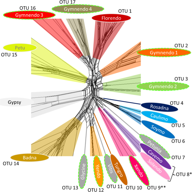
Endogenous viral elements (EVEs) are viral sequences that are integrated in the nuclear genomes of their hosts and are signatures of viral infections that may have occurred millions of years ago. The study of EVEs, coined paleovirology, provides important insights into virus evolution. The Caulimoviridae is the most common group of EVEs in plants, although their presence has often been overlooked in plant genome studies. We have refined methods for the identification of caulimovirid EVEs and interrogated the genomes of a broad diversity of plant taxa, from algae to advanced flowering plants. Evidence is provided that almost every vascular plant (tracheophyte), including the most primitive taxa (clubmosses, ferns and gymnosperms) contains caulimovirid EVEs, many of which represent previously unrecognized evolutionary branches. In angiosperms, EVEs from at least one and as many as five different caulimovirid genera were frequently detected, and florendoviruses were the most widely distributed, followed by petuviruses. From the analysis of the distribution of different caulimovirid genera within different plant species, we propose a working evolutionary scenario in which this family of viruses emerged at latest during Devonian era (approx. 320 million years ago) followed by vertical transmission and by several cross-division host swaps.


Conflict of interest statement

The authors declare that they have no competing interests.

Figures

Figure 1
Figure 1
Augmented diversity of the Caulimoviridae. Core of asequence similarity network constructed using an alignment of amino acid reverse transcriptase (RT) sequences from reference genera, representative endogenous caulimovirids and Ty3/Gypsy LTR retrotransposons. The full network is available in Supplementary Fig. 1. Operational taxonomic units (OTUs) that do not correspond to recognized genera are highlighted by  dashed lime green outlines (referred to as novel OTUs). Each fill color corresponds to a different OTU, except for  novel OTUs that contain only one type of sequence, which are shaded dark grey and named after the host plant genome of origin (Petunia-, Vitis-, and Glycine-virus). *RT clustering at 55% amino acid identity groups, which has led to the lumping of the genera Cavemovirus and Solendovirus in a single OTU (OTU 8). **Sequences grouped in the Xendovirus OTU are paraphyletic after phylogenetic reconstruction (see Fig. 3).
Figure 2
Figure 2
Estimates of copy numbers and densities of endogenous caulimovirids (ECRTs) in different plant genomes. (A) Number of ECRTs found in each plant genome as a function of Log10 genome size, expressed in megabases (assembly gaps excluded). The logarithmic trendline indicates moderate correlation between the number of ECRTs and genome size (R2 = 0.544). (B) Density of ECRTs per megabase in each plant genome as a function of Log10 genome size, expressed in megabases (assembly gaps excluded). In (A) and (B), arrows indicate outliers andcorresponding plant species names.
Figure 3
Figure 3
Phylogeny of the Caulimoviridae, as inferred using  maximum likelihood criteria and a multiple sequence alignment of protease, reverse transcriptase and ribonuclease H1 domain sequences from recognized (black) and putative (red) genera. Ty3/Gypsy LTR retrotransposons were designated as the outgroup. Bootstrap support values below 50% are not shown. Branch nodes were collapsed until only taxa at the genus level were illustrated. Viruses included in the analysis were as follows: Orendovirus - Aegilops tauschii virus and Brachypodium distachyon virus; Tungrovirus - Rice tungro bacilliform virus (Type and West Bengal isolates); Badnavirus - Commelina yellow mottle virus and Banana streak OL virus; Yendovirus - Capiscum annuum virus; Zendovirus - Fragaria vesca virus; Unassigned - Blueberry fruit drop-associated virus (BFDaV); Caulimovirus - Cauliflower mosaic virus and Figwort mosaic virus; Unassigned - Rudbeckia flower distortion virus (RuFDV); Soymovirus - Soybean chlorotic mottle virus and Peanut chlorotic streak virus; Solendovirus - Sweet potato vein clearing virus and Tobacco vein clearing virus; Cavemovirus - Cassava vein mosaic virus and Sweet potato collusive virus; Petuvirus - Petunia vein clearing virus; Rosadnavirus - Rose yellow vein virus; Florendovirus - Fragaria vesca virus and Mimulus guttatus virus; Gymnendovirus 1 - Pinus taeda gymnendovirus 1 and Picea glauca gymnendovirus 1; Gymnendovirus 2 - Pinus taeda gymnendovirus 2, Picea glauca gymnendovirus 2 and Ginkgo biloba gymnendovirus 2; Gymnendovirus 3 - Pinus taeda gymnendovirus 3; Gymnendovirus 4 - Pinus taeda gymnendovirus 4 and Picea glauca gymnendovirus 4; Fernendovirus 1: Cystopteris protrusa fernendovirus 1 contig 1, and the transcript scaffolds BEGM-2004510 from Botrypus virginianus, NOKI-2097008 from Lindsaea linearis, and ENQF-2084799 from Lycopodium annotinum; Fernendovirus 2 - Dipteris conjugata fernendovirus 2 Contigs 2, 4 and 1319. Clade A and B and the last common ancestor (LCA) of the Caulimoviridae are indicated with red arrows. The upper cladogram indicates the evolutionary relationships between major classes of vascular plants. At the intersection between both trees, colored boxes indicate the presence of either endogenous (green) or exogenous (blue) representatives of the Caulimoviridae.
Figure 4
Figure 4
Distribution of endogenous caulimovirid RTs (ECRTs) among the Euphyllophytes. The  cladogram on the left margin represents  the phylogeny of euphyllophyte species investigated in this study; the names of major branches and nodes are indicated. The cladogram on the upper margin, which represents virus phylogeny, is derived from Fig. 3. At the intersection of these two trees, the colored boxes indicate the number of ECRT loci from each virus genus in each plant genome. Abbreviations of virus genera are as follows: Pe (Petuvirus), Gy1 (Gymnendovirus 1), Gy2 (Gymnendovirus 2), Gy3 (Gymnendovirus 3), Gy4 (Gymnendovirus 4), Fe1 (Fernendovirus 1), Fe2 (Fernendovirus 2), Flo (Florendovirus), Soy (Soymovirus), Rud (Rudbeckia flower distortion virus), Cau (Caulimovirus), Blu (Blueberry fruit drop-associated virus), Zen (Zendovirus), Xen (Xendovirus), Yen (Yendovirus), CaS (Cavemovirus + Solendovirus), Ros (Rosadnavirus), Bad (Badnavirus), Tun (Tungrovirus), Ore (Orendovirus).

References

    1. Roossinck MJ. Deep sequencing for discovery and evolutionary analysis of plant viruses. Virus Res. 2017;239:82–86. doi: 10.1016/j.virusres.2016.11.019. - DOI - PubMed
    1. Aiewsakun P, Katzourakis A. Endogenous viruses: Connecting recent and ancient viral evolution. Virology. 2015;479-480:26–37. doi: 10.1016/j.virol.2015.02.011. - DOI - PubMed
    1. Hayward A, Grabherr M, Jern P. Broad-scale phylogenomics provides insights into retrovirus-host evolution. Proc. Natl. Acad. Sci. USA. 2013;110:20146–20151. doi: 10.1073/pnas.1315419110. - DOI - PMC - PubMed
    1. Hayward A, Cornwallis CK, Jern P. Pan-vertebrate comparative genomics unmasks retrovirus macroevolution. Proc. Natl. Acad. Sci. USA. 2015;112:464–469. doi: 10.1073/pnas.1414980112. - DOI - PMC - PubMed
    1. Aiewsakun P, Katzourakis A. Marine origin of retroviruses in the early Palaeozoic Era. Nat Commun. 2017;8:13954. doi: 10.1038/ncomms13954. - DOI - PMC - PubMed

Publication types

LinkOut - more resources