. 2018 Jan 12;8(1):606.
doi: 10.1038/s41598-017-18739-3.

miR-143-3p inhibits the proliferation, migration and invasion in osteosarcoma by targeting FOSL2

Affiliations

miR-143-3p inhibits the proliferation, migration and invasion in osteosarcoma by targeting FOSL2

Xiangran Sun et al. Sci Rep. .

Abstract

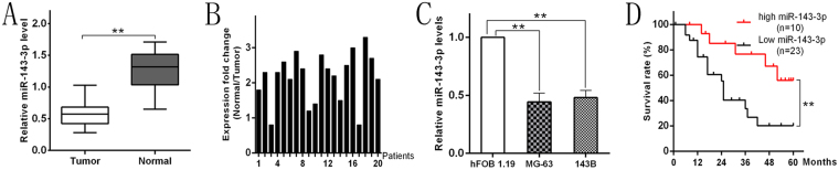
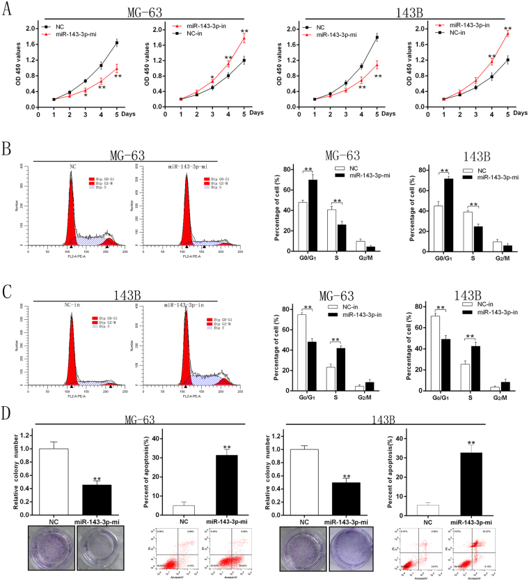
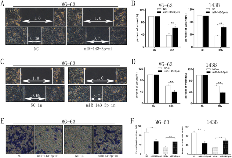
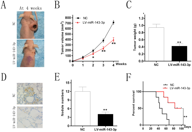
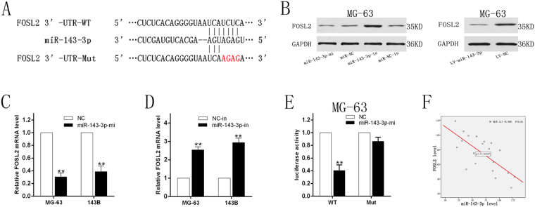
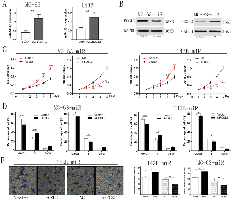
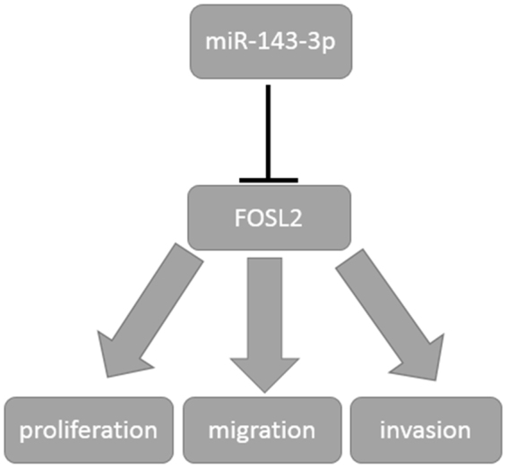
Osteosarcoma (OS) is the most common type of primary malignant bone tumor and mainly occurs in children and adolescent. Because of its early migration and invasion, OS has a poor prognosis. It has been reported that mircoRNAs (miRNAs) play a crucial role in the occurrence and development of multiple tumors. In this study, we identified the aberrant-expression of miR-143-3p in osteosarcoma and examined the role of miR-143-3p in OS development. Further, we searched the miR-143-3p target gene and verified its accuracy by luciferase experiments. Finally, we explored the relationship between miR-143-3p and FOS-Like antigen 2 (FOSL2). Our data indicated that miR-143-3p expression was substantially lower in OS tissues and cell-line compared with normal tissues, and was lower in patients with poor prognosis. In addition miR-143-3p inhibited OS cell proliferation and metastasis while promoting apoptosis. We next showed that FOSL2 was directly targeted by miR-143-3p and could reverse the inhibition caused by miR-143-3p. Finally, we found FOSL2 expression in OS cells was significantly higher compared with normal cells and negatively correlated with miR-143-3p. Thus, miR-143-3p directly and negatively targets FOSL2 to affect OS characteristics. This provides a new target for the treatment of OS and deserves further study.


Conflict of interest statement

The authors declare that they have no competing interests.

Figures

Figure 1
Figure 1
miR-143-3p downregulation in OS tissues and cell lines is associated with poor prognosis. (A) The miR-143-3p expression is significantly decreased in OS tissues. (B) miR-143-3p expression relative to adjacent normal tissues for 20 patients. (C) The miR-143-3p expression in MG-63 and 143B cell lines are significantly lower compare to hFOB 1.19 cell line determined by qRT-PCR. (D) Kaplan-Meier curves were performed for survival in all 33 patients.
Figure 2
Figure 2
miR-143-3p inhibits proliferation of OS cells. (A) MG-63 and 143B cell lines were transfected with miR-143-3p mimics and miR-NC and the cell viability determined with the CCK-8 assay. (B) The results of flow cytometry analysis of MG-63 and 143B cell lines after transfected with miR-144 mimics and miR-NC. (C) The results of flow cytometry analysis of 143B cell lines after transfected with miR-143-3p-in and NC-in. (D) Results of colony formation and apoptosis cells of MG-63and 143B cell lines transfected with miR-143-3p mimics and miR-NC.
Figure 3
Figure 3
miR-143-3p expression inhibits migration and invasion of OS cells in vitro. (A,B) Wound-closure assay for MG-63 and 143B cell lines transfected with miR-144 mimics and miR-NC. (C,D) Wound-closure assay for MG-63 and 143B cell lines transfected with miR-143-3p-in and NC-in. (E) Images of the Transwell invasion assay of MG-63 cell line transfected with miR-144 mimics, miR-NC, miR-143-3p-in and NC-in. (F) Quantification of Transwell invasion assay for MG-63 and143B cell lines.
Figure 4
Figure 4
miR-143-3p inhibit tumor growth and metastasis in vivo. (A) Photographs of tumors (B) Curve of tumor volume growth for the nude mice. (C) Tumor of weight. (D) KI-67 staining section of lung tissue. (E) Number of lung metastatic nodules of each group. (F) Survival curve of the two groups mice injected the cell line respectively which could stably express the miR-143-3p mimics and miR-NC.
Figure 5
Figure 5
miR-143-3p directly targets FOSL2. (A) The sequences of the putative miR-143-3p binding sites in wild type and mutant (red) FOSL2-3′UTR. (B) Determination of FOSL2 protein in MG-63 cell after transfection with miR-144 mimics, inhibitors and miR-NC, NC-in or miR-144 lentivirus infection. (C) Determination of FOSL2 mRNA in MG-63 and 143B cell lines after transfected with miR-144 mimics and miR-NC. (D) FOSL2 mRNA in MG-63 and 143B cell lines after transfected with miR-143-3p-in and NC-in. (E) The relative luciferase activity of luciferase reports with wild type or mutant FOSL2-3′UTR were determined in MG-63 cell line, which were transfected with the miR-143-3p mimics or miR-NC. Statistical significance was observed between the wild type and the mutant groups. (F) Spearman’s correlation analysis between miR-143-3p expression and FOSL2 mRNA level.
Figure 6
Figure 6
FOSL2 downregulation is a critical step in regulation of OS properties by miR-143-3p. (A) The miR-143-3p expression after transfected with lentivirus vectors. (B) (left) MG-63-miR cell (stably express miR-143-3p) were transfected with empty pcDNA3.1 vector, FOSL2-containing plasmid; (right) MG-63-miR cell were transfected with siRNA against FOSL2. (C) Cell viability of the two cell lines were measured by CCK-8 assay. The results showed that cell viability of ectopic FOSL2 group was higher than the control group. And the cell viability of siFOSL2 group was lower than NC group. (D) Cell cycle distribution were measured by flow cytometry analysis. The effects of G1/S phase blocking was weakened by ectopic FOSL2 and enhanced by siFOSL2. (E) Image and quantification of cell invasion ability were measured by Transwell assay.
Figure 7
Figure 7
Schematic diagram of the regulation of miR143-3p to FOSL2. miR-143-3p could inhibit the proliferation, migration and invasion of OS by regulating the expression of FOSL2 negatively.

References

    1. Yu W, et al. A review and outlook in the treatment of osteosarcoma and other deep tumors with photodynamic therapy: from basic to deep. Oncotarget. 2017;8:39833. - PMC - PubMed
    1. Bielack SS, Hecker-Nolting S, Blattmann C, Kager L. Advances in the management of osteosarcoma. F1000 Res. 2016;5:2767. doi: 10.12688/f1000research.9465.1. - DOI - PMC - PubMed
    1. Friebele JC, Peck J, Pan X, Abdel-Rasoul M, Mayerson JL. Osteosarcoma: A Meta-Analysis and Review of the Literature. Am J Orthop (Belle Mead NJ) 2015;44:547. - PubMed
    1. Chang L, Shrestha S, LaChaud G, Scott MA, James AW. Review of microRNA in osteosarcoma and chondrosarcoma. Med Oncol. 2015;32:613. - PubMed
    1. Taran SJ, Taran R, Malipatil NB. Pediatric Osteosarcoma: An Updated Review. Indian J Med Paediatr Oncol. 2017;38:33. doi: 10.4103/0971-5851.203513. - DOI - PMC - PubMed

Publication types