. 2018 Apr;27(4):848-860.
doi: 10.1002/pro.3374. Epub 2018 Jan 30.

Structure, function and evolution of the hemerythrin-like domain superfamily

Affiliations

Structure, function and evolution of the hemerythrin-like domain superfamily

Claudia Alvarez-Carreño et al. Protein Sci. 2018 Apr.

Abstract

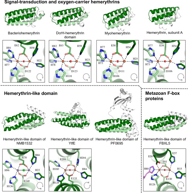
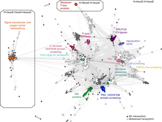
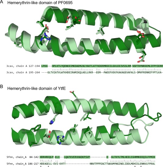
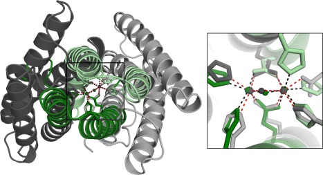
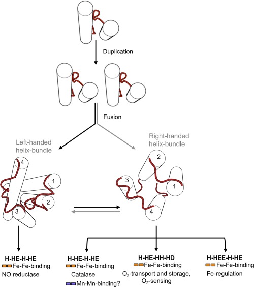
Hemerythrin-like proteins have generally been studied for their ability to reversibly bind oxygen through their binuclear nonheme iron centers. However, in recent years, it has become increasingly evident that some members of the hemerythrin-like superfamily also participate in many other biological processes. For instance, the binuclear nonheme iron site of YtfE, a hemerythrin-like protein involved in the repair of iron centers in Escherichia coli, catalyzes the reduction of nitric oxide to nitrous oxide, and the human F-box/LRR-repeat protein 5, which contains a hemerythrin-like domain, is involved in intracellular iron homeostasis. Furthermore, structural data on hemerythrin-like domains from two proteins of unknown function, PF0695 from Pyrococcus furiosus and NMB1532 from Neisseria meningitidis, show that the cation-binding sites, typical of hemerythrin, can be absent or be occupied by metal ions other than iron. To systematically investigate this functional and structural diversity of the hemerythrin-like superfamily, we have collected hemerythrin-like sequences from a database comprising fully sequenced proteomes and generated a cluster map based on their all-against-all pairwise sequence similarity. Our results show that the hemerythrin-like superfamily comprises a large number of protein families which can be classified into three broad groups on the basis of their cation-coordinating residues: (a) signal-transduction and oxygen-carrier hemerythrins (H-HxxxE-HxxxH-HxxxxD); (b) hemerythrin-like (H-HxxxE-H-HxxxE); and, (c) metazoan F-box proteins (H-HExxE-H-HxxxE). Interestingly, all but two hemerythrin-like families exhibit internal sequence and structural symmetry, suggesting that a duplication event may have led to the origin of the hemerythrin domain.

Keywords: hemerythrin-like superfamily subgroups; nonheme iron protein; oxygen-binding protein; up-and-down bundle.


Figures

Figure 1
Figure 1
Ribbon diagram of distant hemerythrin homologues. In all structures, helices 1 and 2 are colored in dark green; helices 3 and 4 are colored in light green. Non‐homologous regions are shown in white. A disordered region in the tertiary structure of hemerythrin in FBXL5 is highlighted in purple. Gray arrows indicate the sequential arrangement of the helices in the hemerythrin fold.
Figure 2
Figure 2
Cluster map of hemerythrin homologs. Cluster map of 6599 hemerythrin sequences in two‐dimensional space at a P value cutoff of 1e‐10. Dotted lines enclose three large groups formed at a P value cutoff of 1e‐13. Each dot represents a sequence; dots are colored by groups of sequences with known domain organization. (1) Q9PIQ3 and (2) Q0P932 from Campylobacter jejuni; (3) Q60AX2 from Methylococcus capsulatus, and Q726F3 from Desulfovibrio vulgaris; (4) Q9KSP0 from Vibrio cholerae; (5) Q9RJ01 from Streptomyces coelicolor; (6) Q92Z60 from Rhizobium meliloti; (7) Q8YS92 from Nostoc sp.; (8) P69506 from Escherichia coli; (9) Q7WX96 from Cupriavidus necator; (10) Q8U2Y3 from Pyrococcus furiosus; (11) A0KMZ0 from Aeromonas hydrophila; (12) Q9JYL1 from Neisseria meningitidis; (13) Q9UKA1 from Homo sapiens; (14) A0QXI3, A0R5J3, A0QV17 from Mycobacterium smegmatis; (15) Q8LPQ5 from Arabidopsis thaliana; (16) V9G2Z0 from Oryza sativa; and (17) Rv2633c from Mycobacterium tuberculosis.
Figure 3
Figure 3
Repeat units of the hemerythrin‐like domain. Internal sequence symmetry was identified by HHpredID with a probability >90. Structural superimposition of the repeat units in (A) PF0695 from Pyrococcus furiosus (PDB ID: 3CAX), and in (B) YtfE from E. coli (PDB ID: 5FNN). Sequence alignments are based on the structural superimposition of the repeat units. Aligned residues are connected by a dot. Exact matches are connected by a colon. Amino acids that participate in cation‐coordination are marked with an asterisk.
Figure 4
Figure 4
Local structural similarity between hemerythrin and Q4MWP8 from Bacillus cereus G9241. Ribbon diagram of two evolutionarily unrelated protein structures, Q4MWP8 from Bacillus cereus G9241 (shown in gray, PDB ID: 3DBY) and hemerythrin‐like domain of DcrH (shown in green, PDB ID: 2AWC), aligned with MetalS2. The inset shows the superposition of the metal‐binding sites.
Figure 5
Figure 5
Fold change in the evolution of the hemerythrin‐like domain superfamily. Schematic representation of the evolution of hemerythrins depicting possible scenarios for the origination of different handedness of the hemerythrin fold.

References

    1. Terwilliger NB (1998) Functional adaptations of oxygen‐transport proteins. J Exp Biol 201:1085–1098. - PubMed
    1. Rouault TA (2009) An ancient gauge for iron. Science 326:676–677. - PMC - PubMed
    1. Salahudeen AA, Thompson JW, Ruiz JC, Ma H‐W, Kinch LN, Li Q, Grishin NV, Bruick RK (2009) An E3 ligase possessing an iron‐responsive hemerythrin domain is a regulator of iron homeostasis. Science 326:722–726. - PMC - PubMed
    1. Vashisht AA, Zumbrennen KB, Huang X, Powers DN, Durazo A, Sun D, Bhaskaran N, Persson A, Uhlen M, Sangfelt O, Spruck C, Leibold EA, Wohlschlegel JA (2009) Control of iron homeostasis by an iron‐regulated ubiquitin ligase. Science 326:718–721. - PMC - PubMed
    1. Selote D, Samira R, Matthiadis A, Gillikin JW, Long TA (2015) Iron‐binding E3 ligase mediates iron response in plants by targeting basic helix‐loop‐helix transcription factors. Plant Physiol 167:273–286. - PMC - PubMed

Publication types

LinkOut - more resources