Review
. 2018 Feb;56(2):10.1002/dvg.23091.
doi: 10.1002/dvg.23091. Epub 2018 Jan 25.

Generating retinoic acid gradients by local degradation during craniofacial development: One cell's cue is another cell's poison

Affiliations
Review

Generating retinoic acid gradients by local degradation during craniofacial development: One cell's cue is another cell's poison

Aditi Dubey et al. Genesis. 2018 Feb.

Abstract

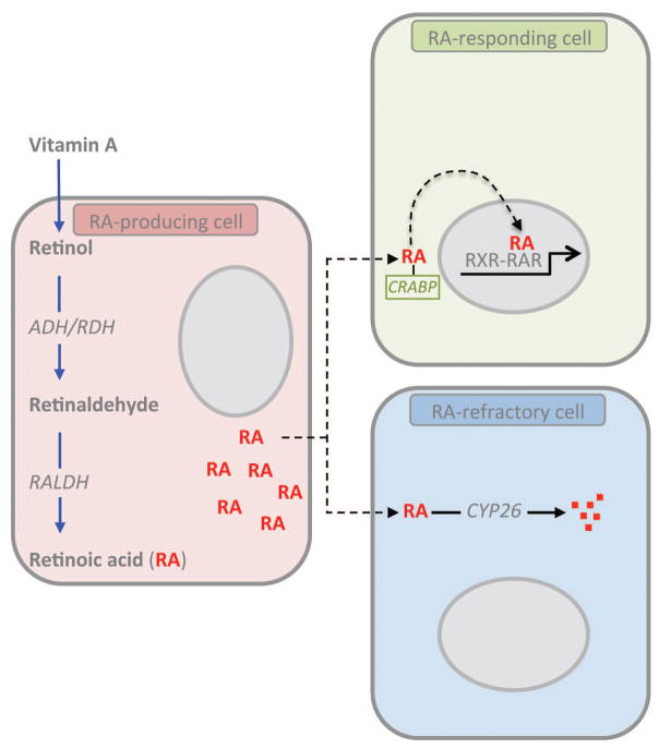
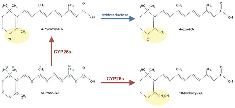
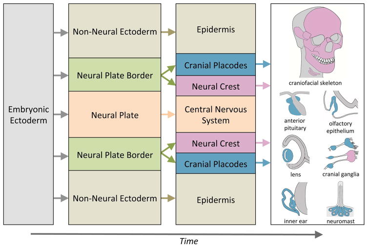
Retinoic acid (RA) is a vital morphogen for early patterning and organogenesis in the developing embryo. RA is a diffusible, lipophilic molecule that signals via nuclear RA receptor heterodimeric units that regulate gene expression by interacting with RA response elements in promoters of a significant number of genes. For precise RA signaling, a robust gradient of the morphogen is required. The developing embryo contains regions that produce RA, and specific intracellular concentrations of RA are created through local degradation mediated by Cyp26 enzymes. In order to elucidate the mechanisms by which RA executes precise developmental programs, the kinetics of RA metabolism must be clearly understood. Recent advances in techniques for endogenous RA detection and quantification have paved the way for mechanistic studies to shed light on downstream gene expression regulation coordinated by RA. It is increasingly coming to light that RA signaling operates not only at precise concentrations but also employs mechanisms of degradation and feedback inhibition to self-regulate its levels. A global gradient of RA throughout the embryo is often found concurrently with several local gradients, created by juxtaposed domains of RA synthesis and degradation. The existence of such local gradients has been found especially critical for the proper development of craniofacial structures that arise from the neural crest and the cranial placode populations. In this review, we summarize the current understanding of how local gradients of RA are established in the embryo and their impact on craniofacial development.

Keywords: Cyp26; Raldh; craniofacial; degradation; gradient; neural crest; placode; retinoic acid.


Figures

Figure 1
Figure 1. Retinoic acid production and signaling
A schematic diagram of the three-types of cells involved in RA signaling. RA-producing cells (pink) uptake retinol synthesized from Vitamin A and undergo a series of enzymatic steps culminating in the synthesis of RA by RALDH. This RA can alter gene expression in the RA-producing cell (autocrine signaling) or diffuse to neighboring cells (paracrine signaling). Neighboring cells may be RA-responsive (green) or RA-refractory (blue). RA responsive cells uptake RA through cellular retinoid binding proteins such as CRABP and allow signaling through RAR/RXR heterodimers in the nucleus. These heterodimers interact with RA-responsive elements (RAREs) in the promoters of genes and result in gene activation or repression. RA-refractory cells express RA catabolizing Cyp26 enzymes that convert RA into polar metabolites that are cleared by the cell. Typically, RA-producing and RA-refractory cells are mutually exclusive.
Figure 2
Figure 2. Retinoic acid metabolism and degradation
A schematic representation of catabolism of RA by Cyp26 enzymes. RA contains a β-ionone ring (C1–C6) conjugated to a polyene hydrocarbon chain (C7–C15). Cyp26 enzymes act mainly on C4–C5 and C18 (highlighted in yellow) of the β-ionone ring of all-trans RA (bottom left) to generated polar metabolites. The main metabolites generated by all three Cyp26 enzymes are 4-hydroxy RA (top left), which gets converted to 4-oxo RA by an unknown oxidoreductase (blue arrow, top right), and 18-hydroxy RA (bottom right).
Figure 3
Figure 3. Major subdivisions of the embryonic ectoderm and contributions of the neural crest and cranial placodes to craniofacial structures
At the end of gastrulation, the embryonic ectoderm is subdivided into three domains: the non-neuronal ectoderm, neural plate and the neural plate border. The non-neural ectoderm and neural plate give rise to the epidermis and central nervous system (CNS), respectively. The neural plate border develops into two cell populations the neural crest and cranial placodes. In the head region the neural crest (red) contributes to the craniofacial skeleton and a subset of cranial ganglia. The cranial placodes (blue) differentiate into the anterior pituitary, optic lens, inner ear, olfactory epithelium and cranial ganglia, as well as neuromasts of the lateral line in anamniotes.

References

    1. Ababon MR, Li BI, Matteson PG, Millonig JH. Quantitative Measurement of Relative Retinoic Acid Levels in E8.5 Embryos and Neurosphere Cultures Using the F9 RARE-Lacz Cell-based Reporter Assay. Journal of Visualized Experiments: JoVE. 2016;(115):54443. - PMC - PubMed
    1. Abe M, Maeda T, Wakisaka S. Retinoic acid affects craniofacial patterning by changing Fgf8 expression in the pharyngeal ectoderm. Development, Growth & Differentiation. 2008;50(9):717–729. - PubMed
    1. Abu-Abed S, Dollé P, Metzger D, Beckett B, Chambon P, Petkovich M. The retinoic acid-metabolizing enzyme, CYP26A1, is essential for normal hindbrain patterning, vertebral identity, and development of posterior structures. Genes & Development. 2001;15(2):226–240. - PMC - PubMed
    1. Abu-Abed S, MacLean G, Fraulob V, Chambon P, Petkovich M, Dollé P. Differential expression of the retinoic acid-metabolizing enzymes CYP26A1 and CYP26B1 during murine organogenesis. Mechanisms of Development. 2002;110(1):173–177. - PubMed
    1. Ahrens K, Schlosser G. Tissues and signals involved in the induction of placodal Six1 expression in Xenopus laevis. Developmental Biology. 2005;288(1):40–59. - PubMed

Publication types

LinkOut - more resources