. 2018 Jul;154(3):434-451.
doi: 10.1111/imm.12893. Epub 2018 Feb 8.

Combination therapy of lovastatin and AMP-activated protein kinase activator improves mitochondrial and peroxisomal functions and clinical disease in experimental autoimmune encephalomyelitis model

Affiliations

Combination therapy of lovastatin and AMP-activated protein kinase activator improves mitochondrial and peroxisomal functions and clinical disease in experimental autoimmune encephalomyelitis model

Inderjit Singh et al. Immunology. 2018 Jul.

Abstract

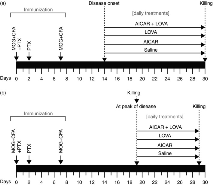
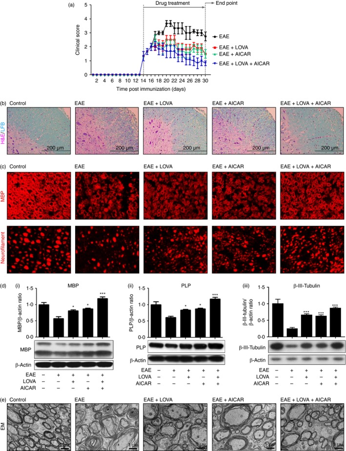
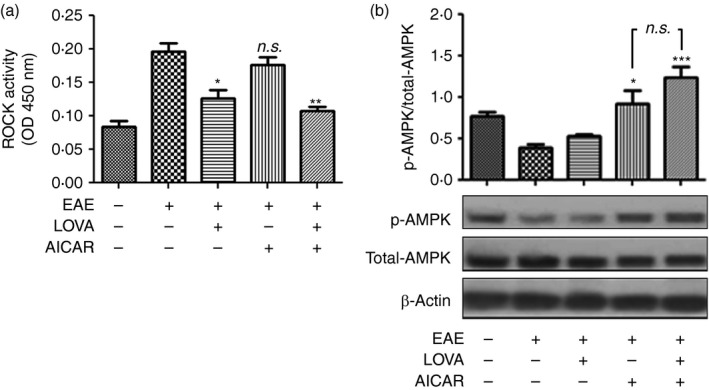
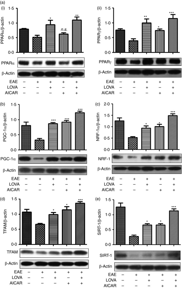
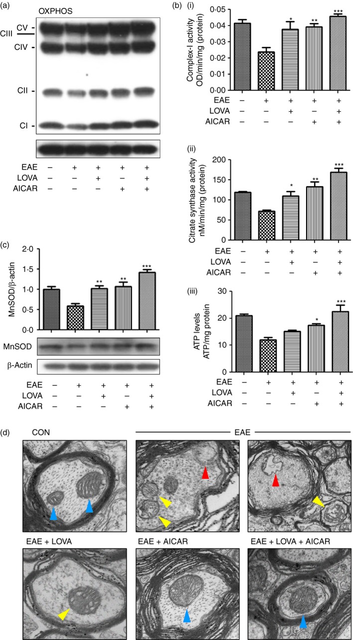
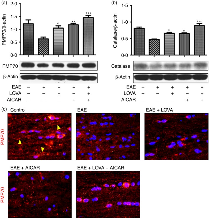
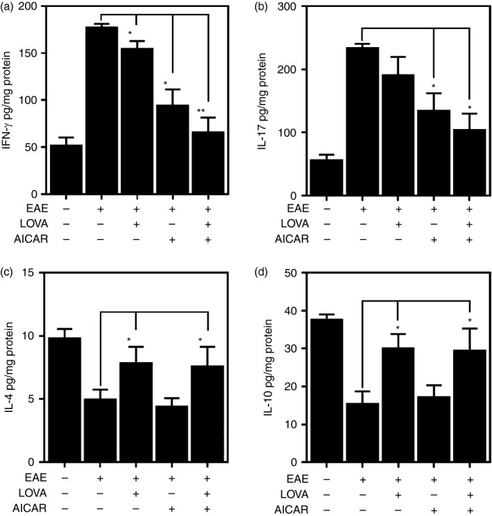
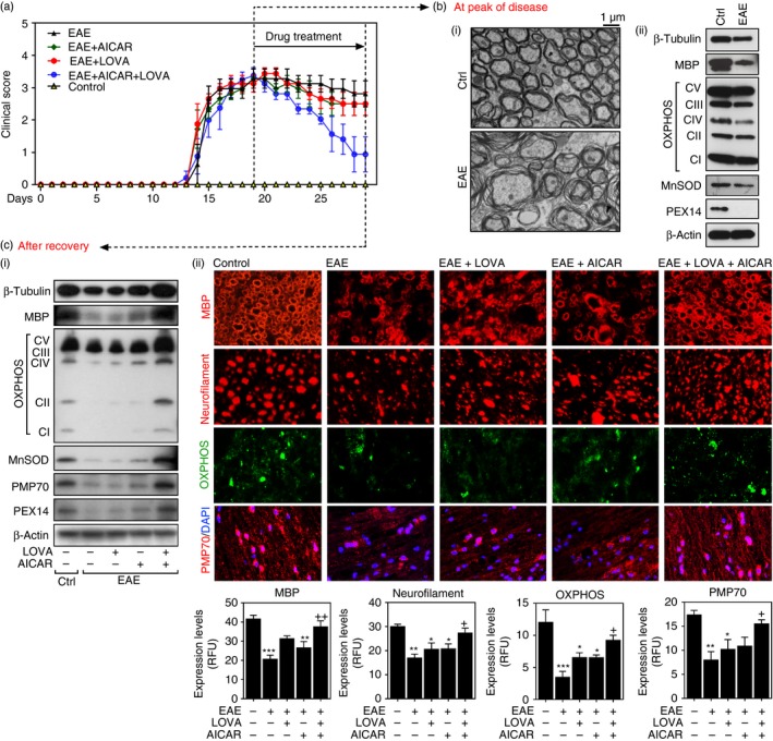
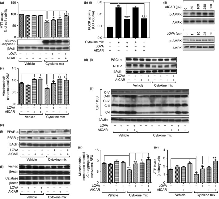
Recent studies report that loss and dysfunction of mitochondria and peroxisomes contribute to the myelin and axonal damage in multiple sclerosis (MS). In this study, we investigated the efficacy of a combination of lovastatin and AMP-activated protein kinase (AMPK) activator (AICAR) on the loss and dysfunction of mitochondria and peroxisomes and myelin and axonal damage in spinal cords, relative to the clinical disease symptoms, using a mouse model of experimental autoimmune encephalomyelitis (EAE, a model for MS). We observed that lovastatin and AICAR treatments individually provided partial protection of mitochondria/peroxisomes and myelin/axons, and therefore partial attenuation of clinical disease in EAE mice. However, treatment of EAE mice with the lovastatin and AICAR combination provided greater protection of mitochondria/peroxisomes and myelin/axons, and greater improvement in clinical disease compared with individual drug treatments. In spinal cords of EAE mice, lovastatin-mediated inhibition of RhoA and AICAR-mediated activation of AMPK cooperatively enhanced the expression of the transcription factors and regulators (e.g. PPARα/β, SIRT-1, NRF-1, and TFAM) required for biogenesis and the functions of mitochondria (e.g. OXPHOS, MnSOD) and peroxisomes (e.g. PMP70 and catalase). In summary, these studies document that oral medication with a combination of lovastatin and AICAR, which are individually known to have immunomodulatory effects, provides potent protection and repair of inflammation-induced loss and dysfunction of mitochondria and peroxisomes as well as myelin and axonal abnormalities in EAE. As statins are known to provide protection in progressive MS (Phase II study), these studies support that supplementation statin treatment with an AMPK activator may provide greater efficacy against MS.

Keywords: autoimmunity; experimental autoimmune encephalomyelitis/multiple sclerosis; neurodegeneration; neuroinflammation.


Figures

Figure 1
Figure 1
Time‐line diagram for experimental autoimmune encephalomyelitis (EAE) induction and drug treatments: Two sets of animal studies were performed: (a) To investigate the effects of 5‐aminoimidazole‐4‐carboxamide ribonucleotide (AICAR), lovastatin (LOVA), and their combination treatments, started after disease onset, on clinical and central nervous system (CNS) diseases of EAE. (b) To investigate the effects of AICAR, LOVA, and their combination treatments, started after peak of the disease, on clinical and CNS diseases of EAE.
Figure 2
Figure 2
Therapeutic effects of lovastatin (LOVA) and 5‐aminoimidazole‐4‐carboxamide ribonucleotide (AICAR) and their combination on clinical and central nervous system (CNS) diseases of experimental autoimmune encephalomyelitis (EAE). EAE was induced in female C57BL/6 mice by immunization with myelin oligodendrocyte glycoprotein (MOG) peptide. At the day of disease onset, the EAE mice were treated with LOVA (1 mg/kg/day/i.p.) or AICAR (100 mg/kg/day/i.p.) or combination of both drugs until day 30 (n = 8). Clinical disease of EAE of each mouse was analysed as described in the Materials and methods (a). At the day 30 post immunization the mice were killed and the CNS disease of EAE was analysed by H&E/LFB staining for the analysis of mononuclear inflammatory cells and myelin status (n = 4) (b), immunofluorescent staining for myelin basic protein (MBP) (myelin) and neurofilament (axons) (n = 4) (c), Western blot analysis for MBP (i) and proteolipid protein (PLP) (ii) and βIII‐tubulin (iii) (n = 4) (d), and electron microscopy (EM) for spinal cords (n = 4) (d). In this study, control mice were also treated with pertussis toxin and complete Freund's adjuvant as vehicle. Each Western blot analysis was repeated three times and all blots were quantified and represented as bar graphs. Data represent mean ± standard error of mean of three independent experiments (six animals per group). *P < 0·05; ***P < 0·001; compared with the vehicle‐treated EAE groups. Panels (b), (c), and (e) are representative among the three independent experiments.
Figure 3
Figure 3
Effects of lovastatin (LOVA) and 5‐aminoimidazole‐4‐carboxamide ribonucleotide (AICAR) and their combination on the activities of Rho‐associated kinase (ROCK) and AMP kinase (AMPK) in the spinal cords of experimental autoimmune encephalomyelitis (EAE) mice. Tissue lysates (n = 4) were extracted from spinal cords of the EAE mice treated with LOVA, AICAR, or LOVA and AICAR combination at the day 30 post‐immunization and analysed for activities of ROCK (a) and AMPK (b). AMPK activity was analysed by ratio of phospho‐AMPK (p‐AMPK) versus total AMPK following the Western blot analysis using antibody specific to phospho‐AMPK (Thr172) or pan‐AMPK (total AMPK). β‐Actin was used for internal loading control. Each analysis was repeated three times and the data are represented as a bar graph. Data represent mean ± standard error of mean of three independent experiments (six animals per group). *P < 0·05; **P < 0·01; ***P < 0·001; compared with the vehicle‐treated EAE groups. n.s. stands for not significant.
Figure 4
Figure 4
Effects of lovastatin (LOVA) and 5‐aminoimidazole‐4‐carboxamide ribonucleotide (AICAR) and their combination on the expression of peroxisomal and mitochondrial biogenesis factors in the spinal cords of experimental autoimmune encephalomyelitis (EAE) mice. Tissue lysates (n = 4) were extracted from spinal cords of the EAE mice treated with LOVA, AICAR, or LOVA and AICAR in combination at day 30 post immunization and analysed for Western blot analysis for expressions of peroxisome proliferator‐activated receptor α (PPAR α) (a‐i), PPAR γ (a‐ii), peroxisome proliferator‐activated receptor‐γ coactivator‐1α (PGC‐1α) (b), nuclear respiratory factor 1 (NRF‐1) (c) mitochondrial transcription factor A (TFAM) (d), and sirtuin 1 (SIRT1) (e). β‐Actin was used for internal loading control. Data represent mean ± standard error of mean of three independent experiments (six animals per group). Each Western blot analysis was repeated three times and all blots were quantified and represented as bar graphs. *P < 0·05; **P < 0·01; ***P < 0·001; compared with the vehicle‐treated EAE groups. n.s. stands for not significant.
Figure 5
Figure 5
Effects of lovastatin (LOVA) and 5‐aminoimidazole‐4‐carboxamide ribonucleotide (AICAR) and their combination on the function and structure of mitochondria in the spinal cords of experimental autoimmune encephalomyelitis (EAE) mice. Tissue lysates (n = 4) were extracted from spinal cords of the EAE mice treated with LOVA, AICAR, or LOVA and AICAR combination at the day 30 post immunization and analysed for expressions of mitochondrial oxidative phosphorylation (OXPHOS) (a), enzyme activities for mitochondrial complex‐I (b‐i) and citrate synthase (b‐ii), tissue levels of ATP (b‐iii), expression of mitochondrial manganese superoxide dismutase (MnSOD) (c). In OXPHOS Western blot analysis, CICVI stand for NUDFB8/Complex‐I NADH dehydrogenase 1β8 (CI), SDHB/complex‐II succinate ubiquinone oxidoreductase (CII), UQCRC2/complex‐III Ubiquinol cytochrome c oxidoreductase (CIII), MTCO1/complex‐IV cytochrome c oxidase subunit 1 (CIV), and ATP5A/complex V ATP synthase 5A (CV). β‐Actin was used for internal loading control. Data represent mean ± standard error of mean of three independent experiments (six animals per group). Each analysis was repeated three times and represented as a bar graph. Panels (a) and (d) are representative among 12 images from four mice. *P < 0·05; **P < 0·01; ***P < 0·001; compared with the vehicle‐treated EAE groups. Effect of LOVA, AICAR or LOVA + AICAR combination on EAE‐induced alteration in mitochondrial structure was analysed by electron microscopy. Blue arrowheads represent the healthy mitochondrial structure. Yellow arrowheads represent damaged mitochondrial structure with cristae disarrangement and partial cristolysis. Red arrowheads represent mitochondrial lysis. In this study, control mice were also treated with pertussis toxin and complete Freund's adjuvant as vehicle. Panels (a) and (d) are representative among the three independent experiments.
Figure 6
Figure 6
Effects of lovastatin (LOVA) and 5‐aminoimidazole‐4‐carboxamide ribonucleotide (AICAR) and their combination on the expression of peroxisomal proteins in the spinal cords of (experimental autoimmune encephalomyelitis) (EAE) mice. Tissue lysates (n = 4) were extracted from spinal cords of the EAE mice treated with LOVA, AICAR or LOVA and AICAR combination at the day 30 post immunization and analysed for Western blot analysis for expressions of 70 000 MW peroxisomal membrane protein (PMP70) (a) and catalase (b). β‐Actin was used for internal loading control. Each Western analysis was repeated three times and all blots were quantified and represented as bar graph. Data represents mean ± standard error of mean of three independent experiments (six animals per group). *P < 0·05; **P < 0·01; ***P < 0·001; compared with the vehicle‐treated EAE groups. Next, PMP70 expression in peroxisomes was analysed by immunofluorescent staining of the spinal cord section (c). Panel (c) is the representative among the three independent experiments. The punctuated red/orange dots represent intact peroxisomes (yellow arrowheads).
Figure 7
Figure 7
Release of pro‐inflammatory and anti‐inflammatory cytokines by central nervous system (CNS) mononuclear cells isolated from experimental autoimmune encephalomyelitis (EAE) mice treated with lovastatin (LOVA), 5‐aminoimidazole‐4‐carboxamide ribonucleotide (AICAR) or LOVA and AICAR combination. EAE was induced in female C57BL/6 mice by immunization with myelin oligodendrocyte glycoprotein (MOG) peptide. At the day of disease onset, the EAE mice were treated with LOVA or AICAR or a combination of both drugs until day 30. Mononuclear cells were isolated from spinal cords and brains from these mice (n = 4) and stimulated with MOG peptide and release of interferon (IFN). Data represent mean ± standard error mean (n = 4 animals). *P < 0·05; **P < 0·01; compared to EAE groups.
Figure 8
Figure 8
Neurorepair effects of lovastatin (LOVA) and 5‐aminoimidazole‐4‐carboxamide ribonucleotide (AICAR) and their combination on clinical and central nervous system (CNS) disease of experimental autoimmune encephalomyelitis (EAE). EAE was induced in female C57BL/6 mice by immunization with MOG peptide. At the peak of disease (day 19 post immunization), the EAE mice were treated with lovastatin (LOVA; 1 mg/kg/day/i.p.) or AICAR (100 mg/kg/day/i.p.) or a combination of both drugs until day 29 (n = 12 for control and EAE groups and n = 8 for AICAR, LOVA and AICAR+LOVA treated groups). Clinical disease of EAE of each mouse was analysed as described under Materials and methods (a). At the peak of the disease, the control and EAE mice (n = 4) were killed and myelin structure in the spinal cord was analysed by electron microscopy (b‐i) and the spinal cord expressions of βIII‐tubulin (for axons), MBP (for myelin), OXPHOS and MnSOD (for mitochondria), PEX14 and (for peroxisomes) were analysed by Western blot analysis (b‐ii). Panel (b) is the representative among the three independent experiments. For Western analysis, the spinal cord samples (n = 4) were homogenized and equal amounts of proteins were pooled. The levels of β‐actin were used for internal loading control for Western blot analysis. At day 29 post immunization, the control and EAE mice and EAE mice treated with LOVA, AICAR or LOVA + AICAR combination (n = 8 per each group) were killed and the spinal cord expression of βIII‐tubulin, MBP, OXPHOS, MnSOD, 70 000 MW peroxisomal membrane protein (PMP70) and PEX14 was analysed by Western blot analysis (c‐i). For Western blot analysis, the spinal cord samples (n = 4) were homogenized and equal amounts of proteins were pooled. Status of myelin (MBP/coronal sections), axons (neurofilament/coronal sections), mitochondria (OXPHOS/coronal sections), and peroxisomes (PMP70/sagittal sections) was also analysed by immunofluorescent staining of spinal cord sections (c‐ii). DAPI was used for the staining of nuclei. The bar graphs represent immunofluorescence quantified using imagepro plus. Data represent mean ± standard error of mean (n = 4 animals). *P < 0·05; **P < 0·01; ***P < 0·001; compared with the vehicle‐treated control groups. + P < 0·05; ++ P < 0·01; compared with the vehicle‐treated EAE groups.
Figure 9
Figure 9
Effect of lovastatin (LOVA) and 5‐aminoimidazole‐4‐carboxamide ribonucleotide (AICAR) combination on inflammatory oligodendrocyte (OL) death and mitochondrial and peroxisomal biogenesis in cell culture model. Effects of LOVA (10 μm), AICAR (200 μm), or their combination on pro‐inflammatory cytokines interleukin‐17 (IL‐17; 25 ng/ml), tumour necrosis factor‐α (TNFα; 10 ng/ml), and IL‐1β (10 ng/ml for 24 hr) induced loss of cell viability (MTT assay) and caspase‐3 activation (cleaved caspase‐3) were analysed in differentiated MO3.13 oligodendrocyte‐like cells (a). Effects of LOVA and AICAR on pro‐inflammatory cytokine‐induced ROCK activation (b‐i) and AMPK activation (b‐ii) were analysed as described in Materials and methods. Effect of LOVA and AICAR combination on pro‐inflammatory cytokine‐induced losses of mitochondrial copy number (ratio of mitochondrial DNA versus chromosomal DNA) (c), transcription factors for mitochondrial biogenesis (PGC1α, and NRF‐1) (d‐i), expressions of OXPHOS (d‐ii), mitochondrial membrane potential (d‐iii), cellular ATP levels (d‐iv), PPAR expression (e‐i), peroxisomal proteins [70 000 MW peroxisomal membrane protein (PMP70) and catalase] (e‐ii) were analysed as described in Materials and methods. Each analysis was repeated three times and represented as a bar graph.

Similar articles

Cited by

References

    1. Compston A, Coles A. Multiple sclerosis. Lancet 2002; 359:1221–31. - PubMed
    1. Michel L, Larochelle C, Prat A. Update on treatments in multiple sclerosis. Presse Med 2015; 44:e137–51. - PubMed
    1. Mahad D, Ziabreva I, Lassmann H, Turnbull D. Mitochondrial defects in acute multiple sclerosis lesions. Brain 2008; 131:1722–35. - PMC - PubMed
    1. Zambonin JL, Zhao C, Ohno N, Campbell GR, Engeham S, Ziabreva I et al Increased mitochondrial content in remyelinated axons: implications for multiple sclerosis. Brain 2011; 134:1901–13. - PMC - PubMed
    1. Nikic I, Merkler D, Sorbara C, Brinkoetter M, Kreutzfeldt M, Bareyre FM et al A reversible form of axon damage in experimental autoimmune encephalomyelitis and multiple sclerosis. Nat Med 2011; 17:495–9. - PubMed

Publication types

MeSH terms