

Interaction of ceramides and tear lipocalin
- PMID: 29331331
- PMCID: PMC5835416
- DOI: 10.1016/j.bbalip.2018.01.004
Interaction of ceramides and tear lipocalin
Abstract
The distribution of lipids in tears is critical to their function. Lipids in human tears may retard evaporation by forming a surface barrier at the air interface. Lipids complexed with the major lipid binding protein in tears, tear lipocalin, reside in the bulk (aqueous) and may have functions unrelated to the surface. Many new lipids species have been revealed through recent mass spectrometric studies. Their association with lipid binding proteins has not been studied. Squalene, (O-acyl) omega-hydroxy fatty acids (OAHFA) and ceramides are examples. Even well-known lipids such as wax and cholesteryl esters are only presumed to be unbound because extracts of protein fractions of tears were devoid of these lipids. Our purpose was to determine by direct binding assays if the aforementioned lipids can bind tear lipocalin. Lipids were screened for ability to displace DAUDA from tear lipocalin in a fluorescence displacement assay. Di- and tri-glycerides, squalene, OAHFA, wax and cholesterol esters did not displace DAUDA from tear lipocalin. However, ceramides displaced DAUDA. Apparent dissociation constants for ceramide-tear lipocalin complexes using fluorescent analogs were measured consistently in the submicromolar range with 3 methods, linear spectral summation, high speed centrifugal precipitation and standard fluorescence assays. At the relatively small concentrations in tears, all ceramides were complexed to tear lipocalin. The lack of binding of di- and tri-glycerides, squalene, OAHFA, as well as wax and cholesterol esters to tear lipocalin is consonant with residence of these lipids near the air interface.
Keywords: (O-acyl) omega-hydroxy fatty acids (OAHFA); Ceramide; Cholesterol esters; DAUDA; Di- and tri-acylglycerols; Dry eye; LCN1; Linear spectral summation; Lipid binding; Lipocalin-1; Squalene; Tear lipocalin; Tears; Wax esters.
Copyright © 2018 Elsevier B.V. All rights reserved.
Figures
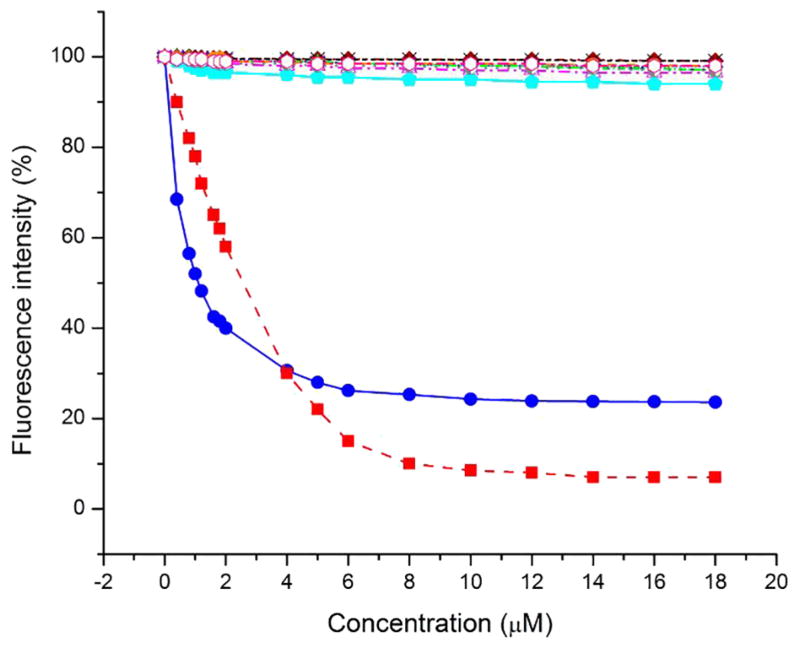
 ), stearic acid (
 ), cholesteryl stearate (
 ), cholesteryl oleate (
 ), stearyl behenate (
 ), behenyl stearate (-x-), behenyl oleate (
 ), squalene (
 ), (O-oleoyl)-16-hydroxypalmitic acid (
 ) 1,2-distearin (
 ), 1,3-distearin (
 ), tristearin (
 ).
).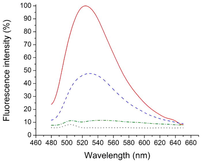
 ), 1 μM C12-NBD ceramide (
 ), 1 μM C6-NBD ceramide bound to 1 μM tear lipocalin(
 ) and 1 μM C12-NBD ceramide 1 μM tear lipocalin (
 ).
).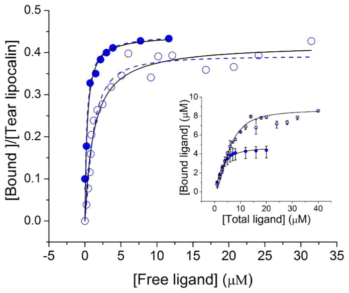
 ) and C12-NBD ceramide (
 ) to 10 μM or 20 μM tear lipocalin, respectively. Concentration of bound ligand-protein complex (supernatant) was determined by absorption spectra after centrifugal separation from unbound insoluble precipitant. Curve fit to a hyperbola (—) Kd= 0.32 μM, n=0.44 for C6-NBD ceramide and Kd= 1.23 μM and n=0.42 for C12-NBD ceramide; and the Hill equation (
) Kd= 0.29 μM, n=0.45 for C6-NBD ceramide and Kd= 1.06 μM and n=0.39 for C12-NBD ceramide. Inset: concentration of total versus bound ligand concentration. Error bars shows the range from 3 experiments.
) to 10 μM or 20 μM tear lipocalin, respectively. Concentration of bound ligand-protein complex (supernatant) was determined by absorption spectra after centrifugal separation from unbound insoluble precipitant. Curve fit to a hyperbola (—) Kd= 0.32 μM, n=0.44 for C6-NBD ceramide and Kd= 1.23 μM and n=0.42 for C12-NBD ceramide; and the Hill equation (
) Kd= 0.29 μM, n=0.45 for C6-NBD ceramide and Kd= 1.06 μM and n=0.39 for C12-NBD ceramide. Inset: concentration of total versus bound ligand concentration. Error bars shows the range from 3 experiments.
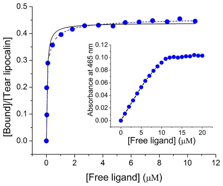
 ). Absorbance composite spectra of mixtures were fit to the sum of varying pure free and pure bound spectra. Curve was fit to hyperbola (—) Kd= 0.06 μM, n=0.45, and to Hill equation (
) Kd= 0.07 μM, n=0.47. Inset, concentration dependent absorption of C6-NBD ceramide suspended in 10 mM sodium phosphate, pH 7.3.
). Absorbance composite spectra of mixtures were fit to the sum of varying pure free and pure bound spectra. Curve was fit to hyperbola (—) Kd= 0.06 μM, n=0.45, and to Hill equation (
) Kd= 0.07 μM, n=0.47. Inset, concentration dependent absorption of C6-NBD ceramide suspended in 10 mM sodium phosphate, pH 7.3.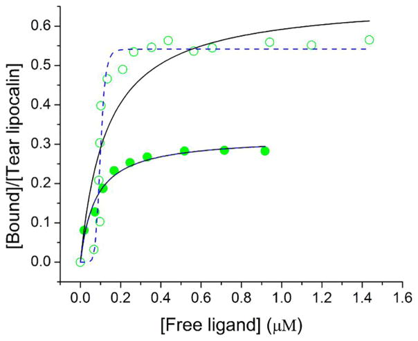
 ) and C12-NBD ceramide (
 ) to tear lipocalin by fluorescence. Curve fit to hyperbola (—) Kd= 0.08 μM and n=0.32 for C6-NBD ceramide binding and Kd= 0.13 μM and n=0.67 for C12-NBD ceramide, and to the Hill equation (
) Kd= 0.08 μM and n=0.32 for C6-NBD ceramide and Kd= 0.1 μM and n=0.21 for c12-NBD ceramide.
) to tear lipocalin by fluorescence. Curve fit to hyperbola (—) Kd= 0.08 μM and n=0.32 for C6-NBD ceramide binding and Kd= 0.13 μM and n=0.67 for C12-NBD ceramide, and to the Hill equation (
) Kd= 0.08 μM and n=0.32 for C6-NBD ceramide and Kd= 0.1 μM and n=0.21 for c12-NBD ceramide.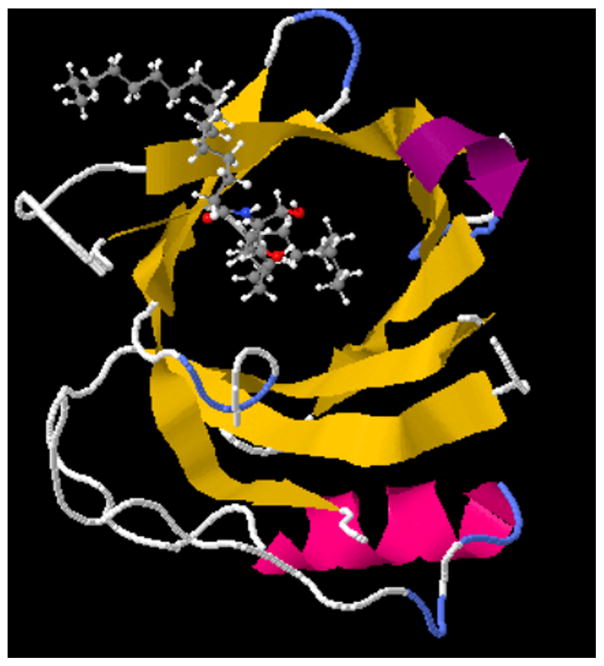
Similar articles
-
Functional cavity dimensions of tear lipocalin.Curr Eye Res. 2000 Oct;21(4):824-32. doi: 10.1076/ceyr.21.4.824.5551. Curr Eye Res. 2000. PMID: 11120574
-
Tear lipocalin captures exogenous lipid from abnormal corneal surfaces.Invest Ophthalmol Vis Sci. 2010 Apr;51(4):1981-7. doi: 10.1167/iovs.09-4622. Epub 2009 Dec 3. Invest Ophthalmol Vis Sci. 2010. PMID: 19959641 Free PMC article.
-
A novel fluorescent lipid probe for dry eye: retrieval by tear lipocalin in humans.Invest Ophthalmol Vis Sci. 2013 Feb 19;54(2):1398-410. doi: 10.1167/iovs.12-10817. Invest Ophthalmol Vis Sci. 2013. PMID: 23361507 Free PMC article.
-
Tear film lipids.Exp Eye Res. 2013 Dec;117:4-27. doi: 10.1016/j.exer.2013.05.010. Epub 2013 Jun 12. Exp Eye Res. 2013. PMID: 23769846 Free PMC article. Review.
-
Tear lipocalin: structure and function.Ocul Surf. 2011 Jul;9(3):126-38. doi: 10.1016/s1542-0124(11)70022-2. Ocul Surf. 2011. PMID: 21791187 Free PMC article. Review.
Cited by
-
Metabolomic changes in tear fluid following zinc biofortification in the BiZiFED nutritional study: a feasibility study.Front Mol Biosci. 2024 Sep 10;11:1421699. doi: 10.3389/fmolb.2024.1421699. eCollection 2024. Front Mol Biosci. 2024. PMID: 39318550 Free PMC article.
-
Ligand binding studies by high speed centrifugal precipitation and linear spectral summation using ultraviolet-visible absorption spectroscopy.MethodsX. 2018 Apr 17;5:345-351. doi: 10.1016/j.mex.2018.04.007. eCollection 2018. MethodsX. 2018. PMID: 30050754 Free PMC article.
-
Concentration dependent cholesteryl-ester and wax-ester structural relationships and meibomian gland dysfunction.Biochem Biophys Rep. 2020 Jan 30;21:100732. doi: 10.1016/j.bbrep.2020.100732. eCollection 2020 Mar. Biochem Biophys Rep. 2020. PMID: 32042930 Free PMC article.
-
Yeast Svf1 binds ceramides and contributes to sphingolipid metabolism at the ER cis-Golgi interface.J Cell Biol. 2023 May 1;222(5):e202109162. doi: 10.1083/jcb.202109162. Epub 2023 Mar 10. J Cell Biol. 2023. PMID: 36897280 Free PMC article.
-
Human Meibum Cholesteryl and Wax Ester Variability With Age, Sex, and Meibomian Gland Dysfunction.Invest Ophthalmol Vis Sci. 2019 May 1;60(6):2286-2293. doi: 10.1167/iovs.19-26812. Invest Ophthalmol Vis Sci. 2019. PMID: 31112994 Free PMC article.
References
-
- Andrews JS. Human tear film lipids. I. Composition of the principal non-polar component. Exp Eye Res. 1970;10:223–7. - PubMed
-
- Young WH, Hill RM. Tear cholesterol levels and contact lens adaptation. Am J Optom Arch Am Acad Optom. 1973;50:12–6. - PubMed
-
- van Haeringen NJ, Glasius E. Cholesterol in human tear fluid. Exp Eye Res. 1975;20:271–4. - PubMed
-
- Stuchell, et al. Lipid composition of human tears. ARVO Abstr. 1984;25:320.
-
- Saatçi AO, Irkeç M, Unlü N. Tear cholesterol levels in blepharitis. Ophthalmic Res. 1990;22:269–70. - PubMed
Publication types
MeSH terms
Substances
Grants and funding
LinkOut - more resources
Full Text Sources
Other Literature Sources
