

Elastin in lung development and disease pathogenesis
- PMID: 29331337
- PMCID: PMC6041195
- DOI: 10.1016/j.matbio.2018.01.005
Elastin in lung development and disease pathogenesis
Abstract
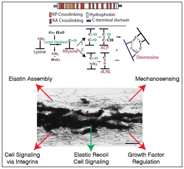
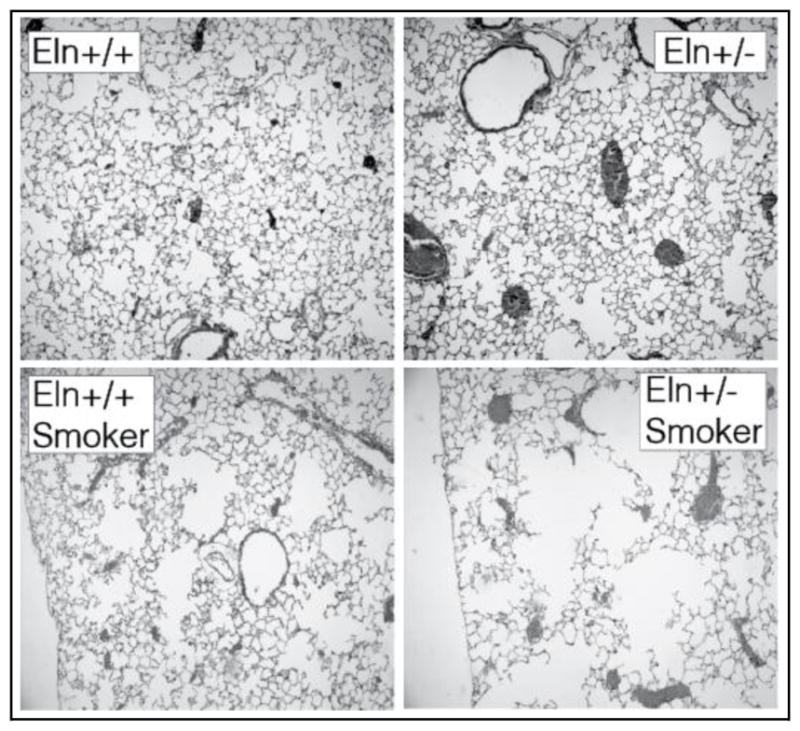
Elastin is expressed in most tissues that require elastic recoil. The protein first appeared coincident with the closed circulatory system, and was critical for the evolutionary success of the vertebrate lineage. Elastin is expressed by multiple cell types in the lung, including mesothelial cells in the pleura, smooth muscle cells in airways and blood vessels, endothelial cells, and interstitial fibroblasts. This highly crosslinked protein associates with fibrillin-containing microfibrils to form the elastic fiber, which is the physiological structure that functions in the extracellular matrix. Elastic fibers can be woven into many different shapes depending on the mechanical needs of the tissue. In large pulmonary vessels, for example, elastin forms continuous sheets, or lamellae, that separate smooth muscle layers. Outside of the vasculature, elastic fibers form an extensive fiber network that originates in the central bronchi and inserts into the distal airspaces and visceral pleura. The fibrous cables form a looping system that encircle the alveolar ducts and terminal air spaces and ensures that applied force is transmitted equally to all parts of the lung. Normal lung function depends on proper secretion and assembly of elastin, and either inhibition of elastin fiber assembly or degradation of existing elastin results in lung dysfunction and disease.
Keywords: Elastic fiber; Elastin; Emphysema; Fibrillin; Lung development; Microfibril.
Copyright © 2018 Elsevier B.V. All rights reserved.
Figures
References
-
- Liem KF. Form and function of lungs: The evolution of air breathing mechanisms. Amer Zool. 1988;28(2):739–759.
-
- Loosli C, Potter E. Pre- and postnatal development of the respiratory portion of the human lung with special reference to the elastic fibers. Am Rev Respir Dis. 1959;80(1 Part 2):5–23. - PubMed
-
- Mariani TJ, Reed JJ, Shapiro SD. Expression profiling of the developing mouse lung: insights into the establishment of the extracellular matrix. Am J Respir Cell Mol Biol. 2002;26(5):541–548. - PubMed
-
- Mariani TJ, Sandeful S, Pierce RA. Elastin in lung development. Exp Lung Res. 1997;23:131–145. - PubMed
Publication types
MeSH terms
Substances
Grants and funding
LinkOut - more resources
Full Text Sources
Other Literature Sources
Medical
Miscellaneous
