

Sharp-wave ripples as a signature of hippocampal-prefrontal reactivation for memory during sleep and waking states
- PMID: 29331447
- PMCID: PMC6039287
- DOI: 10.1016/j.nlm.2018.01.002
Sharp-wave ripples as a signature of hippocampal-prefrontal reactivation for memory during sleep and waking states
Abstract
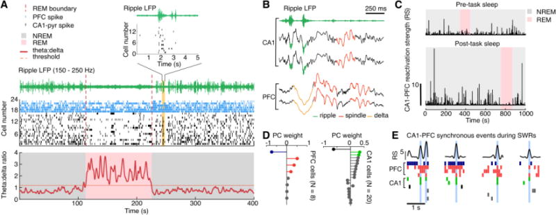
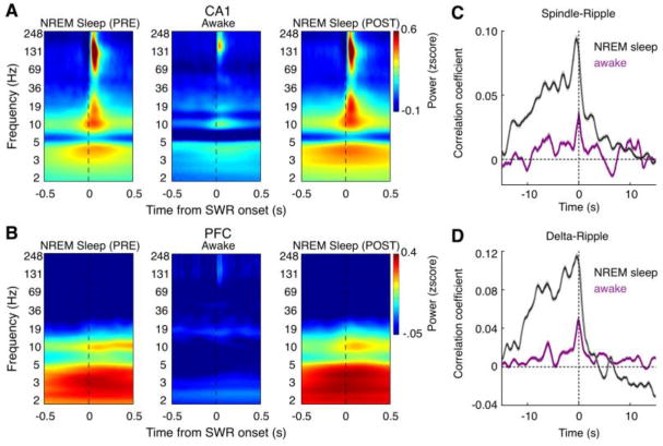
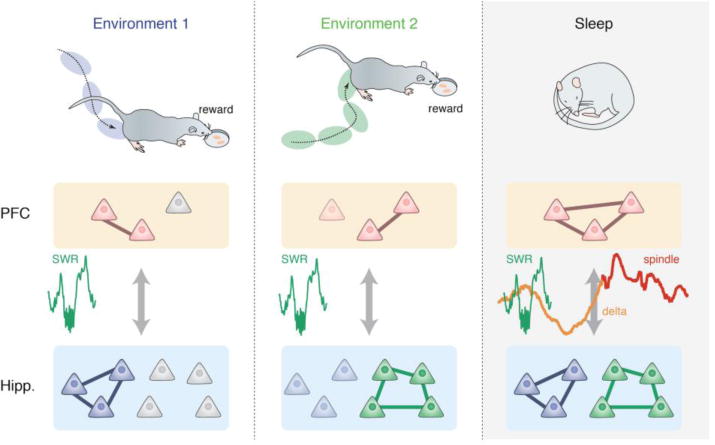
It is widely believed that memories that are encoded and retrieved during waking behavior are consolidated during sleep. Recent studies on the interactions between the hippocampus and the prefrontal cortex have greatly advanced our understanding of the physiological bases of these memory processes. Although hippocampal-prefrontal network activity differs in many aspects during waking and sleep states, here we review evidence that hippocampal sharp-wave ripples (SWRs) emerge as a common neurophysiological pattern in both states, facilitating communication between these two regions via coordinated reactivation of stored memory information. We further consider whether sleep and awake reactivation mediate similar memory processes or have different mnemonic functions, and the mechanistic role of this cross-regional dialogue in learning and memory. Finally, we provide an integrated view of how these two forms of reactivation might work together to support spatial learning and memory.
Keywords: Consolidation; Hippocampus; Memory; Prefrontal cortex; Reactivation; Sharp-wave ripples (SWRs).
Copyright © 2018 Elsevier Inc. All rights reserved.
Conflict of interest statement
Figures
Similar articles
-
Hippocampal-Prefrontal Reactivation during Learning Is Stronger in Awake Compared with Sleep States.J Neurosci. 2017 Dec 6;37(49):11789-11805. doi: 10.1523/JNEUROSCI.2291-17.2017. Epub 2017 Oct 31. J Neurosci. 2017. PMID: 29089440 Free PMC article.
-
Hippocampal sharp-wave ripples in waking and sleeping states.Curr Opin Neurobiol. 2015 Dec;35:6-12. doi: 10.1016/j.conb.2015.05.001. Epub 2015 May 23. Curr Opin Neurobiol. 2015. PMID: 26011627 Free PMC article. Review.
-
Age-associated changes in waking hippocampal sharp-wave ripples.Hippocampus. 2020 Jan;30(1):28-38. doi: 10.1002/hipo.23005. Epub 2018 Nov 11. Hippocampus. 2020. PMID: 29981255 Free PMC article.
-
The role of replay and theta sequences in mediating hippocampal-prefrontal interactions for memory and cognition.Hippocampus. 2020 Jan;30(1):60-72. doi: 10.1002/hipo.22821. Epub 2018 Jan 11. Hippocampus. 2020. PMID: 29251801 Free PMC article. Review.
-
Validating the theoretical bases of sleep reactivation during sharp-wave ripples and their association with emotional valence.Hippocampus. 2020 Jan;30(1):19-27. doi: 10.1002/hipo.23143. Epub 2019 Jul 23. Hippocampus. 2020. PMID: 31334590 Review.
Cited by
-
Multiple time-scales of decision-making in the hippocampus and prefrontal cortex.Elife. 2021 Mar 8;10:e66227. doi: 10.7554/eLife.66227. Elife. 2021. PMID: 33683201 Free PMC article.
-
Inhibition is a prevalent mode of activity in the neocortex around awake hippocampal ripples in mice.Elife. 2023 Jan 16;12:e79513. doi: 10.7554/eLife.79513. Elife. 2023. PMID: 36645126 Free PMC article.
-
Hippocampo-cortical circuits for selective memory encoding, routing, and replay.Neuron. 2023 Jul 5;111(13):2076-2090.e9. doi: 10.1016/j.neuron.2023.04.015. Epub 2023 May 16. Neuron. 2023. PMID: 37196658 Free PMC article.
-
Nightmares and the Cannabinoids.Curr Neuropharmacol. 2020;18(8):754-768. doi: 10.2174/1570159X18666200114142321. Curr Neuropharmacol. 2020. PMID: 31934840 Free PMC article. Review.
-
Reconfiguration of the cortical-hippocampal interaction may compensate for Sharp-Wave Ripple deficits in APP/PS1 mice and support spatial memory formation.PLoS One. 2020 Dec 31;15(12):e0243767. doi: 10.1371/journal.pone.0243767. eCollection 2020. PLoS One. 2020. PMID: 33382724 Free PMC article.
References
Publication types
MeSH terms
Grants and funding
LinkOut - more resources
Full Text Sources
Other Literature Sources
Medical
