. 2018 Mar:516:86-101.
doi: 10.1016/j.virol.2018.01.002. Epub 2018 Jan 11.

Classification and evolution of human papillomavirus genome variants: Alpha-5 (HPV26, 51, 69, 82), Alpha-6 (HPV30, 53, 56, 66), Alpha-11 (HPV34, 73), Alpha-13 (HPV54) and Alpha-3 (HPV61)

Affiliations

Classification and evolution of human papillomavirus genome variants: Alpha-5 (HPV26, 51, 69, 82), Alpha-6 (HPV30, 53, 56, 66), Alpha-11 (HPV34, 73), Alpha-13 (HPV54) and Alpha-3 (HPV61)

Zigui Chen et al. Virology. 2018 Mar.

Abstract

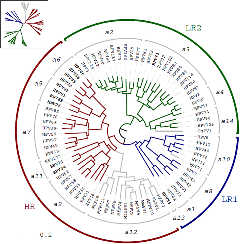
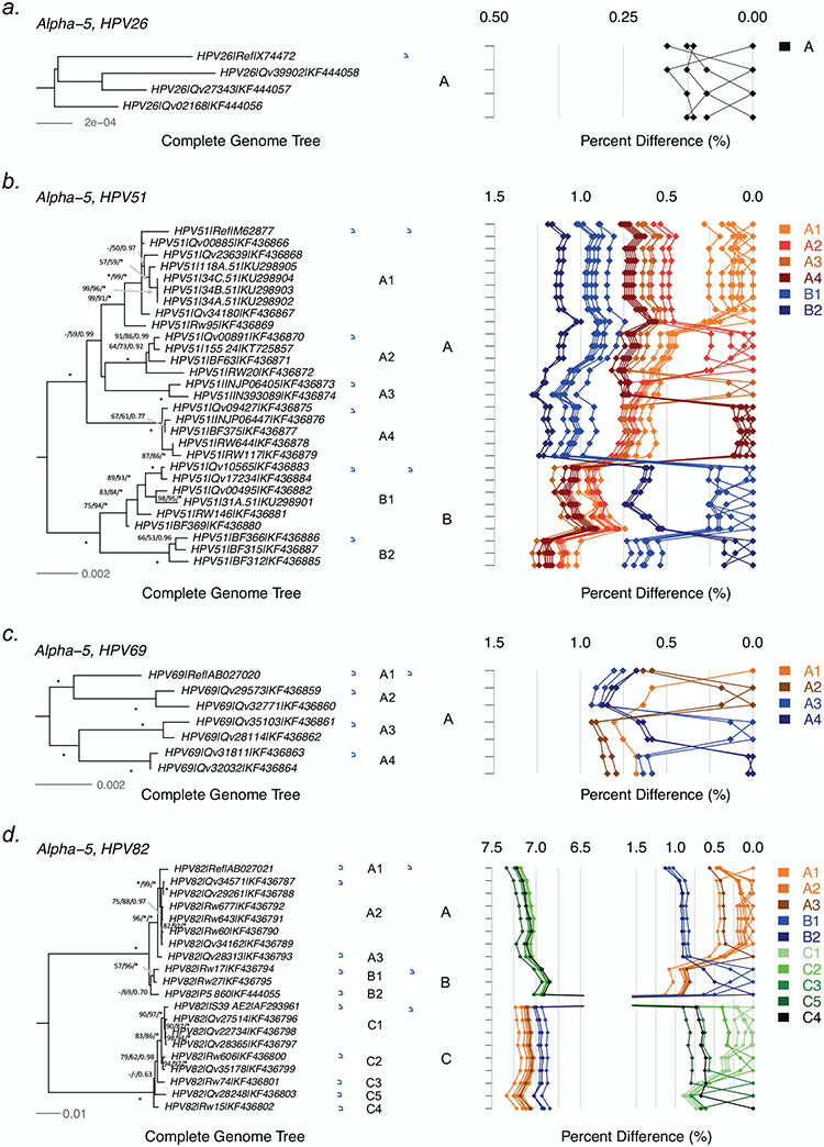
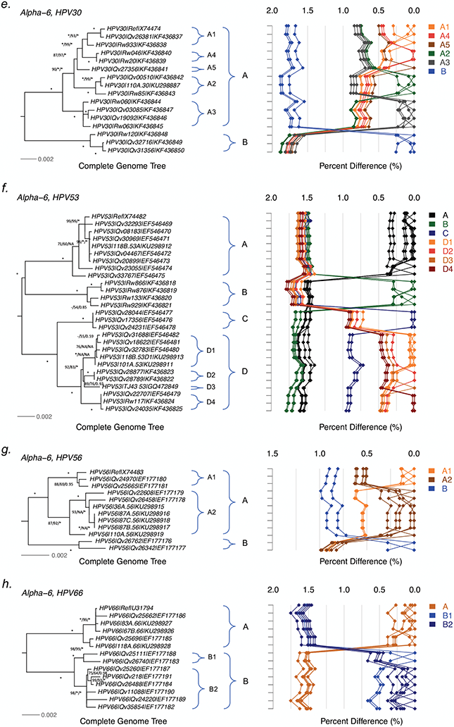
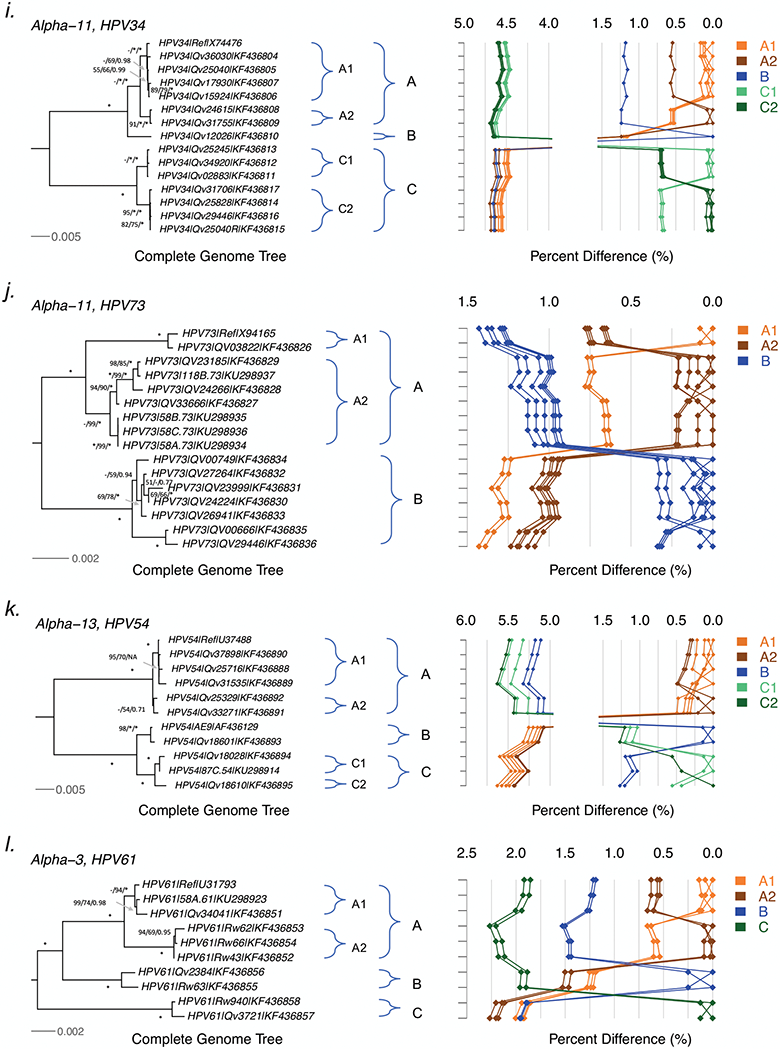
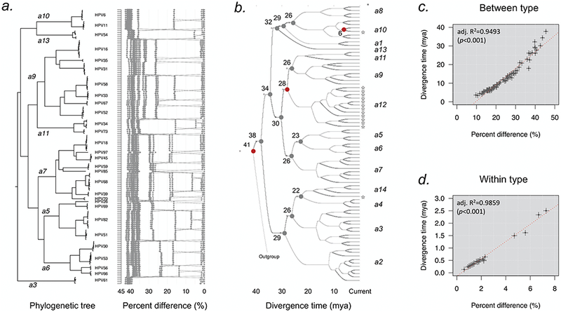
HPV variants from the same type can be classified into lineages and sublineages based on the complete genome differences and the phylogenetic topologies. We examined nucleotide variations of twelve HPV types within the species Alpha-5 (HPV26, 51, 69, 82), Alpha-6 (HPV30, 53, 56, 66), Alpha-11 (HPV34, 73), Alpha-13 (HPV54) and Alpha-3 (HPV61) by analyzing 1432 partial sequences and 181 complete genomes from multiple geographic populations. The inter-lineage and inter-sublineage mean differences of HPV variants ranged between 0.9-7.3% and 0.3-0.9%, respectively. The heterogeneity and phylogenies of HPV isolates indicate an independent evolutionary history for each type. The noncoding regions were the most variable regions whereas the capsid proteins were relatively conserved. Certain variant lineages and/or sublineages were geographically-associated. These data provide the basis to further classify HPV variants and should foster future studies on the evolution of HPV genomes and the associations of HPV variants with cancer risk.

Keywords: Cervical cancer; Classification; Evolution; Human papillomavirus; Variant.


Figures

Figure 1.
Figure 1.
Phylogenetic tree of Alphapapillomavirus. The maximum likelihood tree was constructed using RAxML based on the nucleotide sequence alignment of concatenated 6 ORFs (E6, E7, E1, E2, L2 and L1) and the URR region. Saimiri sciureus papillomavirus types 1/2/3 (SscPV1/2/3) were used as the outgroup taxa. The numbers just within the outer brackets represent the species groups (e.g. ‘α6’ contains HPV30, 53, 56 and 66). At least 3 ancestral papillomaviruses are responsible for the current heterogeneous groups of genital HPV genomes including LR1 (α1, 8, 10 and 13), LR2 (α 2, 3, 4 and 14) and HR (α 5, 6, 7, 9 and 11). The HPV type variants sequenced in this study are indicated in bold. The types joined by grey lines represent non-human primate PVs within Alphapapillomavirus. HR = High-risk; LR = low-risk.
Figure 2.
Figure 2.
Tree topologies and pairwise comparisons of complete genome variants from individual types. Phylogenetic trees were inferred from a global alignment of complete genome nucleotide sequences of the following variants from each of these types: (a) HPV26, (b) HPV69, (c) HPV51, (d) HPV82, (e) HPV30, (f) HPV53, (g) HPV56, (h) HPV66, (i) HPV34, (j) HPV73, (k) HPV54 and (l) HPV61. Numbers on or near branches indicate support indices in the following order: maximum likelihood (ML) bootstrap percentages using RAxML and PhyML, and Bayesian credibility value percentage using MrBayes. An asterisk (*) indicates 100% agreement between methods. “NA” indicates disagreement between a method and the reference RAxML tree at a given node. Thus, one tree is shown, but three different methods of tree construction were used to estimate the support of the provided tree, as explained above. Distinct variant lineages (i.e., termed A, B, and C) are classified according to the topology and nucleotide sequence differences from > 1% to < 10%; distinct sublineages (e.g., termed A1 and A2) were also inferred from the tree topology and nucleotide sequence differences in the > 0.5% to < 1% range. The bar indicates the nucleotide substitution of unit changes (i.e., 0.002) per site. The percent nucleotide differences for each isolate compared to all other isolates (i.e., 1 × 1 comparisons) are shown in the panel to the right of the phylogeny. Values for each comparison of a given isolate are connected by lines and the comparison to self is indicated by the 0.0% difference point. Different colored lines are used to distinguish each lineage and sublineage.
Figure 2.
Figure 2.
Tree topologies and pairwise comparisons of complete genome variants from individual types. Phylogenetic trees were inferred from a global alignment of complete genome nucleotide sequences of the following variants from each of these types: (a) HPV26, (b) HPV69, (c) HPV51, (d) HPV82, (e) HPV30, (f) HPV53, (g) HPV56, (h) HPV66, (i) HPV34, (j) HPV73, (k) HPV54 and (l) HPV61. Numbers on or near branches indicate support indices in the following order: maximum likelihood (ML) bootstrap percentages using RAxML and PhyML, and Bayesian credibility value percentage using MrBayes. An asterisk (*) indicates 100% agreement between methods. “NA” indicates disagreement between a method and the reference RAxML tree at a given node. Thus, one tree is shown, but three different methods of tree construction were used to estimate the support of the provided tree, as explained above. Distinct variant lineages (i.e., termed A, B, and C) are classified according to the topology and nucleotide sequence differences from > 1% to < 10%; distinct sublineages (e.g., termed A1 and A2) were also inferred from the tree topology and nucleotide sequence differences in the > 0.5% to < 1% range. The bar indicates the nucleotide substitution of unit changes (i.e., 0.002) per site. The percent nucleotide differences for each isolate compared to all other isolates (i.e., 1 × 1 comparisons) are shown in the panel to the right of the phylogeny. Values for each comparison of a given isolate are connected by lines and the comparison to self is indicated by the 0.0% difference point. Different colored lines are used to distinguish each lineage and sublineage.
Figure 3.
Figure 3.
Divergence time estimation of Alphapapillomavirus HPV types and variants. (a) Phylogenetic tree and pairwise comparisons of representative Alphapapillomavirus lineages and sublineages show multiple strata of genomic differences. A maximum likelihood (ML) tree was constructed using RAxML inferred from the global alignment of complete genome nucleotide sequences. The percent nucleotide sequence differences are shown in the panel to the right of the phylogenetic tree as described in Fig. 2. The Alpha-5 (HPV26, 51, 69, 82), Alpha-6 (HPV30, 53, 56, 66), Alpha-11 (HPV34, 73), Alpha-13 (HPV54) and Alpha-3 (HPV61) variants from this report, and Alpha-7 (HPV18, 39, 45, 59, 68, 70, 85, 97), Alpha-9 (HPV16, 31, 33, 35, 52, 58, 67) and Alpha-10 (HPV6, 11) variants from previous publications were included. (b) Divergence time estimation of HPV types using a Bayesian MCMC method. The nodes in the tree show split times of distinct species groups, with gray bars indicating the 95% highest posterior density for the corresponding divergence age. The nodes in red represent three main co-divergence events between viruses and host speciation (human and chimpanzee, human-chimpanzee and macaque, and New World and New World monkeys). The stars indicate non-human primate PVs within Alphapapillomavirus. (c) Correlation between genomic diversity (X-axis) and divergence time (Y-axis) of HPV types and species (between type). The divergence time was cited from each node in Figure 3b, and the percent difference was the maximum pairwise diversity of PV types within each node calculated based on the global alignment. (d) Correlation between genomic diversity (X-axis) and divergence time (Y-axis) of HPV variants (within type). The divergence time was the initial split time of variants of each type estimated using a Bayesian MCMC method, and the percent difference inferred from the maximum pairwise diversity of variant of each type.

References

    1. Baele G, Lemey P, Bedford T, Rambaut A, Suchard MA, Alekseyenko AV, 2012. Improving the accuracy of demographic and molecular clock model comparison while accommodating phylogenetic uncertainty. Mol Biol Evol 29, 2157–2167. - PMC - PubMed
    1. Bernard HU, Burk RD, Chen Z, van Doorslaer K, zur Hausen H, de Villiers EM, 2010. Classification of papillomaviruses (PVs) based on 189 PV types and proposal of taxonomic amendments. Virology 401, 70–79. - PMC - PubMed
    1. Burk RD, Chen Z, Van Doorslaer K, 2009. Human papillomaviruses: genetic basis of carcinogenicity. Public Health Genomics 12, 281–290. - PMC - PubMed
    1. Burk RD, Harari A, Chen Z, 2013. Human papillomavirus genome variants. Virology 445, 232–243. - PMC - PubMed
    1. Catarino R, Petignat P, Dongui G, Vassilakos P, 2015. Cervical cancer screening in developing countries at a crossroad: Emerging technologies and policy choices. World J Clin Oncol 6, 281–290. - PMC - PubMed

Publication types