. 2018 Feb;20(2):207-217.
doi: 10.1016/j.neo.2017.12.001. Epub 2018 Jan 12.

IDH1R132H Promotes Malignant Transformation of Benign Prostatic Epithelium by Dysregulating MicroRNAs: Involvement of IGF1R-AKT/STAT3 Signaling Pathway

Affiliations

IDH1R132H Promotes Malignant Transformation of Benign Prostatic Epithelium by Dysregulating MicroRNAs: Involvement of IGF1R-AKT/STAT3 Signaling Pathway

Lili Zhang et al. Neoplasia. 2018 Feb.

Abstract

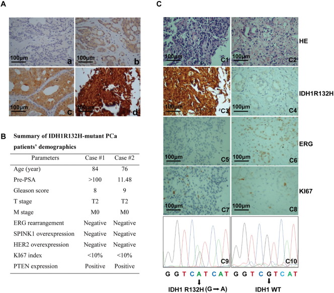
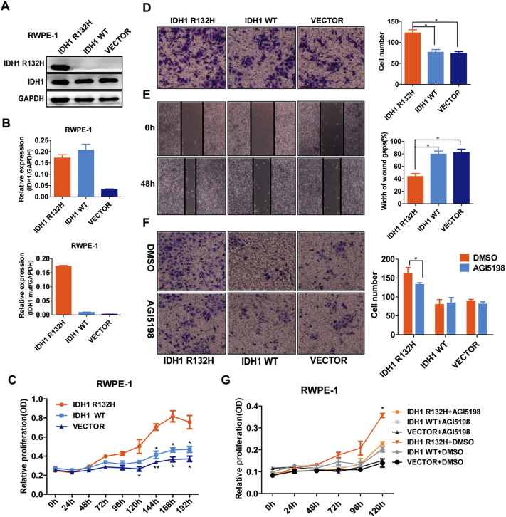
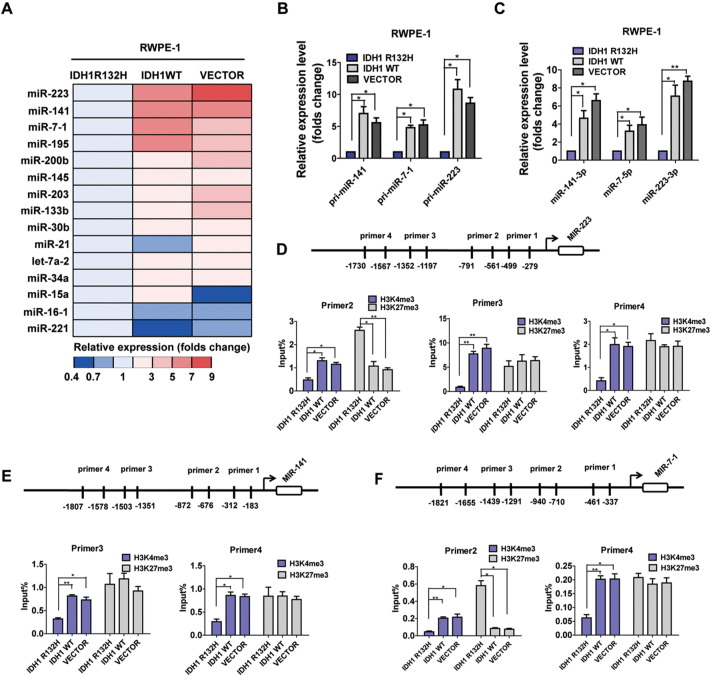
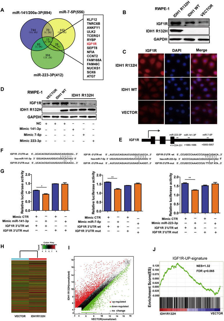
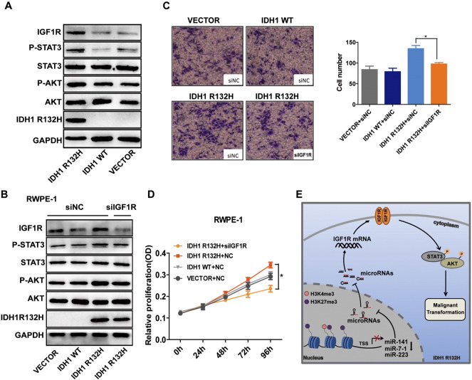
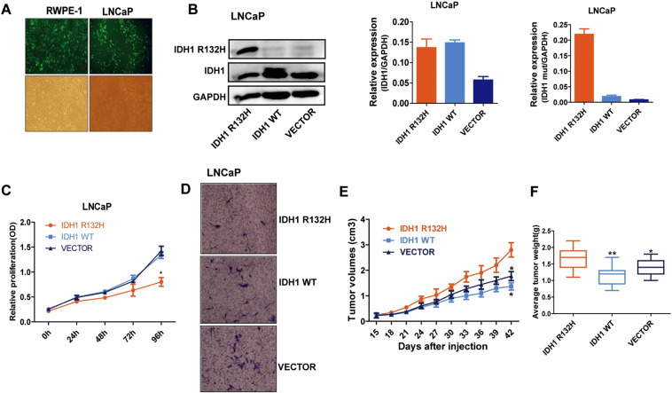
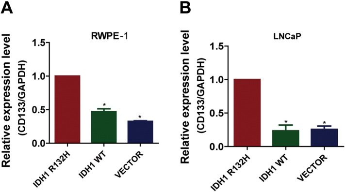
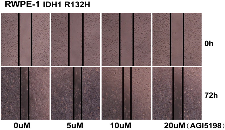
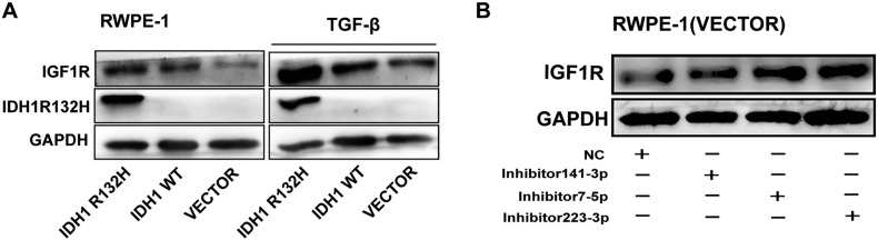
Risk stratification using molecular features could potentially help distinguish indolent from aggressive prostate cancer (PCa). Mutations in isocitrate dehydrogenase (IDH) acquire an abnormal enzymatic activity, resulting in the production of 2-hydroxyglutarate and alterations in cellular metabolism, histone modification, and DNA methylation. Mutant IDH1 has been identified in various human malignancies, and IDH1R132H constituted the vast majority of mutational events of IDH1. Most recent studies suggested that IDH1 mutations define a methylator subtype in PCa. However, the function of IDH1R132H in PCa development and progression is largely unknown. In this study, we showed that the prevalence of IDH1R132H in Chinese PCa patients is 0.6% (2/336). Of note, IDH1R132H-mutant PCa patients lacked other canonical genomic lesions (e.g., ERG rearrangement, PTEN deletion) that are common in most other PCa patients. The in vitro experiment suggested that IDH1R132H can promote proliferation of benign prostate epithelial cell RWPE-1 when under the situation of low cytokine. It could also promote migration capacity of RWPE-1 cells. Mechanistically, IDH1R132H was an important regulator of insulin-like growth factor 1receptor (IGF1R) by downregulating a set of microRNAs (miR-141-3p, miR-7-5p, miR-223-3p). These microRNAs were repressed by the alteration of epigenetic modification to decrease the enrichment of active marker H3K4me3 or to increase repressive marker H3K27me3 at their promoters. Collectively, we proposed a novel model for an IDH1R132H-microRNAs-IGF1R regulatory axis, which might provide insight into the function of IDH1R132H in PCa development.


Figures

Figure 1
Figure 1
Mutational analysis of IDH1R132H in PCa patients. (A) Representative IHC images were shown in a-d. Sections were stained with antibody against IDH1R132H in PCa patients (n = 336). Staining intensity was classified into four scales [a: negative staining (0+); b: weak staining (1+); c: moderate staining (2+); d: strong staining(3+)]. (B) Clinicopathological characteristic of IDH1R132H-positive PCa patients. The status of ERG rearrangement, SPINK1 overexpression, HER2 overexpression, PTEN deletion, and KI67 index were performed in our early studies. (C) Representative images of molecular pathological features of IDH1R132H-positive and -negative cases. For IHC, the slides were incubated with IDH1R132H antibody (c3, c4). The IHC of ERG and KI67 (c5-c8) and hematoxylin-eosin (HE) staining (c1, c2) were performed in our previous research. PCR-based gene sequencing of PCa patients. G to A mutation confirmed IDH1R132H mutation (c9, c10). Scale bar, 100 μm.
Figure 2
Figure 2
IDH1R132H promotes malignant transformation of prostate benign epithelial cell RWPE-1. (A, B) Western blot and qRT-PCR analysis of protein and mRNA expression levels of wild-type IDH1 and IDH1R132H in RWPE-1cells, following stably expressing empty vector, wild-type, or mutant IDH1R132H. (C) IDH1R132H enhances the cytokine-independent growth of benign prostatic epithelial cells. The growth of stable RWPE-1 cells expressing IDH1R132H, IDH1WT, and VECTOR was examined by MTS assay. Cells were cultured under cytokine-poor conditions (1/4 normal dose EGF and BPE). (D, E) IDH1R132H promotes migration of benign prostatic epithelial cells. The migration of IDH1R132H on RWPE-1 cells was examined by wound-healing assay and Transwell migration assay. (F, G) The growth and migration of RWPE-1 cells. Cells were treated with 20 μM AGI-5198 or DMSO and analyzed using MTS and Transwell migration assay. All data represent mean ± SD of at least three independent replicates. *P < .05, **P < .01.
Figure 3
Figure 3
IDH1R132H downregulates miR-141-3p/miR-7-5p/miR-223-3p in RWPE-1 Cells. (A) qRT-PCR was used to assess the expression color heat map of miRNAs precursors in RWPE-1 cells which were transfected with IDH1R132H, IDH1WT, and VECTOR. The results are shown as folds number. (B, C) Expressions of primary precursor and mature miR-141-3p, miR-223-3p, and miR-7-5p were assessed by qRT-PCR in RWPE-1 cells. The results are shown as folds number. (D, E, F) ChIP-qPCR was performed to evaluate the enrichment of H3K4me3 and H3K27me3 in different promoter regions of miR-223, miR-141, and miR-7-1 in RWPE-1 cells. All data represent mean ± SD of at least three independent replicates. P values: *P < .05, **P < .01.
Figure 4
Figure 4
IDH1R132H-downregulated miRNAs lead to increased IGF1R expression. (A) The predicted target gene analysis of miR-141-3p, miR-223-3p, and miR-7-5p was performed by TargetScan software. (B, C) Western blot and immunofluorescence were used to examine IGF1R protein levels in RWPE-1 cells expressing IDH1R132H, IDH1WT, and VECTOR. (D) The protein levels of IGF1R were assessed in IDH1R132H-mutant RWPE-1 cells after treatment with miR-141-3p, miR-223-3p, and miR-7-5p mimics or negative control (Western blot). (E) Diagram indicates the recognition sites for miR-141-3p, miR-223-3p, and miR-7-5p in IGF1R 3′-UTR region, respectively. (F) WT- and mutated recognition sites of miR-141-3p, miR-223-3p, and miR-7-5p in IGF1R 3′-UTR region. (G) LUC reporter assay was performed in HEK293T cells. Cells were transiently co-transfected with wt- or mutated IGF1R 3′-UTR region together with corresponding miRNA mimics, respectively. After incubation for 48 hours, luciferase activities were measured. (H, I) Heat map and scatter diagram were used to indicate the different gene expression of IDH1R132H and VECTOR in RWPE-1 cells. (G) IGF1R-upregulated (GSE5225) gene signatures were further analyzed by GSEA in mRNA microarray of RWPE-1 cells expressing IDH1R132H and VECTOR. FDR q = 0.065. All histograms represent mean ± SD of at least three independent replicates. P values: *P < .05, **P < .01.
Figure 5
Figure 5
IGF1R is required for IDH1R132H-mediated malignant transformation. (A) Western blot analysis of indicted genes in RWPE-1 cells transfected with IDH1R132H, IDH1WT, or VECTOR. (B) Representative Western blot showed the protein levels of indicated genes in IDH1R132H-mutant RWPE-1 cells after knocking down IGF1R. (C, D) MTS and Transwell assays were used to determine the ability of proliferation and migration after silencing IGF1R in IDH1R132H-mutant RWPE-1 cells. (E) Schematic illustration of IDH1R132H-stimulated RWPE-1 cells’ malignant transformation. IDH1R132H reduces the expression of miR-141-3p, miR-223-3p, and miR-7-5p by altering histone modifications in their promoter region; the reduction of miRNAs eventually leads to the activation of the IGF1R-AKT/STAT3 signaling, which leads to the malignant transformation of RWPE-1 cells. Data are means of biological triplicate and mean ± SD. P values: *P < .05.
Figure S1
Figure S1
IDH1R132H decreased proliferation and migration in LNCAP cells. (A) The representative transfection efficiency of virus observation in RWPE-1 and LNCAP cells under fluorescence microscope. (B) The wild-type and mutant IDH1 protein and mRNA expression levels of LNCAP cells stably expressing IDH1R132H, wild-type, and empty vector were tested by Western blot and qRT-PCR. (C, D) MTS and Transwell migration assays were used to identify the abilities of proliferation and migration in IDH1R132H, IDH1WT, and VECTOR-expressing LNCAP cells. (E) Growth curves were used to test tumors formation of subcutaneous xenograft assay. (F) The weight of tumors was measured at harvest time (n = 6 per group). Data in C and D represent mean ± SD of at least three independent replicates. P values: *P < .05
Figure S2
Figure S2
IDH1R132H promotes the expression of CD133. (A, B) qRT-PCR was performed to detect the expression of CD133 in RWPE-1 and LNCAP cells. All data represent mean ± SD of at least three independent replicates. P values: *P < .05
Figure S3
Figure S3
Twenty micomolars of AGI5198 inhibits migration of IDH1R132H RWPE-1 cells. IDH1R132H-mutant RWPE-1 cells were treated with increasing concentrations of AGI-5198 and corresponding DMSO, and were analyzed by wound-healing assay after 72 hours of treatment. All data represent three independent replicates.
Figure S4
Figure S4
IDH1R132H-downregulated miRNAs lead to increased IGF1R expression. (A) Western blot analysis of IGF1R protein expression in stable RWPE-1 cells treated with TGF-β. (B) The protein levels of IGF1R were assessed by Western blot analysis in RWPE-1-VECTOR cells transfected with miR-141-3p, miR-223-3p, miR-7-5p inhibitor, or negative control. WB was performed independently three times.

References

    1. Zhao L, Yu N, Guo T, Hou Y, Zeng Z, Yang X, Hu P, Tang X, Wang J, Liu M. Tissue biomarkers for prognosis of prostate cancer: a systematic review and meta-analysis. Cancer Epidemiol Biomarkers Prev. 2014;23:1047–1054. - PubMed
    1. Attard G, Parker C, Eeles RA, Schroder F, Tomlins SA, Tannock I, Drake CG, de Bono JS. Prostate cancer. Lancet. 2016;387:70–82. - PubMed
    1. Cancer Genome Atlas Research N The molecular taxonomy of primary prostate cancer. Cell. 2015;163:1011–1025. - PMC - PubMed
    1. Yen KE, Bittinger MA, Su SM, Fantin VR. Cancer-associated IDH mutations: biomarker and therapeutic opportunities. Oncogene. 2010;29:6409–6417. - PubMed
    1. Cairns RA, Mak TW. Oncogenic isocitrate dehydrogenase mutations: mechanisms, models, and clinical opportunities. Cancer Discov. 2013;3:730–741. - PubMed

Publication types

MeSH terms