Whole-mount Enteroid Proliferation Staining
- PMID: 29333475
- PMCID: PMC5760989
- DOI: 10.21769/BioProtoc.1837
Whole-mount Enteroid Proliferation Staining
Abstract
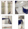
Small intestinal organoids, otherwise known as enteroids, have become an increasingly utilized model for intestinal biology in vitro as they recapitulate the various epithelial cells within the intestinal crypt (Mahe et al., 2013; Sato et al., 2009). Assessment of growth dynamics within these cultures is an important step to understanding how alterations in gene expression, treatment with protective and toxic agents, and genetic mutations alter properties essential for crypt growth and survival as well as the stem cell properties of the individual cells within the crypt. This protocol describes a method of visualization of proliferating cells within the crypt in three dimensions (Barrett et al., 2015). Whole-mount proliferation staining of enteroids using EdU incorporation enables the researcher to view all proliferating cells within the enteroid as opposed to obtaining growth information in thin slices as would be seen with embedding and sectioning, ensuring a true representation of proliferation from the stem cell compartment to the terminally differentiated cells of the crypt.
Figures


References
-
- Barrett CW, Reddy VK, Short SP, Motley AK, Lintel MK, Bradley AM, Freeman T, Vallance J, Ning W, Parang B, Poindexter SV, Fingleton B, Chen X, Washington MK, Wilson KT, Shroyer NF, Hill KE, Burk RF, Williams CS. Selenoprotein P influences colitis-induced tumorigenesis by mediating stemness and oxidative damage. J Clin Invest. 2015;125(7):2646–2660. - PMC - PubMed
-
- Sato T, Vries RG, Snippert HJ, van de Wetering M, Barker N, Stange DE, van Es JH, Abo A, Kujala P, Peters PJ, Clevers H. Single Lgr5 stem cells build crypt-villus structures in vitro without a mesenchymal niche. Nature. 2009;459(7244):262–265. - PubMed
Grants and funding
LinkOut - more resources
Full Text Sources
Other Literature Sources

