. 2018 Jan 10;4(6):ENEURO.0277-17.2017.
doi: 10.1523/ENEURO.0277-17.2017. eCollection 2017 Nov-Dec.

Activation of the Medial Prefrontal Cortex Reverses Cognitive and Respiratory Symptoms in a Mouse Model of Rett Syndrome

Affiliations

Activation of the Medial Prefrontal Cortex Reverses Cognitive and Respiratory Symptoms in a Mouse Model of Rett Syndrome

C James Howell et al. eNeuro. .

Abstract

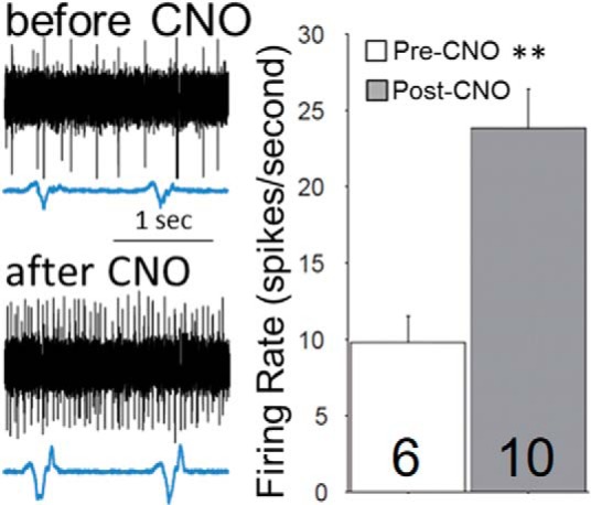
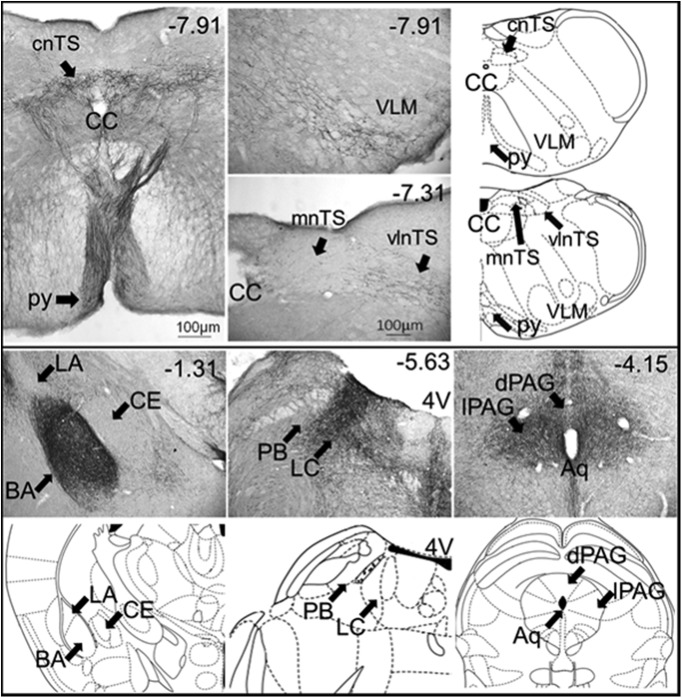
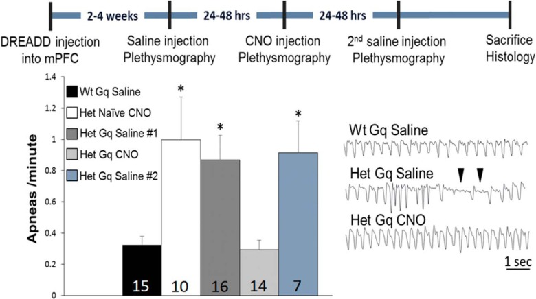
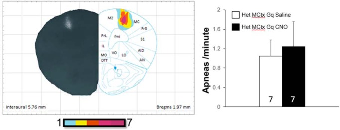
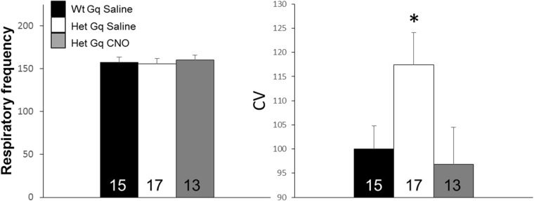
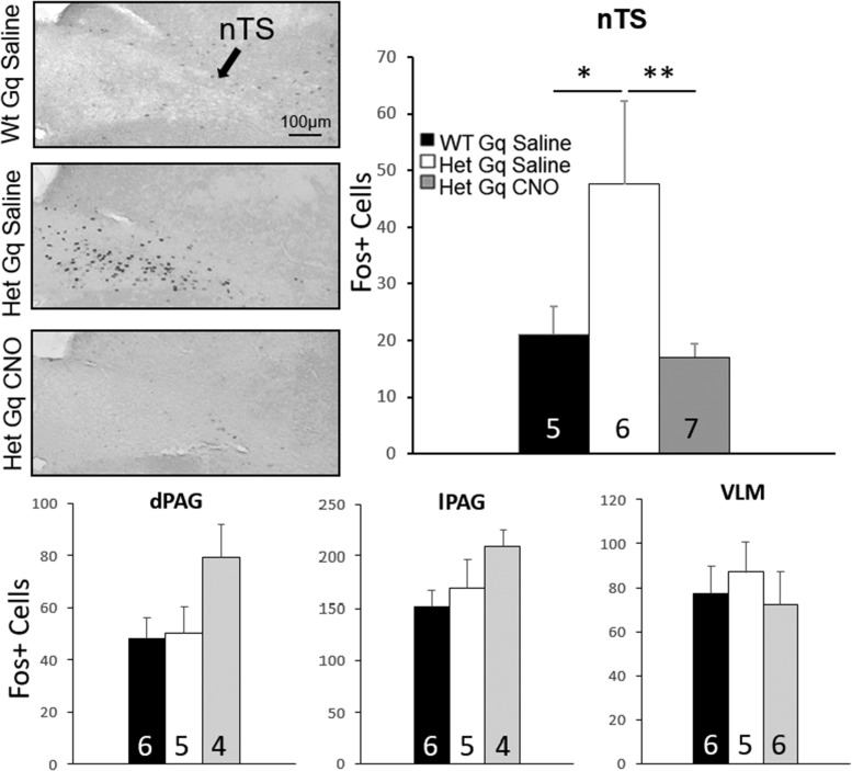
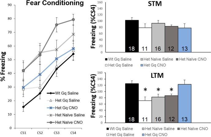
Rett syndrome (RTT) is a severe neurodevelopmental disorder caused by loss-of-function mutations in the gene encoding methyl-CpG-binding protein 2 (MeCP2; Amir et al., 1999), a transcriptional regulatory protein (Klose et al., 2005). Mouse models of RTT (Mecp2 mutants) exhibit excitatory hypoconnectivity in the medial prefrontal cortex (mPFC; Sceniak et al., 2015), a region critical for functions that are abnormal in RTT patients, ranging from learning and memory to regulation of visceral homeostasis (Riga et al., 2014). The present study was designed to test the hypothesis that increasing the activity of mPFC pyramidal neurons in heterozygous female Mecp2 mutants (Hets) would ameliorate RTT-like symptoms, including deficits in respiratory control and long-term retrieval of auditory conditioned fear. Selective activation of mPFC pyramidal neurons in adult animals was achieved by bilateral infection with an AAV8 vector expressing excitatory hm3D(Gq) DREADD (Designer Receptors Exclusively Activated by Designer Drugs) (Armbruster et al., 2007) under the control of the CamKIIa promoter. DREADD activation in Mecp2 Hets completely restored long-term retrieval of auditory conditioned fear, eliminated respiratory apneas, and reduced respiratory frequency variability to wild-type (Wt) levels. Reversal of respiratory symptoms following mPFC activation was associated with normalization of Fos protein levels, a marker of neuronal activity, in a subset of brainstem respiratory neurons. Thus, despite reduced levels of MeCP2 and severe neurological deficits, mPFC circuits in Het mice are sufficiently intact to generate normal behavioral output when pyramidal cell activity is increased. These findings highlight the contribution of mPFC hypofunction to the pathophysiology of RTT and raise the possibility that selective activation of cortical regions such as the mPFC could provide therapeutic benefit to RTT patients.

Keywords: DREADD; Mecp2; autism spectrum disorder; hypofrontality; mPFC; memory.


Figures

Figure 1.
Figure 1.
DREADD-Gq expression in the mPFC. The distribution of DREADD-Gq-labeled neurons was plotted from low-magnification micrographs of mCherry labeling in serial coronal sections through the forebrain in 14 animals; the plots were then overlaid to produce heat maps of the injection sites in which the color coding represents the extent of overlap in the distribution of labeled neurons in all 14 animals. These heat maps are shown on the right side of each figure, with representative hemi-brain sections shown on the opposite side (note that all animals received bilateral injections). The faint labeling seen lateral to the mPFC at 1.77 and 1.41 mm rostral to bregma is mCherry expression in the axons of infected neurons. Bottom right, Representative photomicrograph of mCherry-labeled neurons in a coronal section through the mPFC of an infected animal. Anatomic labels for this and all subsequent figures as follows: AC, anterior cingulate; AID, dorsal agranular insular cortex; AIV, ventral agranular insular cortex; CPu, caudate putamen; DI, dysgranular insular cortex; DP, dorsal peduncular cortex; DTT, dorsal tenia tecta; fmi, forceps minor of the corpus callosum; Fr3, frontal cortex area 3; IL, infralimbic cortex; LO, lateral orbital cortex; M1, primary motor cortex; M2, secondary motor cortex; MO, medial orbital cortex; PrL, prelimbic cortex; S1, primary somatosensory cortex; S1DZ, somatosensory cortex dysgranular zone; S1FL, forelimb region of the somatosensory cortex; S1J, jaw region of the somatosensory cortex; VO, ventral orbital cortex. Inset shows a high-magnification view of an infected neuron.
Figure 2.
Figure 2.
Activation of mPFC pyramidal neurons by DREADD-Gq increases neuronal activity in vivo. Left, Representative extracellular traces of spontaneous activity in the mPFC of mPFC-DREADD-Gq Het animals before and after CNO treatment. Right, Summary data from two animals; the total number of recorded cells is shown within each bar. **p < 0.01 by Wilcoxon rank sum test.
Figure 3.
Figure 3.
Photomicrographs showing mCherry-positive fibers in coronal sections through brain regions important for respiratory control and cue-dependent fear memory following DREADD-Gq infection of the mPFC. Numbers in the top right of each micrograph correspond to the distance from bregma (mm). 4V, fourth ventricle; Aq, aqueduct of Sylvius; BA, basal amygdala; CC, central canal; CE, central amygdala; cnTS, commissural nTS; LC, locus coeruleus; LA, lateral amygdala; mnTS, medial nTS; PB, parabrachial nucleus; py, pyramidal tract; vlnTS, ventrolateral nTS (abbreviations from Franklin and Paxinos, 2012).
Figure 4.
Figure 4.
DREADD-Gq activation of pyramidal neurons in the mPFC eliminates the apneic breathing phenotype in Mecp2 mutants. The timeline of DREADD injection, CNO (or saline) treatment and plethysmographic recordings is shown at the top of the figure. The bar graph shows summary data illustrating the apneas/min for each experimental group with group sizes included within each bar. *p < 0.05 by ANOVA with Bonferroni post hoc test. Bottom right, Representative respiratory traces; ▼ indicates typical respiratory pauses scored as apneas.
Figure 5.
Figure 5.
Activation of pyramidal neurons in the motor cortex by DREADD-Gq does not alter respiration. Left, The distribution of DREADD-Gq-labeled neurons was plotted from low-magnification micrographs of mCherry labeling in coronal sections through the forebrain in all animals tested; the plots were then overlaid to produce a heat map of the injection sites in which the color coding represents the extent of overlap in the distribution of labeled neurons in all seven animals. Note that all animals received bilateral injections. Right, Animals expressing DREADD-Gq in the motor cortex were subjected to two consecutive days of plethysmographic recording of respiration, 1 h following either saline treatment (day 1) or CNO treatment (day 2). The bar graph shows summary data illustrating the apneas/min on day 1 (saline) and day 2 (CNO), with group sizes included within each bar.
Figure 6.
Figure 6.
DREADD-Gq activation of pyramidal neurons in the mPFC reduces respiratory frequency variability to Wt levels without impacting the average frequency of respiration. Left, Summary data showing the mean instantaneous respiratory frequency for each experimental group. Right, Summary data showing the coefficient of variation (CV) for instantaneous respiratory frequency for each experimental group. Group sizes are shown within each bar. *p < 0.05 by ANOVA with LSD post hoc test.
Figure 7.
Figure 7.
Activation of mPFC pyramidal neurons by DREADD-Gq impacts neuronal function in respiratory subnuclei of the nTS. Animals were sacrificed 90 min after either saline CNO injection and processed for Fos immunostaining. Photomicrographs show representative sections through the caudal nTS from 3 different animals, each of which received bilateral injections of DREADD-Gq in the mPFC. CNO treatment restored Fos levels in the nTS of mPFC-DREADD-Gq Het mice to Wt levels. *p < 0.05, **p < 0.01, ANOVA with LSD post hoc test. No significant differences were observed between treatment groups in the dPAG, lPAG or VLM.
Figure 8.
Figure 8.
Activation of mPFC pyramidal neurons by DREADD-Gq rescues long-term expression of cue-dependent fear memory. Left, Conditioning to CS-US pairings across four trials demonstrates similar responses in all groups using % freezing (immobility) during the CS presentations as an index of fear learning. Right, Short-term (4 h after conditioning) and long-term (24 h after conditioning) memory retrieval were tested by exposure to the CS alone, with the animal in a test chamber that was distinguished from the conditioning chamber by novel environmental features (see Materials and Methods). Data are presented as a percentage of the freezing to CS4 during conditioning ([FreezingTone(LTM or STM) * 100]/FreezingCS4). *p < 0.05 by ANOVA with LSD post hoc test.

Similar articles

Cited by

References

    1. Abdala AP, Toward MA, Dutschmann M, Bissonnette JM, Paton JF (2016) Deficiency of GABAergic synaptic inhibition in the Kölliker-Fuse area underlies respiratory dysrhythmia in a mouse model of Rett syndrome. J Physiol 594:223–237. 10.1113/JP270966 - DOI - PMC - PubMed
    1. Adhikari A, Lerner TN, Finkelstein J, Pak S, Jennings JH, Davidson TJ, Ferenczi E, Gunaydin LA, Mirzabekov JJ, Ye L, Kim SY, Lei A, Deisseroth K (2015) Basomedial amygdala mediates top-down control of anxiety and fear. Nature 527:179–185. 10.1038/nature15698 - DOI - PMC - PubMed
    1. Akbarian S (2003) The neurobiology of Rett syndrome. Neuroscientist 9:57–63. 10.1177/1073858402239591 - DOI - PubMed
    1. Alexander GM, Rogan SC, Abbas AI, Armbruster BN, Pei Y, Allen JA, Nonneman RJ, Hartmann J, Moy SS, Nicolelis MA, McNamara JO, Roth BL (2009) Remote control of neuronal activity in transgenic mice expressing evolved G protein-coupled receptors. Neuron 63:27–39. 10.1016/j.neuron.2009.06.014 - DOI - PMC - PubMed
    1. Alexandrov VG, Ivanova TG, Alexandrova NP (2007) Prefrontal control of respiration. J Physiol Pharmacol 58 [Suppl 5]:17–23. - PubMed

Publication types

MeSH terms

LinkOut - more resources