Dopamine Development in the Mouse Orbital Prefrontal Cortex Is Protracted and Sensitive to Amphetamine in Adolescence
- PMID: 29333488
- PMCID: PMC5762649
- DOI: 10.1523/ENEURO.0372-17.2017
Dopamine Development in the Mouse Orbital Prefrontal Cortex Is Protracted and Sensitive to Amphetamine in Adolescence
Erratum in
-
Correction: Hoops et al., Dopamine Development in the Mouse Orbital Prefrontal Cortex is Protracted and Sensitive to Amphetamine in Adolescence (eNeuro January/February 2018, 5(1) e0372-17.2017 1-9 https://doi.org/10.1523/ENEURO.0372-17.2017.eNeuro. 2018 Feb 26;5(1):ENEURO.0026-18.2018. doi: 10.1523/ENEURO.0026-18.2018. eCollection 2018 Jan-Feb. eNeuro. 2018. PMID: 29488511 Free PMC article.
Abstract
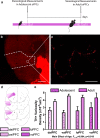
The prefrontal cortex (PFC) is divided into subregions, including the medial and orbital prefrontal cortices. Dopamine connectivity in the medial PFC (mPFC) continues to be established throughout adolescence as the result of the continuous growth of axons that innervated the nucleus accumbens (NAcc) prior to adolescence. During this period, dopamine axons remain vulnerable to environmental influences, such as drugs used recreationally by humans. The developmental trajectory of the orbital prefrontal dopamine innervation remains almost completely unstudied. Nonetheless, the orbital PFC (oPFC) is critical for some of the most complex functions of the PFC and is disrupted by drugs of abuse, both in adolescent humans and rodents. Here, we use quantitative neuroanatomy, axon-initiated viral-vector recombination, and pharmacology in mice to determine the spatiotemporal development of the dopamine innervation to the oPFC and its vulnerability to amphetamine in adolescence. We find that dopamine innervation to the oPFC also continues to increase during adolescence and that this increase is due to the growth of new dopamine axons to this region. Furthermore, amphetamine in adolescence dramatically reduces the number of presynaptic sites on oPFC dopamine axons. In contrast, dopamine innervation to the piriform cortex is not protracted across adolescence and is not impacted by amphetamine exposure during adolescence, indicating that dopamine development during adolescence is a uniquely prefrontal phenomenon. This renders these fibers, and the PFC in general, particularly vulnerable to environmental risk factors during adolescence, such as recreational drug use.
Keywords: drug use; guidance cue; orbitofrontal cortex; piriform cortex.
Figures


Similar articles
-
Amphetamine in adolescence disrupts the development of medial prefrontal cortex dopamine connectivity in a DCC-dependent manner.Neuropsychopharmacology. 2015 Mar 13;40(5):1101-12. doi: 10.1038/npp.2014.287. Neuropsychopharmacology. 2015. PMID: 25336209 Free PMC article.
-
DCC Receptors Drive Prefrontal Cortex Maturation by Determining Dopamine Axon Targeting in Adolescence.Biol Psychiatry. 2018 Jan 15;83(2):181-192. doi: 10.1016/j.biopsych.2017.06.009. Epub 2017 Jun 16. Biol Psychiatry. 2018. PMID: 28720317 Free PMC article.
-
DCC-related developmental effects of abused- versus therapeutic-like amphetamine doses in adolescence.Addict Biol. 2020 Jul;25(4):e12791. doi: 10.1111/adb.12791. Epub 2019 Jun 13. Addict Biol. 2020. PMID: 31192517 Free PMC article.
-
Synaptic Cytoskeletal Plasticity in the Prefrontal Cortex Following Psychostimulant Exposure.Traffic. 2015 Sep;16(9):919-40. doi: 10.1111/tra.12295. Epub 2015 Jun 1. Traffic. 2015. PMID: 25951902 Free PMC article. Review.
-
Rewiring the future: drugs abused in adolescence may predispose to mental illness in adult life by altering dopamine axon growth.J Neural Transm (Vienna). 2024 May;131(5):461-467. doi: 10.1007/s00702-023-02722-6. Epub 2023 Dec 1. J Neural Transm (Vienna). 2024. PMID: 38036858 Free PMC article. Review.
Cited by
-
DCC gene network in the prefrontal cortex is associated with total brain volume in childhood.J Psychiatry Neurosci. 2020 Nov 18;46(1):E154-E163. doi: 10.1503/jpn.200081. J Psychiatry Neurosci. 2020. PMID: 33206040 Free PMC article.
-
Emerging ecophenotype: reward anticipation is linked to high-risk behaviours after sexual abuse.Soc Cogn Affect Neurosci. 2022 Nov 2;17(11):1035-1043. doi: 10.1093/scan/nsac030. Soc Cogn Affect Neurosci. 2022. PMID: 35438797 Free PMC article.
-
Future considerations for pediatric cancer survivorship: Translational perspectives from developmental neuroscience.Dev Cogn Neurosci. 2019 Aug;38:100657. doi: 10.1016/j.dcn.2019.100657. Epub 2019 May 24. Dev Cogn Neurosci. 2019. PMID: 31158802 Free PMC article. Review.
-
The Netrin-1/DCC Guidance Cue Pathway as a Molecular Target in Depression: Translational Evidence.Biol Psychiatry. 2020 Oct 15;88(8):611-624. doi: 10.1016/j.biopsych.2020.04.025. Epub 2020 May 11. Biol Psychiatry. 2020. PMID: 32593422 Free PMC article. Review.
-
Mesocorticolimbic Dopamine Pathways Across Adolescence: Diversity in Development.Front Neural Circuits. 2021 Sep 8;15:735625. doi: 10.3389/fncir.2021.735625. eCollection 2021. Front Neural Circuits. 2021. PMID: 34566584 Free PMC article. Review.
References
-
- Andersen SL (2003) Trajectories of brain development: point of vulnerability or window of opportunity? Neurosci Biobehav Rev 27:3–18. - PubMed
-
- Änggård E, Gunne LM, Jönsson LE, Niklasson F (1970) Pharmacokinetic and clinical studies on amphetamine dependent subjects. Eur J Clin Pharmacol 3:3–11.
Publication types
MeSH terms
Substances
Grants and funding
LinkOut - more resources
Full Text Sources
Other Literature Sources
Miscellaneous
