. 2017:2017:PO.16.00038.
doi: 10.1200/PO.16.00038. Epub 2017 Jun 14.

Next-Generation Rapid Autopsies Enable Tumor Evolution Tracking and Generation of Preclinical Models

Affiliations

Next-Generation Rapid Autopsies Enable Tumor Evolution Tracking and Generation of Preclinical Models

David J Pisapia et al. JCO Precis Oncol. 2017.

Abstract

Purpose: Patients with cancer who graciously consent for autopsy represent an invaluable resource for the study of cancer biology. To advance the study of tumor evolution, metastases, and resistance to treatment, we developed a next-generation rapid autopsy program integrated within a broader precision medicine clinical trial that interrogates pre- and postmortem tissue samples for patients of all ages and cancer types.

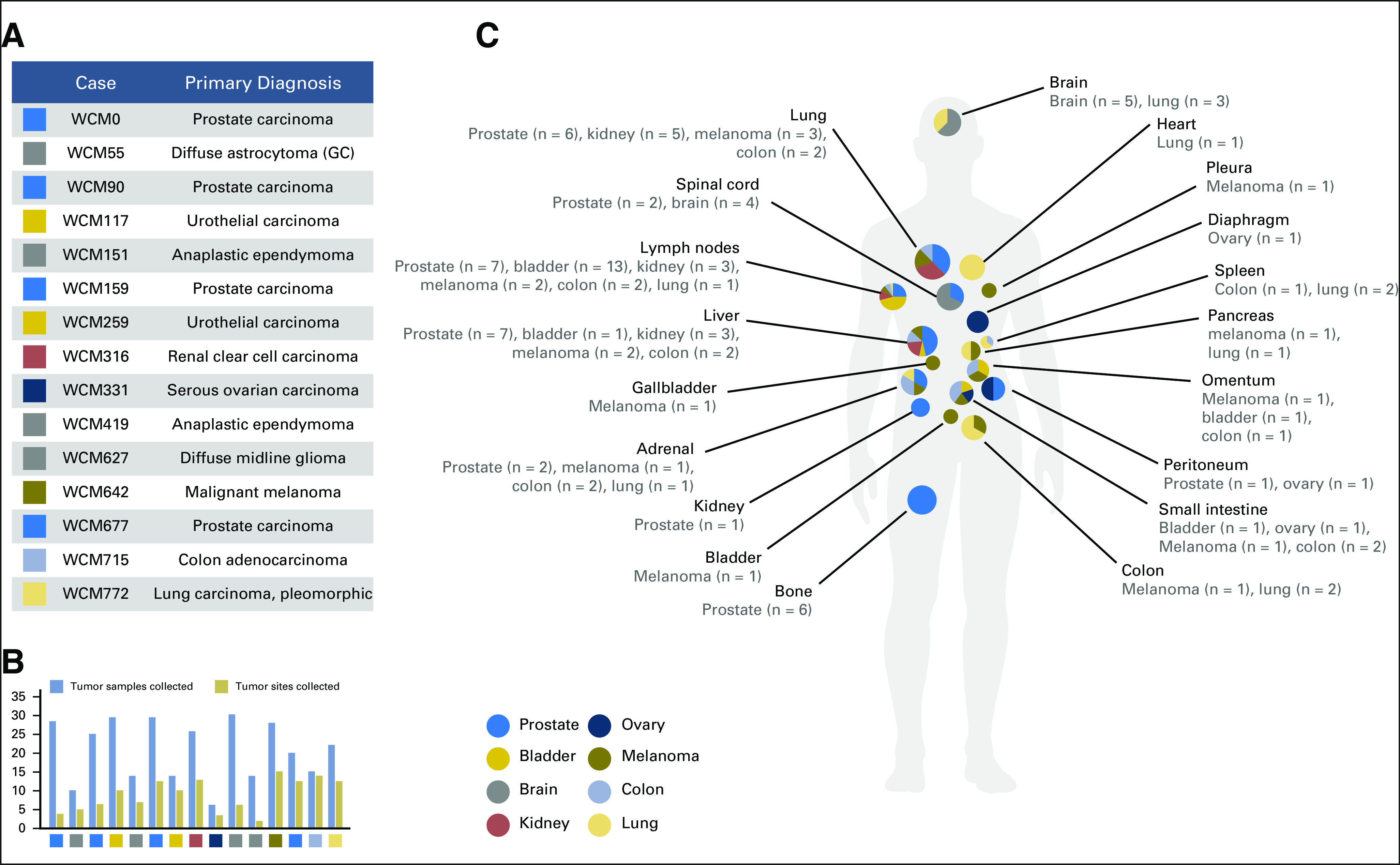
Materials and methods: One hundred twenty-three (22%) of 554 patients who consented to the clinical trial also consented for rapid autopsy. This report comprises the first 15 autopsies, including patients with metastatic carcinoma (n = 10), melanoma (n = 1), and glioma (n = 4). Whole-exome sequencing (WES) was performed on frozen autopsy tumor samples from multiple anatomic sites and on non-neoplastic tissue. RNA sequencing (RNA-Seq) was performed on a subset of frozen samples. Tissue was also used for the development of preclinical models, including tumor organoids and patient-derived xenografts.

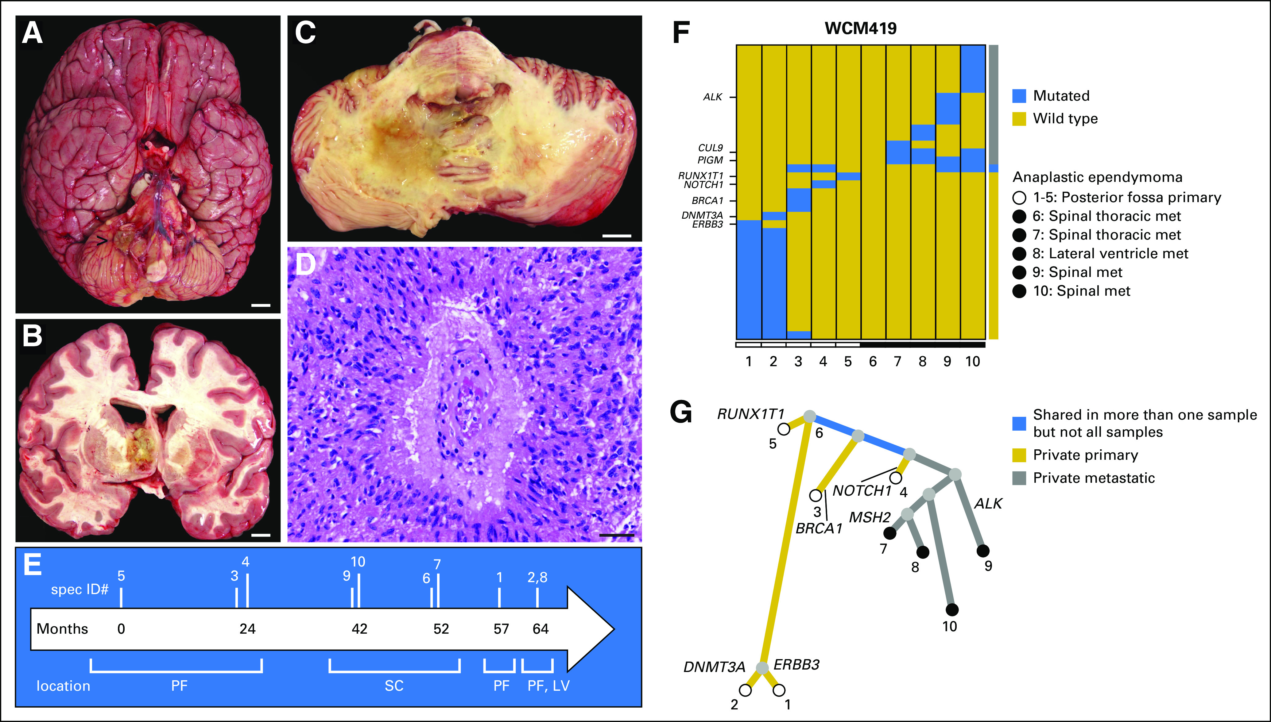
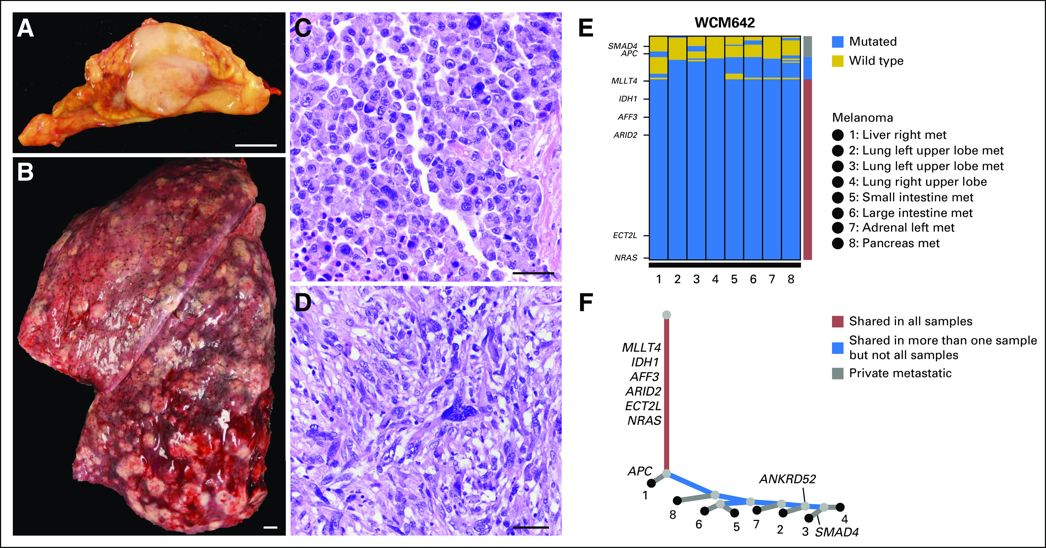
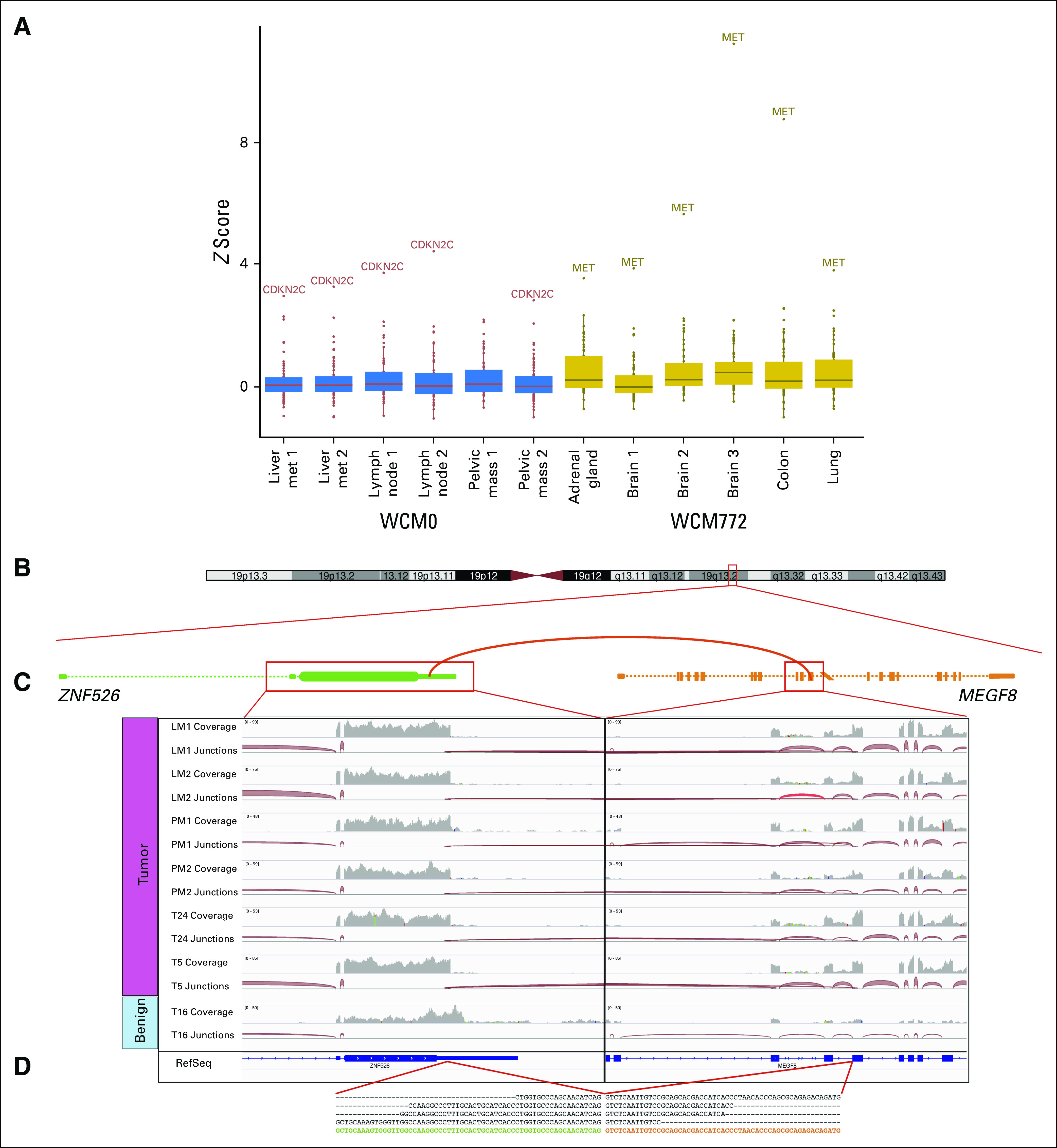
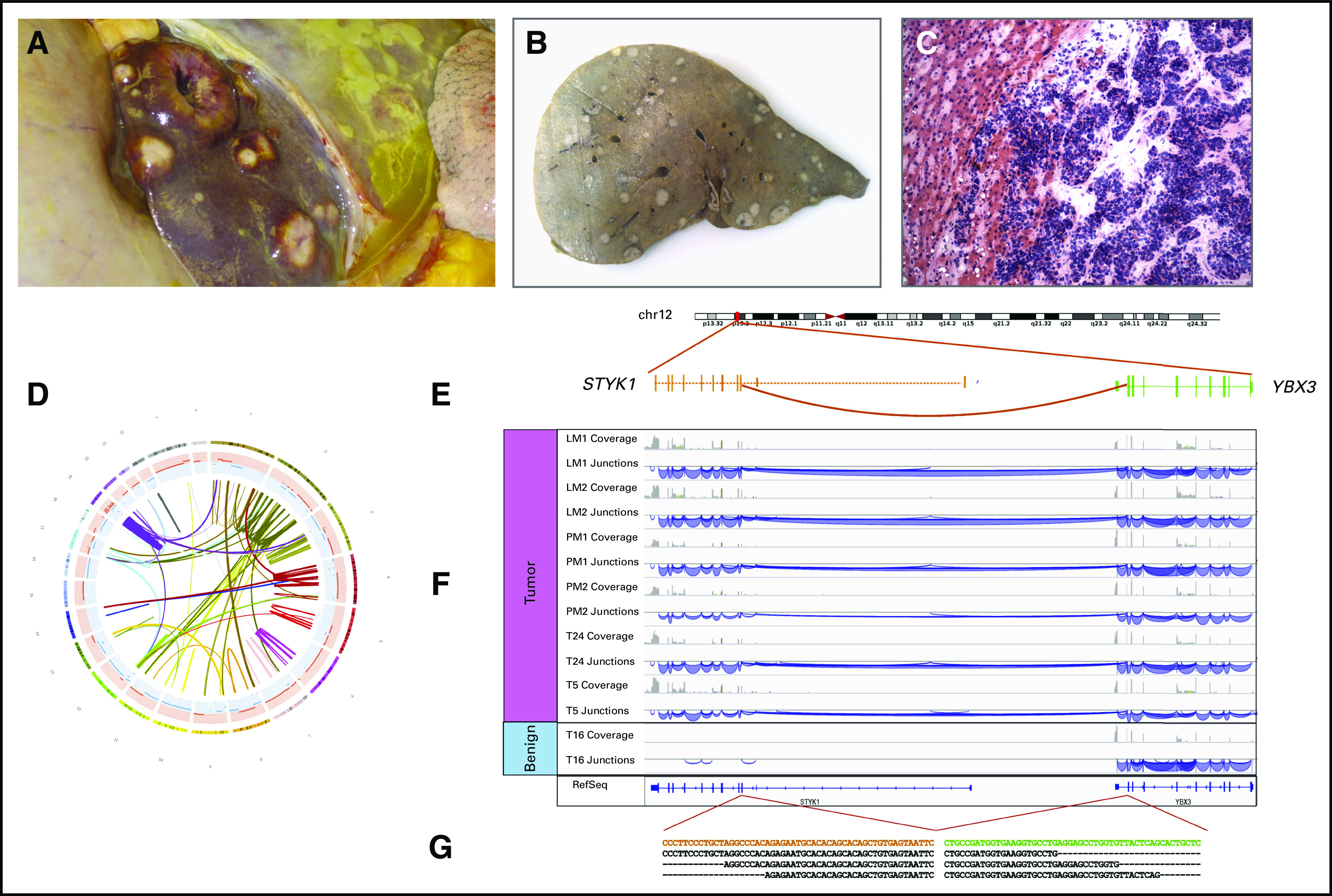
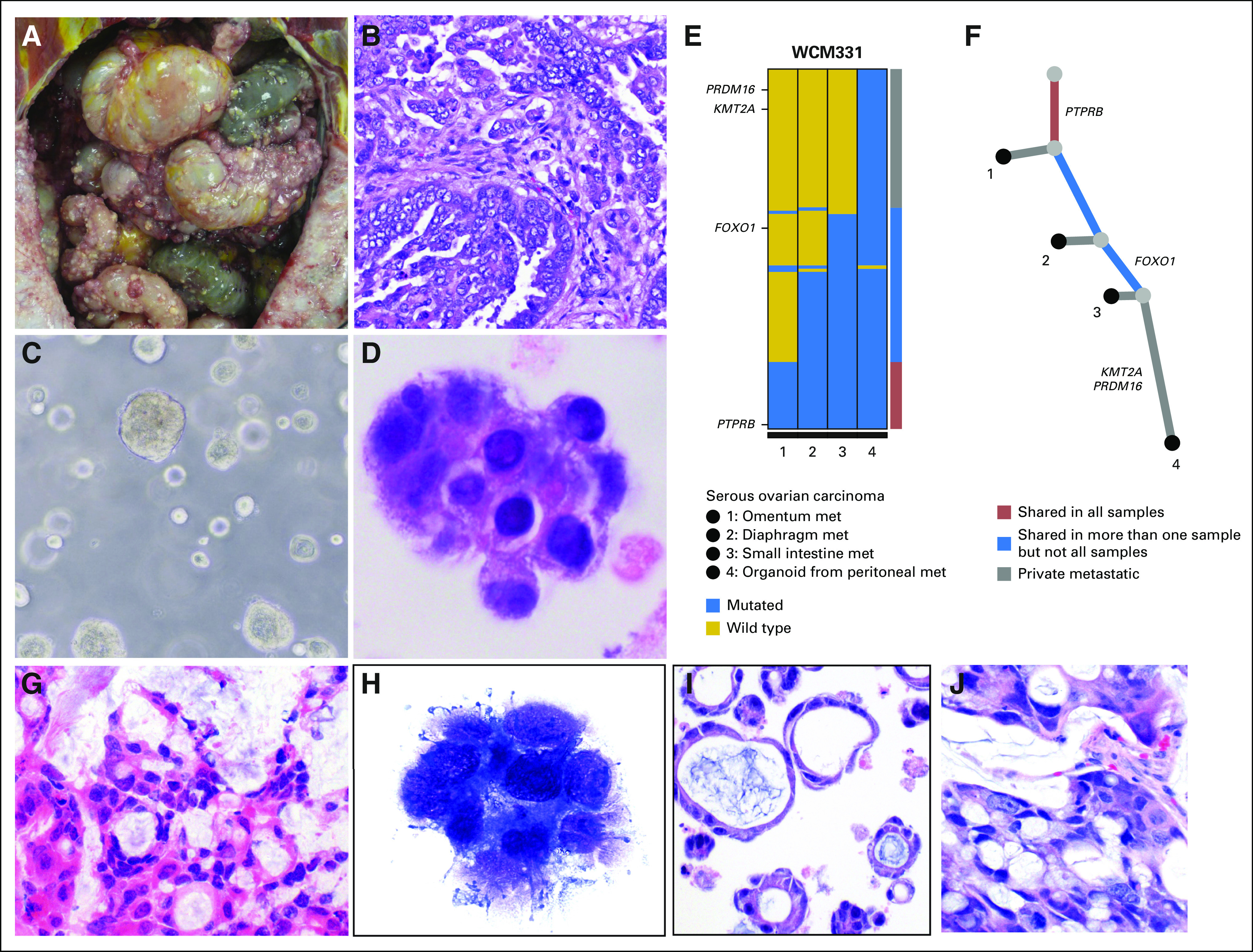
Results: Three hundred forty-six frozen samples were procured in total. WES was performed on 113 samples and RNA-Seq on 72 samples. Successful cell strain, tumor organoid, and/or patient-derived xenograft development was achieved in four samples, including an inoperable pediatric glioma. WES data were used to assess clonal evolution and molecular heterogeneity of tumors in individual patients. Mutational profiles of primary tumors and metastases yielded candidate mediators of metastatic spread and organotropism including CUL9 and PIGM in metastatic ependymoma and ANKRD52 in metastatic melanoma to the lung. RNA-Seq data identified novel gene fusion candidates.

Conclusion: A next-generation sequencing-based autopsy program in conjunction with a pre-mortem precision medicine pipeline for diverse tumors affords a valuable window into clonal evolution, metastasis, and alterations underlying treatment. Moreover, such an autopsy program yields robust preclinical models of disease.


Conflict of interest statement

Next-Generation Rapid Autopsies Enable Tumor Evolution Tracking and Generation of Preclinical Models

The following represents disclosure information provided by authors of this manuscript. All relationships are considered compensated. Relationships are self-held unless noted. I = Immediate Family Member, Inst = My Institution. Relationships may not relate to the subject matter of this manuscript. For more information about ASCO's conflict of interest policy, please refer to www.asco.org/rwc or po.ascopubs.org/site/ifc.

David J. Pisapia

No relationship to disclose

Steven Salvatore

No relationship to disclose

Chantal Pauli

No relationship to disclose

Erika Hissong

No relationship to disclose

Ken Eng

No relationship to disclose

Davide Prandi

No relationship to disclose

Verena-Wilbeth Sailer

No relationship to disclose

Brian D. Robinson

Stock and Other Ownership Interests: Metastat,

Consulting or Advisory Role: Progenics Pharmaceuticals

Patents, Royalties, Other Intellectual Property: Methods for diagnosing and treating prostate cancer

Kyung Park

No relationship to disclose

Joanna Cyrta

No relationship to disclose

Scott T. Tagawa

Consulting or Advisory Role: Medivation, Astellas Pharma, Dendreon, Janssen, Bayer, Genentech, Sanofi, Endocyte, Immunomedics

Speakers' Bureau: Amgen

Research Funding: Eli Lilly (Inst), Sanofi (Inst), Janssen (Inst), Astellas Pharma (Inst), Progenics (Inst), Millennium (Inst), Amgen (Inst), Bristol-Myers Squibb (Inst), Dendreon (Inst), Rexahn Pharmaceuticals (Inst), Bayer (Inst), Genentech (Inst), Newlink Genetics (Inst), Inovio Pharmaceuticals (Inst), AstraZeneca (Inst), Immunomedics (Inst), Novartis (Inst), AVEO (Inst), Rexahn Pharmaceuticals (Inst), Boehringer Ingelheim (Inst), Merck (Inst), Stem CentRx (Inst)

Travel, Accommodations, Expenses: Sanofi

Myriam Kossai

No relationship to disclose

Jacqueline Fontugne

No relationship to disclose

Robert Kim

No relationship to disclose

Alexandros Sigaras

Employment: Weill Cornell Medical College

Rema Rao

No relationship to disclose

Danielle Pancirer

No relationship to disclose

Bishoy Faltas

No relationship to disclose

Rohan Bareja

No relationship to disclose

Ana M. Molina

Honoraria: ASCO

Consulting or Advisory Role: Eisai, Exelixis, Novartis

David M. Nanus

Consulting or Advisory Role: Genentech

Prajwal Rajappa

No relationship to disclose

Mark M. Souweidane

Consulting or Advisory Role: Aesculap

Travel, Accommodations, Expenses: Aesculap

Jeffrey Greenfield

No relationship to disclose

Anne-Katrin Emde

No relationship to disclose

Nicolas Robine

No relationship to disclose

Olivier Elemento

No relationship to disclose

Andrea Sboner

No relationship to disclose

Francesca Demichelis

Patents, Royalties, Other Intellectual Property: Co-inventor on a patent filed by the University of Michigan and the Brigham and Women’s Hospital covering the diagnostic and therapeutic fields for ETS fusions in prostate cancer. The diagnostic field has been licensed to Gen-Probe.

Himisha Beltran

Consulting or Advisory Role: Bayer, Janssen Oncology, Genzyme

Research Funding: Astellas Pharma (Inst), Eli Lilly (Inst), Janssen (Inst), Millennium (Inst), Stemcentryx Abbvie

Mark A. Rubin

Research Funding: Eli Lilly, Janssen

Juan Miguel Mosquera

No relationship to disclose

Figures

Fig 1.
Fig 1.
Rapid autopsies of Weill Cornell Medicine (WCM) study cohort. (A) The list of 15 autopsies shows the research identifier (WCM number) and primary diagnosis. (B) Bar chart showing the total number of frozen tumor samples collected and the number of distinctly annotated anatomic sites from which tumor was obtained for each autopsy. (C) Schematic illustration of anatomic sites of metastatic samples that were collected at autopsy. The pie charts represent the breakdown with respect to primary tumor origin, which correspond to the color key on the left side. GC, gliomatosis cerebri pattern of glioma infiltration.
Fig 2.
Fig 2.
Clonal archeology of a patient with anaplastic ependymoma (rapid autopsy WCM419). Multiple sites of disease were available for study, including (A and B) the posterior fossa (primary site; [A] ventral surface of the whole brain with exophytic tumor component indicated by the arrow; [B] metastatic deposits within the supratentorial compartment involving the lateral ventricle and subventricular brain parenchyma, and lesions within the spinal cord (not shown), (C) axial cross-section through the pons and cerebellum). (D) Characteristic tumor histomorphology demonstrates spindled glial cells with fibrillar processes and perivascular pseudorosette formation (hematoxylin and eosin stain). (E) Timeline from diagnosis (month 0) to autopsy (month 64). Vertical lines indicate the relative timing of procedures from which samples were obtained during the course of disease. (F) Whole-exome sequencing was performed on prior surgical resection formalin-fixed paraffin-embedded specimens including primary tumor in posterior fossa (samples 1, 3, 4, and 5) and metastases to spinal cord (samples 6, 7, 9, and 10), as well as on frozen autopsy tissue from two distinct sites, including recurrent primary tumor in the posterior fossa (sample 2) and metastasis to the lateral ventricle (sample 8). Both the color-coded bar on the right and the phylogenetic tree below indicate whether mutations are private to primary or private to metastases or shared in more than one sample. There is no significant mutational overlap between the 10 samples. (G) Reconstruction of evolutionary tree. The length of the branches represents the distance between two tumors on the basis of the number of shared mutations. A complex branching pattern is present in this anaplastic ependymoma. Several mutations (ERBB3, DNMT3A, BRCA1, NOTCH1, and RUNX1T1) in the primary tumor samples are not shared either between them at different time points (0 to 64 months) or with descendent clones. None of the mutations detected was shared by all tumor samples. Scale bars = (A-C), 1 cm; (D), 40 μm. LV, lateral ventricle; met, metastasis; PF, posterior fossa; SC, spinal cord.
Fig 3.
Fig 3.
Molecular heterogeneity in a case of metastatic malignant melanoma (rapid autopsy WCM642). A 50-year-old male patient presented with disseminated disease from primary tumor of unknown origin. A previous biopsy (not shown) showed an unclassified malignant and epithelioid neoplasm. At autopsy, tissue samples from 17 distinct anatomic locations were snap frozen, including (A) adrenal gland and (B) lung. The tumor demonstrated heterogeneous histology with both (C) epithelioid and (D) spindled areas (hematoxylin and eosin stain). Tumor cells demonstrated focal staining for S100 (not shown), which had been negative in the biopsy. (E) Whole-exome sequencing results from eight distinct anatomic sites were obtained. Both the color-coded bar on the right and the phylogenetic tree below indicate whether mutations are private to any metastatic site or are shared in more than one sample or in all samples. Significant mutational overlap is present between all metastatic samples. (F) Reconstruction of the clonal architecture demonstrates that mutations in MLLT4, IDH1, AFF3, ARID2, ECT2L, and NRAS are shared in all samples. Few mutations are shared in more than one sample but not all samples (eg, APC in right liver metastasis (met); SMAD4 in left upper lobe of lung). The primary tumor was not identified and unavailable for sequencing analysis. Scale bars = (A and B), 1 cm; (C and D), 50 μm.
Fig 4.
Fig 4.
Examples of recurrent outliers and identification of novel gene fusion through RNA sequencing analysis. (A) Gene expression outlier analysis across 300 RNA sequencing samples derived from a diversity of tumors within the Institute for Precision Medicine cohort highlights WCM0 (small-cell carcinoma of prostate) and WCM772 (pleomorphic carcinoma of lung), two patients who show CDKN2C and MET as recurrent outliers in distinct anatomic samples, respectively. z scores were calculated for 70 druggable cancer genes, and outliers were selected at a cutoff of z score greater than 2.5 and fragments per kilobase million greater than 50. (B) RNA sequencing analysis by FusionCatcher and FusionSeq allows for the identification of novel gene fusion candidates. Schematic of the fusion between ZNF526 and MEGF8 in WCM0 showing the connected exons. (C) Integrative Genomics Viewer snapshots illustrating the data from six tumor specimens and one benign tissue specimen from the same patient. The coverage track summarizes the number of reads per nucleotide, whereas the junction tracks show how reads are connected by splicing. Note that the tumor samples all have connections between the two genes, whereas the benign specimen has none. (D) The sequence of the junction with some representative sequenced reads. met, metastasis.
Fig 5.
Fig 5.
Whole-genome sequencing of small-cell carcinoma of prostate nominates a novel gene fusion with clinical relevance (rapid autopsy WCM0). A 55-year-old male patient status post prostatectomy presented with local recurrence, metastases in the pelvis and liver, and Cushing syndrome in the month before death. (A and B) Upon opening the abdominal cavity, numerous metastases were present in the liver. (A) Liver in situ with multiple lesions is shown. (B) Section of liver after fixation is shown. (C) On histologic examination, the tumor demonstrated small-cell neuroendocrine morphology (hematoxylin and eosin stain of frozen tissue, ×200 original magnification). (D) Circos plot of chained rearrangements in sample LM2 (liver metastasis). (E) Structural variants called with Clipping Reveals Structure algorithm led to identification of a novel intrachromosomal gene fusion. Schematic of the fusion between YBX3 and STYK1 showing the connected exons. (F) Integrative Genomics Viewer snapshots illustrating the data from six tumor specimens and one benign tissue specimen from the same patient. The coverage track summarizes the number of reads per nucleotide, whereas the junction tracks show how reads are connected by splicing. Note that the tumor samples all have connections between the two genes, whereas the benign specimen has none. (G) The sequence of the junction with some representative sequenced reads. Overexpression of STYK1 may serve as a potential molecular target in castrate-resistant prostate cancer.
Fig 6.
Fig 6.
Tumor organoid and patient-derived xenograft development (rapid autopsies WCM331 and WCM715). (A) In patient WCM331, upon opening the peritoneal cavity, disseminated carcinomatosis was present. (B) Histologic examination confirmed high-grade serous adenocarcinoma of the ovary (hematoxylin and eosin [HE] stain, ×200 original magnification). (C) Viable tumor organoids were successfully produced. Image of patient-derived tumor organoid in a Matrigel scaffold (Corning, Corning, NY). (D) Cytologic comparison of these tumor organoids with representative sections from metastatic autopsy tissue revealed morphologically similar cells (Diff-Quik stain, ×400 original magnification). (E) Whole-exome sequencing was performed on formalin-fixed paraffin-embedded material derived from an omental metastasis biopsied 2 years before autopsy (sample 1), on frozen material from two metastases obtained at autopsy (samples 2 and 3), and on tumor organoid material derived from a peritoneal nodule obtained at autopsy (sample 4). Both the color-coded bar on the right and the phylogenetic tree below indicate whether mutations are private to the metastatic sites or are shared in more than one sample or in all samples. Multiple genetic alterations are shared across these specimens ranging from one (eg, FOXO1) to all samples (eg, PTPRB). This molecular evidence supports that the tumor organoid recapitulates the patient’s metastatic disease. (F) In the absence of material from the primary tumor, partial reconstruction of the clonal evolution of metastatic samples (including the tumor organoid as surrogate of one of the metastasis at autopsy) demonstrates that PTPRB mutation is shared in all samples and FOXO1 mutation is present in two metastases. Some alterations were seen in the tumor organoid only (eg, PRDM16 and KMT2A). (G) In case WCM715, an HE-stained section of a liver metastasis obtained at autopsy showed metastatic mucinous adenocarcinoma of the colon (×200 original magnification). (H) Smear preparation of tumor organoid culture (Diff-Quik stain) to confirm tumor growth (×400 original magnification). (I) HE-stained section of formalin-fixed paraffin-embedded organoid (×200 original magnification). (J) HE-stained section of patient-derived xenograft derived from organoid implantation in mouse demonstrates comparable histomorphology with original liver metastasis and derived tumor organoid (×200 original magnification). met, metastases.

References

    1. Beltran H, Eng K, Mosquera JM, et al. : Whole-exome sequencing of metastatic cancer and biomarkers of treatment response. JAMA Oncol 1:466-474, 2015 - PMC - PubMed
    1. Pauli C, Puca L, Mosquera JM, et al. : An emerging role for cytopathology in precision oncology. Cancer Cytopathol 124:167-173, 2016 - PubMed
    1. Rubin MA, Putzi M, Mucci N, et al: Rapid (“warm”) autopsy study for procurement of metastatic prostate cancer. Clin Cancer Res 6:1038-1045, 2000. - PubMed
    1. Shah RB, Mehra R, Chinnaiyan AM, et al. : Androgen-independent prostate cancer is a heterogeneous group of diseases: Lessons from a rapid autopsy program. Cancer Res 64:9209-9216, 2004 - PubMed
    1. Zarghooni M, Bartels U, Lee E, et al. : Whole-genome profiling of pediatric diffuse intrinsic pontine gliomas highlights platelet-derived growth factor receptor alpha and poly (ADP-ribose) polymerase as potential therapeutic targets. J Clin Oncol 28:1337-1344, 2010 - PubMed

LinkOut - more resources