. 2018 Jan;21(1):e25063.
doi: 10.1002/jia2.25063.

Exogenous oestrogen inhibits genital transmission of cell-associated HIV-1 in DMPA-treated humanized mice

Affiliations

Exogenous oestrogen inhibits genital transmission of cell-associated HIV-1 in DMPA-treated humanized mice

Nirk E Quispe Calla et al. J Int AIDS Soc. 2018 Jan.

Abstract

Introduction: HIV affects more women than any other life-threatening infectious agent, and most infections are sexually transmitted. HIV must breach the female genital tract mucosal barrier to establish systemic infection, and clinical studies indicate virus more easily evades this barrier in women using depot-medroxyprogesterone acetate (DMPA) and other injectable progestins for contraception. Identifying a potential mechanism for this association, we learned DMPA promotes susceptibility of wild-type mice to genital herpes simplex virus type 2 (HSV-2) infection by reducing genital tissue expression of the cell-cell adhesion molecule desmoglein-1 (DSG-1) and increasing genital mucosal permeability. Conversely, DMPA-mediated increases in genital mucosal permeability and HSV-2 susceptibility were eliminated in mice concomitantly administered exogenous oestrogen (E). To confirm and extend these findings, herein we used humanized mice to define effects of systemic DMPA and intravaginal (ivag) E administration on susceptibility to genital infection with cell-associated HIV-1.

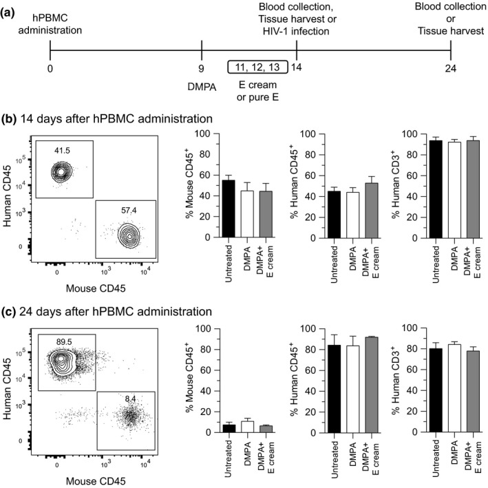
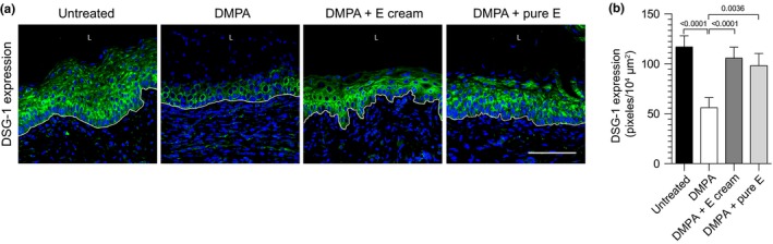
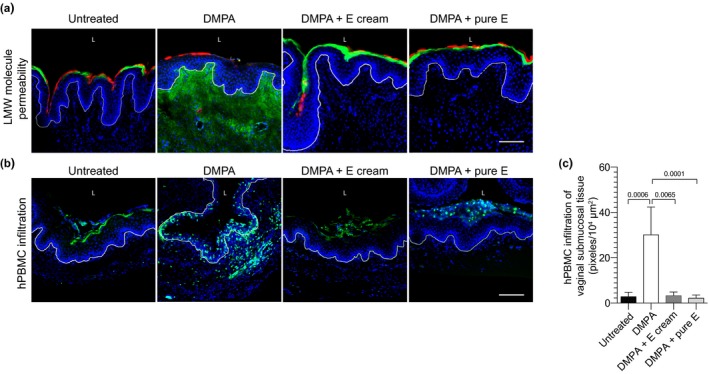
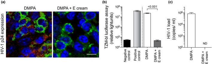
Methods: Effects of DMPA or an intravaginal (ivag) E cream on engraftment of NOD-scid-IL-2Rgcnull (NSG) mice with human peripheral blood mononuclear cells (hPBMCs) were defined with flow cytometry. Confocal microscopy was used to evaluate effects of DMPA, DMPA and E cream, or DMPA and the pharmacologically active component of the cream on vaginal tissue DSG-1 expression and genital mucosal permeability to low molecular weight (LMW) molecules and hPBMCs. In other studies, hPBMC-engrafted NSG mice (hPBMC-NSG) received DMPA or DMPA and ivag E cream before genital inoculation with 106 HIV-1-infected hPBMCs. Mice were euthanized 10 days after infection, and plasma HIV-1 load quantified by qRT-PCR and splenocytes used to detect HIV-1 p24 antigen via immunohistochemistry and infectious virus via TZM-bl luciferase assay.

Results: Whereas hPBMC engraftment was unaffected by DMPA or E treatment, mice administered DMPA and E (cream or the pharmacologically active cream component) displayed greater vaginal tissue expression of DSG-1 protein and decreased vaginal mucosal permeability to LMW molecules and hPBMCs versus DMPA-treated mice. DMPA-treated hPBMC-NSG mice were also uniformly susceptible to genital transmission of cell-associated HIV-1, while no animal concomitantly administered DMPA and E cream acquired systemic HIV-1 infection.

Conclusion: Exogenous E administration reduces susceptibility of DMPA-treated humanized mice to genital HIV-1 infection.

Keywords: DMPA; HIV prevention; genital HIV transmission; humanized mice; oestrogen.


Figures

Figure 1
Figure 1
Exogenous DMPA or E did not affect hPBMC engraftment of NSG mice. (a) Schematic of the study design used to assess effects of administering DMPA and E on hPBMC engraftment, genital mucosal permeability and HIV‐1 susceptibility of NSG mice. To define effects of DMPA and E on engraftment, peripheral blood was obtained 14 days and 24 days after hPBMC administration (untreated NSG mice provided controls). (b,c) Flow cytometric studies identified no between‐group differences in the percentages of murine CD45+ cells and human CD45+ CD3+ cells after hPBMC administration; left panels show representative contour plots; quadrant numbers denote population percentages. Data are from 2 independent experiments with 3 animals per group (bars denote mean ± SD). Statistical analyses performed using one‐way ANOVA with Dunnett's multiple comparisons test. DMPA, depot medroxyprogesterone acetate; E, ivag oestrogen cream; hPBMC‐NSG (hPBMC‐engrafted NOD‐scid‐IL‐2Rgcnull) mice.
Figure 2
Figure 2
DMPA‐mediated reduction in vaginal DSG‐1 protein expression was abrogated in hPBMC‐NSG mice administered DMPA and E. hPBMC‐NSG mice were untreated or treated with DMPA, DMPA and ivag E cream, or DMPA and ivag pure E. As detailed in Methods, vaginal tissue was collected from euthanized mice to quantify DSG‐1 protein expression. (a) Representative confocal microscopic images of vaginal DSG‐1 protein expression; L (vaginal lumen); DAPI (Blue); DSG‐1 (green); white line delimits the vaginal mucosal epithelium; scale bar denotes 100 μm. (b) Quantification of DSG‐1 protein expression showed significantly reduced levels in hPBMC‐NSG mice administered DMPA alone. Data from 2 independent experiments with 3 animals per group (bars denote mean ± SD). Statistical analyses performed using one‐way ANOVA with Dunnett's multiple comparisons test. hPBMC‐NSG (hPBMC‐engrafted NOD‐scid‐IL‐2Rgcnull) mice; DMPA, depot medroxyprogesterone acetate; E, vaginal oestrogen cream; pure E, pharmacologically active component of E cream; DSG‐1, desmoglein‐1.
Figure 3
Figure 3
Combined treatment with DMPA and exogenous E obviated DMPA‐mediated increases in vaginal mucosal permeability. hPBMC‐NSG mice remained untreated or were treated as described in Figure 2. As detailed in Methods, vaginal tissue was excised from euthanized mice to assess permeability to LMW molecules or CFSE‐labelled hPBMCs. (a, b) Representative images illustrate increased permeability to LMW molecules and activated human leukocytes in hPBMC‐NSG mice administered DMPA vs. untreated controls or animals administered DMPA and ivag E; scale bars denote 100 μm. (a) L (vaginal lumen); DAPI (blue) Lucifer Yellow (green); 70 kDa Texas‐Red dextran (red), and (b) L (vaginal lumen); DAPI (blue); CFSE‐labelled hPBMCs (green); white line delimits vaginal mucosal epithelium. (c) Quantifying depth of hPBMC infiltration into vaginal submucosal tissue identified significantly deeper infiltration in mice administered DMPA alone. Displayed data from 2 independent experiments with 3 animals per group (bars denote mean ± SD). Statistical analyses performed using one‐way ANOVA with Dunnett's multiple comparisons test. hPBMC‐NSG (hPBMC‐engrafted NOD‐scid‐IL‐2Rgcnull) mice; DMPA, depot medroxyprogesterone acetate; CFSE, carboxyfluorescein succinimidyl ester; E, oestrogen cream; pure E, pharmacologically active component of the E cream.
Figure 4
Figure 4
Exogenous E reversed susceptibility of DMPA‐treated humanized mice to genital transmission of cell‐associated HIV‐1. hPBMC‐NSG mice were treated with DMPA or DMPA and ivag E cream and genitally inoculated with cell‐associated HIV‐1 (as described in Methods and depicted in Figure 1a). Ten days after genital inoculation, mice were euthanized to assess HIV‐1 infection status. (a) Representative images of immunostaining for HIV‐1 p24 antigen in the spleens of euthanized mice show this viral protein detected only in mice treated with DMPA alone; DAPI (blue); anti‐human CD45 (green); anti‐HIV‐1 p24 antigen (red); scale bar denotes 20 μm. (b) TZM‐bl luciferase assay identified infectious HIV‐1 particles only in spleens from DMPA‐treated mice (splenocytes from uninfected mice and HIV‐1 BaL diluted in media provided negative and positive controls respectively). (c) A qRT‐PCR assay detected HIV‐1 virus only in the serum of hPBMC‐NSG mice administered DMPA alone (ND denotes no virus was detected). Data displayed are from 2 independent experiments with 5 animals per group (bars denote mean ± SD). Statistical analyses were performed using the unpaired Student's t‐test. DMPA, depot medroxyprogesterone acetate; E, vaginal oestrogen cream; hPBMC‐NSG (NOD‐scid‐IL‐2Rgcnull) mice.

Similar articles

Cited by

References

    1. HIV/AIDS JUNPo . Global AIDS Update 2016. UNAIDS Switzerland. 2017. [cited 2017 June 1]. Available from: http://www.unaids.org/en/resources/documents/2016/Global-AIDS-update-2016.
    1. Ramjee G, Daniels B. Women and HIV in Sub‐Saharan Africa. AIDS Res Therapy. 2013;10(1):30. - PMC - PubMed
    1. Leclerc‐Madlala S. Age‐disparate and intergenerational sex in southern Africa: the dynamics of hypervulnerability. AIDS. 2008;22(Suppl 4):S17–25. - PubMed
    1. Polis CB, Phillips SJ, Curtis KM, Westreich DJ, Steyn PS, Raymond E, et al. Hormonal contraceptive methods and risk of HIV acquisition in women: a systematic review of epidemiological evidence. Contraception. 2014;90(4):360–90. - PubMed
    1. Polis CB, Curtis KM, Hannaford PC, Phillips SJ, Chipato T, Kiarie JN, et al. An updated systematic review of epidemiological evidence on hormonal contraceptive methods and HIV acquisition in women. AIDS. 2016;30(17):2665–83. - PMC - PubMed

Publication types

LinkOut - more resources