Review
. 2018 Oct;91(1090):20170664.
doi: 10.1259/bjr.20170664. Epub 2018 Mar 20.

Whole Body MRI and oncology: recent major advances

Affiliations
Review

Whole Body MRI and oncology: recent major advances

Vassiliki Pasoglou et al. Br J Radiol. 2018 Oct.

Abstract

MRI is a very attractive approach for tumour detection and oncological staging with its absence of ionizing radiation, high soft tissue contrast and spatial resolution. Less than 10 years ago the use of Whole Body MRI (WB-MRI) protocols was uncommon due to many limitations, such as the forbidding acquisition times and limited availability. This decade has marked substantial progress in WB-MRI protocols. This very promising technique is rapidly arising from the research world and is becoming a commonly used examination for tumour detection due to recent technological developments and validation of WB-MRI by multiple studies and consensus papers. As a result, WB-MRI is progressively proposed by radiologists as an efficient examination for an expanding range of indications. As the spectrum of its uses becomes wider, radiologists will soon be confronted with the challenges of this technique and be urged to be trained in order to accurately read and report these examinations. The aim of this review is to summarize the validated indications of WB-MRI and present an overview of its most recent advances. This paper will briefly discuss how this examination is performed and which are the recommended sequences along with the future perspectives in the field.


Figures

Figure 1.
Figure 1.
Whole-body MR images in a 75-year-old male with castration-resistant prostate cancer. Coronal T1 weighted MR images (a) and inverted scale DWI images (b-value: 1000 s mm–2) (b) showing a diffuse low signal of the bone marrow on T1 images and a high signal intensity in high b-values DWI (*) indicating diffuse metastatic bone disease, a metastatic lesion of the left lung (arrow) and an abnormal pelvic lymph node (arrow head). Note the discrepancy between T1 and DWI. Homogenous diffuse low signal of the bone marrow on T1 images due to the sclerotic nature of the disease with focal hyper signal lesions on DWI. DWI, diffusion weighted imaging.
Figure 2.
Figure 2.
Whole-body MR images in a 68-year-old female with breast cancer. Bone scintigraphy anterior and posterior views (a), coronal T1 weighted images (b) and inverted scale DWI images (b-value: 1000 s mm–2) (c). The bone scintigraphy is normal, no abnormal foci are observed. WB-MRI study performed 2 days later showing minimum two bone lesions (arrows). Note the last station shading in DWI. DWI, diffusion weighted imaging.
Figure 3.
Figure 3.
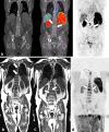
68Ga-PSMA PET CT (a) and WB-MRI (b, c, d) in a 82-year-old patient with high risk for metastasis prostate cancer (a): Clearly visible pathological accumulation of the radiotracer at the level of the spine (arrows) indicating metastatic bone disease and at the level of the pelvis (arrow head) demonstrating the presence of an abnormal lymph node (b–d): Coronal T1 weighted MR images (b, c) and inverted scale DWI images (d, b-value: 1000 s mm–2) (c) with the same findings. Multiple bone marrow metastasis at the level of the spine (arrows) and an abnormal pelvic lymph node (arrow head). Note the splenomegaly as the patient suffers from mild liver failure. DWI, diffusion weighted imaging; PET, positron emission tomography.
Figure 4.
Figure 4.
Whole-body MR images in a 25-year-old male with testicular seminoma. Coronal T1 weighted MR images (a) and and inverted scale DWI images (b-value: 1000 s mm–2) (b) show a pathologic retroperitoneal lymph node (arrow). DWI, diffusion weighted imaging.
Figure 5.
Figure 5.
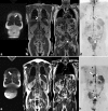
Whole-body MR images in a 68-year-old male with multiple myeloma. Coronal STIR (Short tau inversion recovery) (a) and T1 weighted (b) MR images and inverted scale DWI images (b-value: 1000 s mm–2) (c). The bone marrow shows diffuse hyperintense signal in STIR (a,*), hypointense signal in T1 (b, *) and high signal intensity in high b-value DWI (c,*) indicating diffuse tumoral infiltration (spine, pelvis, femora, humeri and ribs). Two dorsal vertebral fractures are observed (arrows). X-ray of the skull (d) demonstrating multiple lytic lesions (arrows).
Figure 6.
Figure 6.
Whole-body 3D MR images in a 70-year-old male with prostate cancer (a) Coronal reconstructed image showing bone marrow metastasis of right iliac bone (arrows) and an abnormal iliac lymph node (arrow head) (b) Sagittal reconstructed image showing multiple bone marrow metastasis of the spine (arrows) (c1 and c2). Axial reconstructed images showing bone marrow metastasis of a left rib (arrows in c1) and an abnormal para-aortic lymph node (arrow in c2).
Figure 7.
Figure 7.
Progressive disease in a patient on androgen deprivation therapy whole body MRI images in a 69-year-old patient with prostate cancer. (a–d): Baseline examination and (e–h): 6 months follow up (a–c, e–g): Coronal T1 weighted MR images (d, h): Reconstructed maximal intensity projection image from inverted scale DWI images (b-value: 1000 s mm–2) (a–d): Sternal (arrows) and dorsal spine (arrow heads) bone marrow metastasis and a normal sized left iliac lymph node (dotted arrow) (e–h): increase in size (e–g) and in high b-value signal intensity (h) of sternal (arrows) and dorsal spine (arrow heads) bone marrow lesions and increase in size of the abnormal lymph node.
Figure 8.
Figure 8.
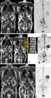
Response to treatment in a patient on androgen deprivation therapy Whole body MRI images in 50-year-old patient with prostate cancer. (a–c): Baseline examination, (d–f): 6 months follow up, (h, i): 1 year follow-up (a, b, d, e, h, i): Coronal T1 weighted MR images (f): dorsal region of (e), magnified (b, g, j): Reconstructed maximal intensity projection image from inverted scale DWI images (b-value: 1000 s mm–2) (a–c): Multiple bone marrow metastasis (arrows) and abnormal pelvic lymph nodes (arrow heads) (d–i): Decrease in size and in signal intensity in T1 sequence (d, e, h, i) of the spine (arrows) and left femoral (dotted arrows). Emergence of peritumoral hyperintense fat around the dorsal lesions (f-arrow) and decrease in signal intensity in T1 sequences (d, e, h, i). Decrease in signal intensity in high b-values DWI for the dorsal lesions (g- arrows) and disappearance of the left femoral lesion (g-dotted arrow). Decrease in size (d, e, h) of the pelvic lymph nodes (arrow heads). DWI, diffusion weighted imaging.
Figure 9.
Figure 9.
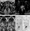
“All in one” WB-MRI protocol for the TNM staging of a high risk for metastasis in a 74-year-old prostate cancer patient. One single step protocol combining a multiparametric MRI of the prostate (a, b) for the T and a WB-MRI protocol (c– f) for the N and M staging. T2 axial images (a) and ADC map (b) of the prostate demonstrating a low signal intensity mass with low ADC values (arrows). No capsular invasion. Coronal T1 weighted WB-MRI images (c, d) and inverted scale DWI images (b-value: 1000 s mm–2) (e, f) showing a metastatic bone marrow lesion of the right ischio-pubic ramous (arrows) and abnormal retro-peritoneal and pelvic lymph nodes (arrow heads). The TNM staging of this patient is T2N1M1.

References

    1. Takahara T, Imai Y, Yamashita T, Yasuda S, Nasu S, Van Cauteren M. Diffusion weighted whole body imaging with background body signal suppression (DWIBS): technical improvement using free breathing, STIR and high resolution 3D display. Radiat Med 2004; 22: 275–82. - PubMed
    1. Padhani AR, Liu G, Koh DM, Chenevert TL, Thoeny HC, Takahara T, et al. . Diffusion-weighted magnetic resonance imaging as a cancer biomarker: consensus and recommendations. Neoplasia 2009; 11: 102–25. doi: 10.1593/neo.81328 - DOI - PMC - PubMed
    1. Lecouvet FE. Whole-body MR imaging: musculoskeletal applications. Radiology 2016; 279: 345–65. doi: 10.1148/radiol.2016142084 - DOI - PubMed
    1. Wu LM, Gu HY, Zheng J, Xu X, Lin LH, Deng X, et al. . Diagnostic value of whole-body magnetic resonance imaging for bone metastases: a systematic review and meta-analysis. J Magn Reson Imaging 2011; 34: 128–35. doi: 10.1002/jmri.22608 - DOI - PubMed
    1. Fischer MA, Nanz D, Hany T, Reiner CS, Stolzmann P, Donati OF, et al. . Diagnostic accuracy of whole-body MRI/DWI image fusion for detection of malignant tumours: a comparison with PET/CT. Eur Radiol 2011; 21: 246–55. doi: 10.1007/s00330-010-1929-x - DOI - PubMed

MeSH terms