. 2018 Feb;36(2):170-178.
doi: 10.1038/nbt.4062. Epub 2018 Jan 15.

Dual gene activation and knockout screen reveals directional dependencies in genetic networks

Affiliations

Dual gene activation and knockout screen reveals directional dependencies in genetic networks

Michael Boettcher et al. Nat Biotechnol. 2018 Feb.

Abstract

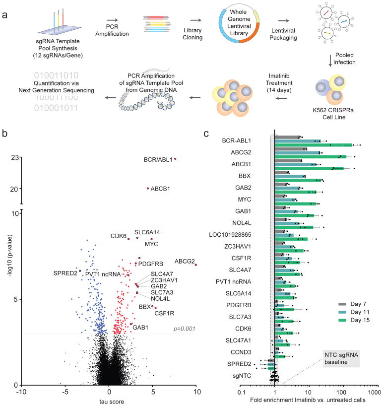
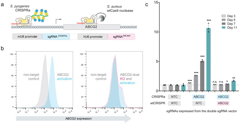
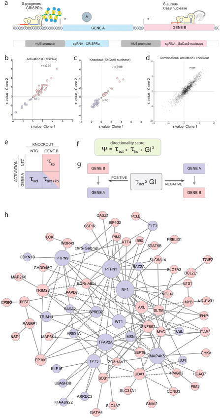
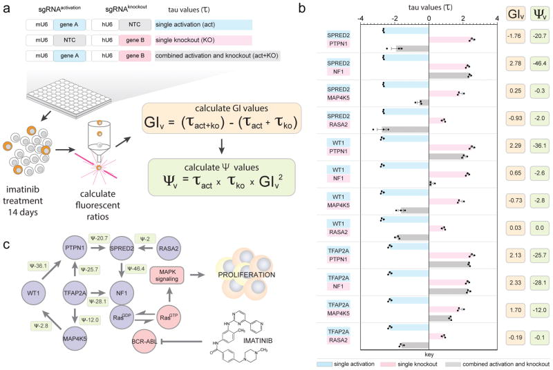
Understanding the direction of information flow is essential for characterizing how genetic networks affect phenotypes. However, methods to find genetic interactions largely fail to reveal directional dependencies. We combine two orthogonal Cas9 proteins from Streptococcus pyogenes and Staphylococcus aureus to carry out a dual screen in which one gene is activated while a second gene is deleted in the same cell. We analyze the quantitative effects of activation and knockout to calculate genetic interaction and directionality scores for each gene pair. Based on the results from over 100,000 perturbed gene pairs, we reconstruct a directional dependency network for human K562 leukemia cells and demonstrate how our approach allows the determination of directionality in activating genetic interactions. Our interaction network connects previously uncharacterized genes to well-studied pathways and identifies targets relevant for therapeutic intervention.


Conflict of interest statement

Competing Financial Interests

The authors declare no competing financial interests.

Figures

Figure 1
Figure 1. Ultra-complex CRISPRa screen identifies hundreds of genes involved in cancer signalling pathways
a, Schematic of genome-scale CRISPRa screening approach (see text for details). b, Overview of CRISPRa screen results. Negative τ values indicate depletion and positive values enrichment of cells following imatinib selection. Significant candidate genes (FDR<0.05, p<0.001) are in colour (blue = depleted, red = enriched). Validated candidate genes are labelled in black. Mann-Whitney U test was used to calculate p-values as described previously. To correct for multiple hypothesis testing, we first performed random sampling with replacement among the set of τ values for non-targeting control sgRNAs and calculated p-values for each sampling. Then, we calculated the false discover rate (FDR) based on the distribution of p-values for all genes in the library and for non-targeting controls generated above. c, Candidate gene validation. Enrichment of candidate sgRNA expressing cells was measured over time. Values represent the mean of three different sgRNAs targeting each gene with s.e.m. Grey shading = two standard deviations of sgNTCs at day 15. All values from separate sgRNAs on days 7, 11 and 15 normalised to baseline or untreated cells are shown in Supplemental Table 3.
Figure 2
Figure 2. The orthogonal CRISPR system
a, Schematic of the orthogonal system on the example of imatinib efflux transporter ABCG2. Combination of CRISPR systems from S.pyogenes (CRISPRa) and S.aureus (Cas9 nuclease) allows the simultaneous activation and knockout of genes in the same cell simply by expressing two appropriate sgRNAs. b, Orthogonal system is able to modulate ABCG2 protein levels. Flow cytometry analysis of ABCG2 levels following CRISPRa mediated activation of ABCG2 without (left) or with (right) SaCas9 nuclease mediated knockout of ABCG2 (grey histogram = sgNTC for both CRISPR systems). A representative result from n>10 independent experiments with similar results is shown. c, Orthogonal system can control imatinib response. Enrichment of imatinib treated cells with activated ABCG2 with/out SaCas9 nuclease mediated knockout of ABCG2. Values represent the mean of independent experiments (n=3) with s.e.m. and statistical significance was determined via two-tailed, homoscedastic t-test with * = p<0.05, ** =p<0.01 and *** = p<0.001.
Figure 3
Figure 3. Orthogonal CRISPR screens can quantify directional genetic interactions
a, Concept of the application of the orthogonal system for directional gene interaction studies. In the same cell, one gene is activated (CRISPRa) while another gene in knocked out (SaCas9 nuclease). b–d, Correlation of τ values from two clonal cell line replicates is shown for b, gene activation, c, gene knockout and d, all possible combinations thereof. Correlation values (r) are Pearson product-moment correlation coefficients. e, Schematic of perturbation data set from each gene pair (blue = depleted, red = enriched, NTC = non-target control sgRNA) f, Formula for calculating Ψ scores. Negative Ψ scores define interactions in which directionality could be inferred. g, To determine which of both interaction partners acts up- or downstream, τactivation values were multiplied with genetic interaction scores. Positive values indicate a downstream function, negative values an upstream function of the activated gene. h, Based on GI and Ψ scores determined by the full orthogonal interaction screen, a genetic interaction model was constructed. For positive regulators of cell fitness, nodes are shown in red and negative regulators in blue. Arrow-shaped edges indicate inferred directional interactions between nodes. Line-shaped edges symbolise genetic interactions where directionality could not be inferred. Node sizes are proportional to the degree of connectivity. In total, 2258 gene:gene combinations that passed the filter criteria were considered for the construction of the directional genetic interaction network.
Figure 4
Figure 4. Validation of a directional Ras-centric genetic sub-network
a, Relative fitness (τ) was measured over 14 days following gene activation, knockout or the combination of both. From those values, genetic interaction (GIv) as well as directionality (Ψv) scores were calculated. NTC = non-target control sgRNA. b, Twelve activation/knockout combinations were re-tested in an arrayed format from which ten were predicted by the orthogonal screen to show a directional genetic interaction. Eight combinations displayed the same trend of directional interactions predicted by the orthogonal screen data while two interactions did not reproduce (see also Supplemental Table 9). Single perturbation, and combinatorial τ values are shown following 14 days of imatinib selection (mean with s.e.m. from technical replicates (n=3)) along with calculated GIv and Ψv scores for each gene:gene combination. c, A directional genetic interaction model was assembled based on validated interactions from b. Arrows indicate the direction of the functional dependencies as explained in the text but do not suggest direct physical interactions. Values represent Ψv scores calculated from τ values in b. Each directional interaction was reproduced three times independently.
Figure 5
Figure 5. Exploiting genetic dependencies for cancer therapy
a, NF1-knockout K562 cells are significantly more sensitive to the AXL kinase inhibitor R428 than NF1-wildtype cells. Cells were treated for 8 days with 500 nM R428 on day 0 and day 4. (mean with s.e.m. from technical replicates (n=6)). b, NF1-knockout K562 cells are significantly more resistant to imatinib but can be re-sensitised by R428 treatment (mean with s.e.m. from technical replicates (n=4 for imatinib and n=2 for imatinib + R428 treated cells). In panel a and b, statistical significance was determined via two-tailed, homoscedastic t-test with * = p<0.05, **=p<0.01 and *** = p<0.001. c, NF1-knockout cells accumulate elevated levels of phosphorylated AXL kinase (p-AXL) which can be reduced by treatment with the AXL kinase inhibitor R428. Quantification of the ratio of band intensity from p-AXL/β-actin, normalised to p-AXL levels in NF1-wt untreated cells is shown. The experiment was performed once. d, TR-FRET assay shows direct interaction between NF1 and AXL in HEK293T cells. Shown is the mean with s.d. from three independent experiments.

Comment in

Similar articles

Cited by

References

    1. Beltrao P, Cagney G, Krogan NJ. Quantitative genetic interactions reveal biological modularity. Cell. 2010;141:739–745. doi: 10.1016/j.cell.2010.05.019. - DOI - PMC - PubMed
    1. Fischer B, et al. A map of directional genetic interactions in a metazoan cell. eLife. 2015;4 doi: 10.7554/eLife.05464. - DOI - PMC - PubMed
    1. Drees BL, et al. Derivation of genetic interaction networks from quantitative phenotype data. Genome biology. 2005;6:R38. doi: 10.1186/gb-2005-6-4-r38. - DOI - PMC - PubMed
    1. St Onge RP, et al. Systematic pathway analysis using high-resolution fitness profiling of combinatorial gene deletions. Nature genetics. 2007;39:199–206. doi: 10.1038/ng1948. - DOI - PMC - PubMed
    1. Avery L, Wasserman S. Ordering gene function: the interpretation of epistasis in regulatory hierarchies. Trends in genetics : TIG. 1992;8:312–316. - PMC - PubMed

Publication types

MeSH terms

Substances