. 2018 Mar;14(3):284-290.
doi: 10.1038/nchembio.2551. Epub 2018 Jan 15.

The molecular basis of subtype selectivity of human kinin G-protein-coupled receptors

Affiliations

The molecular basis of subtype selectivity of human kinin G-protein-coupled receptors

Lisa Joedicke et al. Nat Chem Biol. 2018 Mar.

Abstract

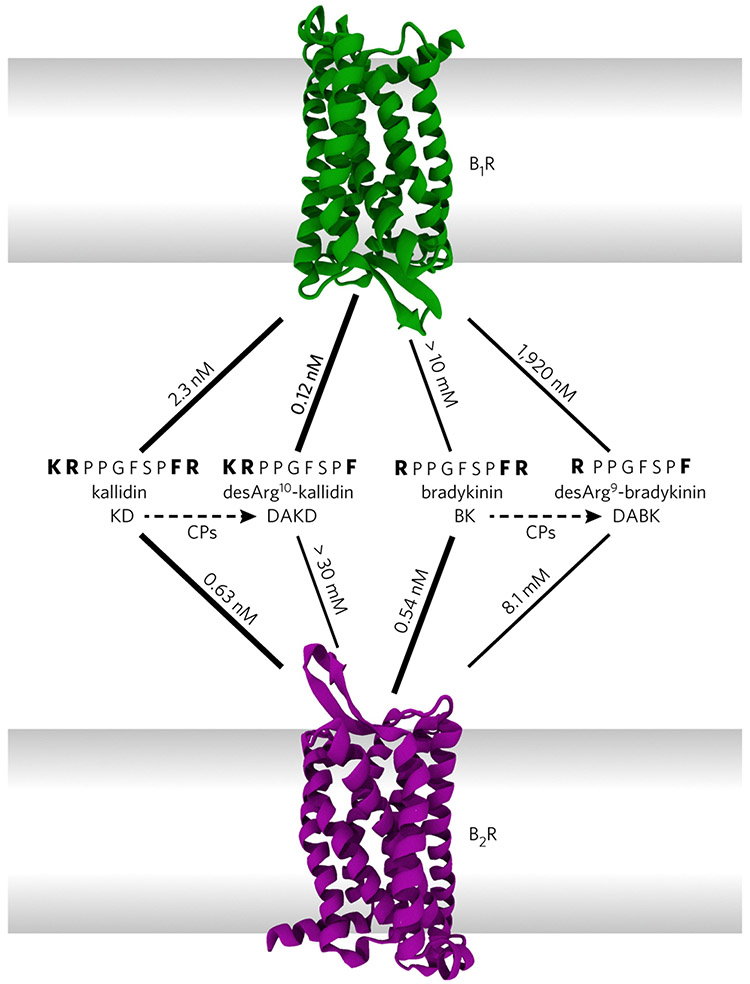
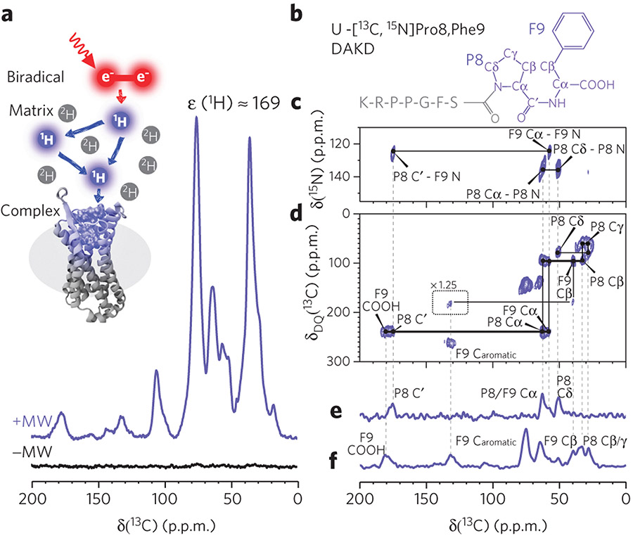
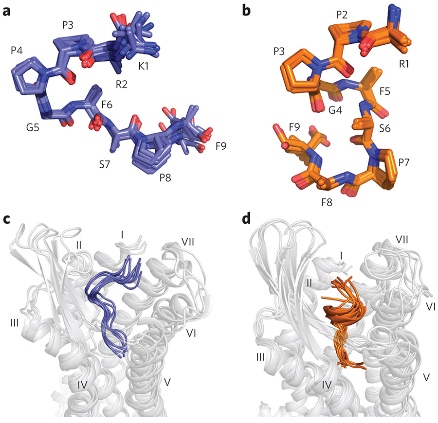
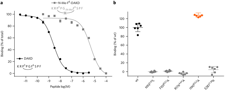
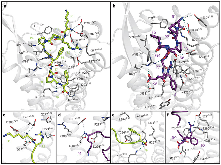
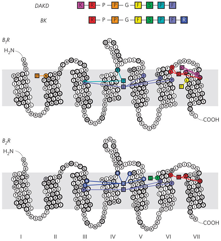
G-protein-coupled receptors (GPCRs) are the most important signal transducers in higher eukaryotes. Despite considerable progress, the molecular basis of subtype-specific ligand selectivity, especially for peptide receptors, remains unknown. Here, by integrating DNP-enhanced solid-state NMR spectroscopy with advanced molecular modeling and docking, the mechanism of the subtype selectivity of human bradykinin receptors for their peptide agonists has been resolved. The conserved middle segments of the bound peptides show distinct conformations that result in different presentations of their N and C termini toward their receptors. Analysis of the peptide-receptor interfaces reveals that the charged N-terminal residues of the peptides are mainly selected through electrostatic interactions, whereas the C-terminal segments are recognized via both conformations and interactions. The detailed molecular picture obtained by this approach opens a new gateway for exploring the complex conformational and chemical space of peptides and peptide analogs for designing GPCR subtype-selective biochemical tools and drugs.


Figures

Figure 1 ∣
Figure 1 ∣. Affinities of kinin peptides for their respective human bradykinin receptors, B1R and B2R.
Kallidin (KD) and bradykinin (BK) derive from kininogen by proteolytic cascades and differ only by an additional N-terminal lysine residue in KD. Both peptides are high-affinity ligands for B2R. Removal of the C-terminal arginine (dashed lines) by carboxypeptidases (CPs) yields desArg10-kallidin (DAKD) and desArg9-bradykinin (DABK). Despite their similarity, only DAKD, but not DABK, binds with high affinity to B1R.
Figure 2 ∣
Figure 2 ∣. Experimental setup and exemplary spectra of B1R in complex with DAKD.
(a) A sample containing the DAKD–B1R complex doped with the biradical AMUPol is subjected to magic-angle sample spinning under continuous wave microwave irradiation, resulting in polarization transfer from electrons via protons to the sites of interest. As a result, a large signal enhancement of the B1R–DAKD complex (purple) is observed in comparison to conventional NMR (black) (see Online Methods). (b) One of the DAKD labeling schemes used here: U-[13C,15N]P8F9 DAKD. The chemical shift assignment was accomplished by following the connection of the signals on TEDOR 15N-13C spectra (c), DQ-SQ 13C-13C (d), TEDOR-filtered 13C-spectra (e), and DQ/REDOR doubly filtered 13C-spectra (f). The pulse program used for f is presented in Supplementary Figure 6. Caromatic, carbons on a phenyl ring. Dashed lines guide the chemical shift connectivity among different spectra.
Figure 3 ∣
Figure 3 ∣. Backbone structures of DAKD in complex with human B1R in comparison to BK bound to human B2R.
Only backbone and Cβ atoms are shown. (a) The backbone structure of DAKD calculated from NMR data features a V-shaped fold with a β-turn-like structure around P3–F6. (b) The NMR-based backbone structure of BK is characterized by an overall S-shape with a 310-helix-like segment (P2–F5) in the middle. (c,d) Rosetta modeling of DAKD in B1R (c) and BK in B2R (d) reproduces the characteristic V-shape of DAKD and the S-shape fold of BK (see text and Supplementary Fig. 10 for further details).
Figure 4 ∣
Figure 4 ∣. Functional characterization of peptide variants and B1R mutants.
(a) Disruption of the central β-turn in DAKD results in a strong decrease in affinity for the B1R. The methylation of the amide nitrogen of F6DAKD disrupts the central β-turn and results in a 1,000-fold decrease of DAKD binding affinity (KI DAKD: 1.11 ± 0.04 nM (circles); DAKD linearized: 2.03 ± 0.17 μM (rectangles)). (b) Verification of key peptide interaction sites predicted from B1R–DAKD models by site-directed mutagenesis (maximal binding of DAKD normalized to wild type; n = 6). I190ECL2A shows an increased binding activity (129%, orange scatter), whereas all other mutations result in a complete loss of DAKD binding. Expression levels of B1R mutants were assessed by western blot (Supplementary Fig. 13).
Figure 5 ∣
Figure 5 ∣. Structural characterization of the B1R–DAKD (green) and B2R–BK (purple) binding pocket.
(a) Top view of DAKD docked to a comparative model of B1R. (b) Top view of BK docked to a comparative model of B2R. (c) Side view of the DAKD and (d) BK N-terminal binding site at TMH VI and VII. (e) Side view of the DAKD and BK (f) C-terminal binding site between TMH3, 5 and 6. The ligand is shown as thick sticks. Receptor residues predicted to be involved in ligand binding are labeled and are shown as thin sticks. Predicted interactions are indicated by dotted cyan lines. Atoms are colored by type (oxygen, red; nitrogen, blue; sulfur, yellow).
Figure 6 ∣
Figure 6 ∣. Representation of key interactions responsible for high affinity binding of DAKD to B1R and of BK to B2R.
B1R discriminates between DAKD and BK mainly via electrostatic interactions at the N terminus, whereas B2R selects via a complex interaction network as a result of different C-terminal structures of the BK and DAKD. The residues conserved among B1R and B2R are shown in bold circles.

Comment in

  • GPCR: Lock and key become flexible.
    Baldus M. Baldus M. Nat Chem Biol. 2018 Feb 14;14(3):201-202. doi: 10.1038/nchembio.2567. Nat Chem Biol. 2018. PMID: 29443979 No abstract available.

References

    1. Kobilka BK G protein coupled receptor structure and activation. Biochim. Biophys. Acta 1768, 794–807 (2007). - PMC - PubMed
    1. Rasmussen SGF et al. Crystal structure of the β2 adrenergic receptor-Gs protein complex. Nature 477, 549–555 (2011). - PMC - PubMed
    1. Rasmussen SGF et al. Structure of a nanobody-stabilized active state of the β2 adrenoceptor. Nature 469, 175–180 (2011). - PMC - PubMed
    1. White JF et al. Structure of the agonist-bound neurotensin receptor. Nature 490, 508–513 (2012). - PMC - PubMed
    1. Isogai S et al. Backbone NMR reveals allosteric signal transduction networks in the β1-adrenergic receptor. Nature 530, 237–241 (2016). - PubMed

Publication types

LinkOut - more resources