. 2018 Feb;176(2):337-350.
doi: 10.1002/ajmg.a.38580.

Thoracic aortic aneurysm in patients with loss of function Filamin A mutations: Clinical characterization, genetics, and recommendations

Affiliations

Thoracic aortic aneurysm in patients with loss of function Filamin A mutations: Clinical characterization, genetics, and recommendations

Ming Hui Chen et al. Am J Med Genet A. 2018 Feb.

Abstract

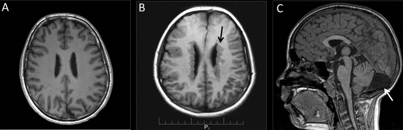
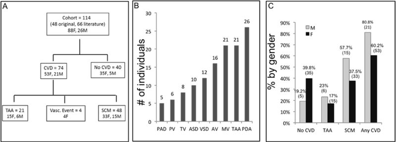
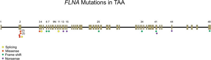
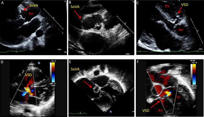
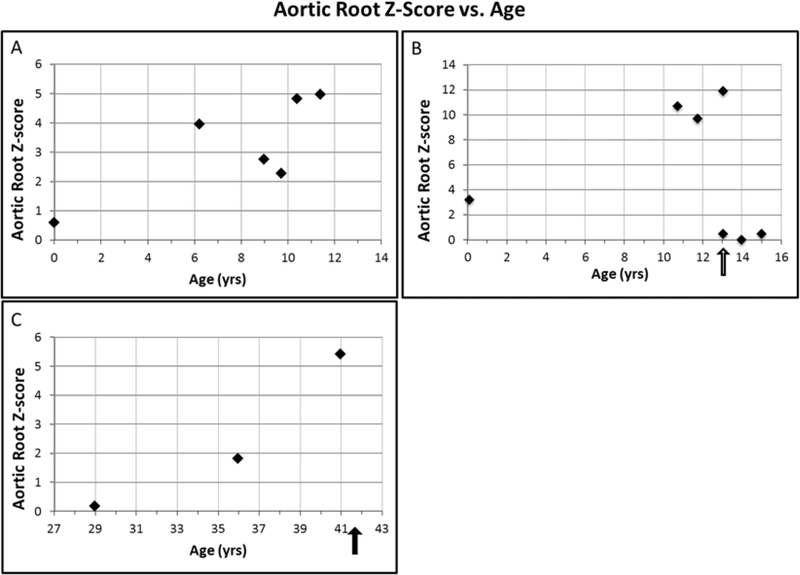
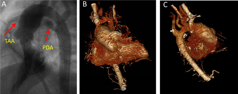
The frequency and gender distribution of thoracic aortic aneurysm as a cardiovascular manifestation of loss-of-function (LOF) X-linked FilaminA (FLNA) mutations are not known. Furthermore, there is very limited cardiovascular morbidity or mortality data in children and adults. We analyzed cardiac data on the largest series of 114 patients with LOF FLNA mutations, both children and adults, with periventricular nodular heterotopia (PVNH), including 48 study patients and 66 literature patients, median age of 22.0 years (88 F, 26 M, range: 0-71 years), with 75 FLNA mutations observed in 80 families. Most (64.9%) subjects had a cardiac anomaly or vascular abnormality (80.8% of males and 60.2% of females). Thoracic aortic aneurysms or dilatation (TAA) were found in 18.4% (n = 21), and were associated with other structural cardiac malformations in 57.1% of patients, most commonly patent ductus arteriosus (PDA) and valvular abnormalities. TAA most frequently involved the aortic root and ascending aorta, and sinus of Valsalva aneurysms were present in one third of TAA patients. Six TAA patients (28.5%) required surgery (median age 37 yrs, range 13-41 yrs). TAA with its associated complications was also the only recorded cause of premature, non-accidental mortality in adults (2 M, 2 F). Two adult patients (1 F, 1 M, median 38.5 yrs), died of spontaneous aortic rupture at aortic dimensions smaller than current recommendations for surgery for other aortopathies. Data from this largest series of LOF FLNA mutation patients underscore the importance of serial follow-up to identify and manage these potentially devastating cardiovascular complications.

Keywords: anomalies; aortic aneurysm; cardiac; filaminA; patent ductus arteriosus; periventricular nodular heterotopia; thoracic.


Figures

Figure 1:
Figure 1:. Representative axial T1-weighted brain Magnetic Resonance Imaging (MR) from normal and PVNH patients.
(A) Patient without PVNH. (B) PVNH patient with nodular heterotopias (arrow) along the lateral ventricles of the brain. (C) Mega cisterna magna (arrow) is seen on sagittal images in some PVNH patients.
Figure 2:
Figure 2:
(A) Study schema (n=114 individuals). 64.9% (n=74) of the cohort had either a vascular aneurysm or event (n=26) or a structural cardiac anomaly (n=48). Twenty-one had a thoracic aortic aneurysm (TAA), with 4/21 patients having both a TAA and a dilated pulmonary artery. One patient with a dilated pulmonary artery alone was not specifically depicted in schema. (B) Frequency of the vascular and structural cardiac abnormalities in individuals. TAA and PDAs were very common in subjects and frequently occurred in conjunction. (C) Gender distribution of cardiovascular malformations and aneurysms: There was no significant difference between males and females, by Fisher’s exact test. Absolute numbers are in parentheses. M = male, F = female. No CVD = No cardiovascular anomalies or abnormalities, TAA = Thoracic aortic aneurysm, SCM= Structural cardiovascular malformation, Vasc = Vascular event (i.e. stroke, myocardial infarction). ASD = Atrial septal defect, AV= Aortic valve abnormality, MV= Mitral valve abnormality, PAD = Pulmonary artery dilatation, PDA = Patent ductus arteriosus, PV = Pulmonary valve abnormality, TV= Tricuspid valve abnormality, VSD = Ventricular septal defect.
Figure 3:
Figure 3:. Genomic locations of exonic FLNA mutations associated with TAA.
A schematic representation of the FLNA gene. The FLNA gene is 25.9 kbp in size and encoded by 49 exons including a newly discovered “poison” exon 9N (Zhang et al, 2016), present in the contig: NT 025965. Exons are depicted by boxes, and introns are shown as black lines between the exons. Exons with relevant mutations are numbered. Types of FLNA mutations occurring in patients with TAA are localized on the FLNA map. The mutation in two families that each show two 2 individuals is indicated by one circle with total family members in parentheses. Five unique missense FLNA mutations (occurring in 7 individuals) associated with TAA localize most commonly to the actin-binding domains of FLNA gene (encoded by exons 2–5), whereas other loss-of-function mutations (nonsense, splicing, frameshift) are seen throughout the gene. One patient had a complete deletion of the FLNA gene.
Figure 4:
Figure 4:. Sinus of Valsalva aneurysms (SoVA) with perimembranous ventricular septal defect in 2 young females.
(A-B) Echocardiography images from 21 year-old female with SoVA involving the right and non-coronary cusps of the aorta (parasternal long-axis and short-axis views). (C-D) Patient also has a perimembranous ventricular septal defect (VSD) with shunt flow, seen on parasternal and apical views. (E-F) Echocardiography images from an 11 year-old female depicting sinus of Valsalva aneurysm and a perimembranous VSD (apical 5-chamber view). Ao = aorta, LV = left ventricle, RV= right ventricle.
Figure 5:
Figure 5:. Aortic root growth vs. age in 3 patients, pediatric and adult.
Aortic size is displayed as Z-scores in all individuals to allow for comparison. (A) Female child (#85) has a normal Z score at birth, but over the next 10 years, her aortic root z-scores have increased to ~5. (B) #37 Female child (#37) was born with a dilated aorta that increased to a Z score of 11.9, before her aneurysm surgery. Root size was normalized by surgery. Black arrow indicates timing of aortic aneurysm surgery. (C) Middle-age adult female (#36) who had normal aortic root Z scores on initial imaging, with a rapid increase in aortic root size between age 35 and 41 years of age. Aortic root size was 4.2 cm (Z score = 5.4) at time of death. Black arrow denotes timing of patient demise.
Figure 6:
Figure 6:. Female with progressive thoracic aortic dilatation requiring surgery.
(A) Dilated aorta and PDA seen on cardiac catheterization images during infancy. (B-C) 3D CT images of the patient’s aorta, obtained a decade later, demonstrate persistent dilatation of the aortic root and ascending aorta without involvement of the distal arch or descending aorta.

References

    1. Chen MH, & Walsh CA (2015). FLNA-Related Periventricular Nodular Heterotopia. In Pagon RA, Adam MP, Ardinger HH, Wallace SE, Amemiya A, Bean LJH, Bird TD, Ledbetter N, Mefford HC, Smith RJH, & Stephens K (Eds.), GeneReviews(R) Seattle (WA).
    1. Chubb H, & Simpson JM (2012). The use of Z-scores in paediatric cardiology. Ann Pediatr Cardiol, 5(2), 179–184. doi:10.4103/0974-2069.99622 - DOI - PMC - PubMed
    1. Clark AR, Sawyer GM, Robertson SP, & Sutherland-Smith AJ (2009). Skeletal dysplasias due to filamin A mutations result from a gain-of-function mechanism distinct from allelic neurological disorders. Hum Mol Genet, 18(24), 4791–4800. doi:10.1093/hmg/ddp442 - DOI - PubMed
    1. de Wit MC, de Coo IF, Lequin MH, Halley DJ, Roos-Hesselink JW, & Mancini GM (2011). Combined cardiological and neurological abnormalities due to filamin A gene mutation. Clin Res Cardiol, 100(1), 45–50. doi:10.1007/s00392-010-0206-y - DOI - PMC - PubMed
    1. de Wit MC, Kros JM, Halley DJ, de Coo IF, Verdijk R, Jacobs BC, & Mancini GM (2009). Filamin A mutation, a common cause for periventricular heterotopia, aneurysms and cardiac defects. J Neurol Neurosurg Psychiatry, 80(4), 426–428. doi:10.1136/jnnp.2008.149419 - DOI - PubMed

Publication types

MeSH terms