. 2018 Mar;12(2):104-116.
doi: 10.1080/17435390.2018.1425497. Epub 2018 Jan 15.

Silver nanoparticles induce neurotoxicity in a human embryonic stem cell-derived neuron and astrocyte network

Affiliations

Silver nanoparticles induce neurotoxicity in a human embryonic stem cell-derived neuron and astrocyte network

Neza Repar et al. Nanotoxicology. 2018 Mar.

Abstract

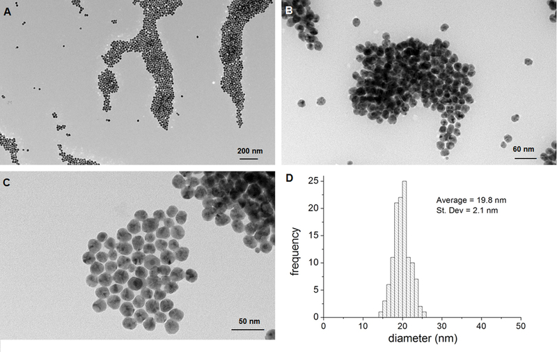
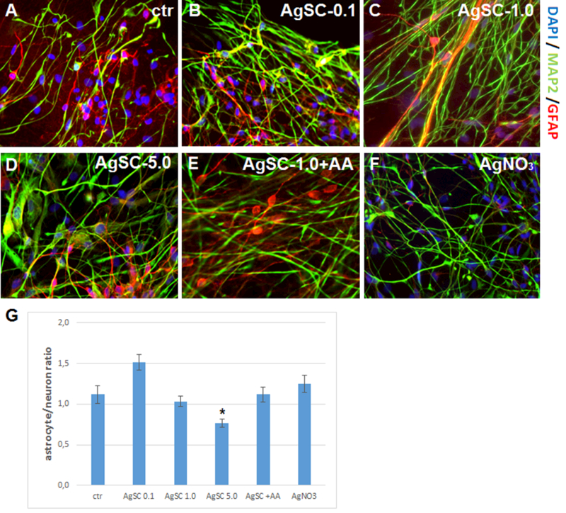
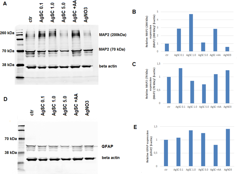
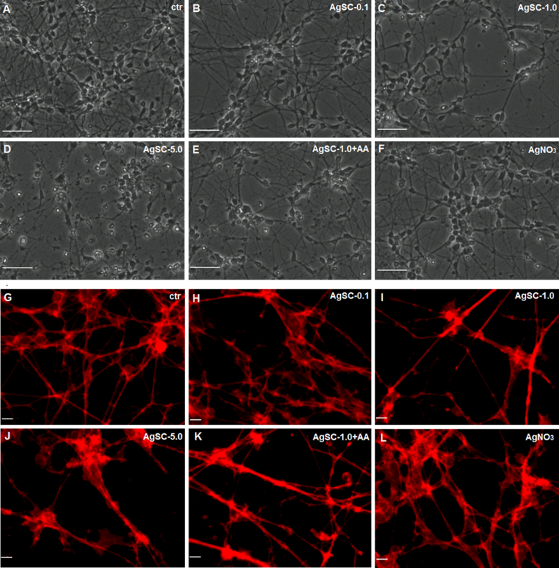
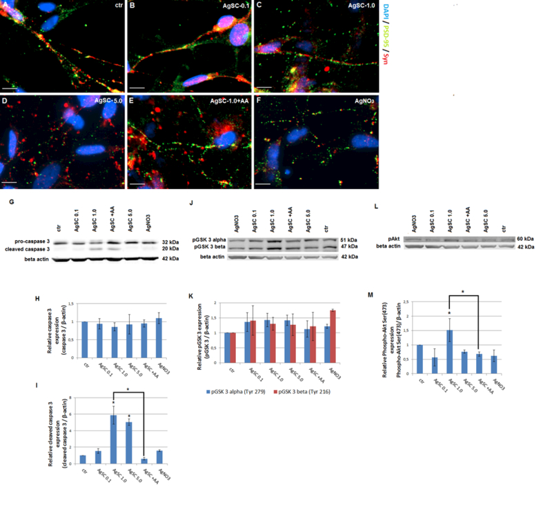
Silver nanoparticles (AgNPs) are among the most extensively used nanoparticles and are found in a variety of products. This ubiquity leads to inevitable exposure to these particles in everyday life. However, the effects of AgNPs on neuron and astrocyte networks are still largely unknown. In this study, we used neurons and astrocytes derived from human embryonic stem cells as a cellular model to study the neurotoxicity that is induced by citrate-coated AgNPs (AgSCs). Immunostaining with the astrocyte and neuron markers, glial fibrillary acidic protein and microtubule-associated protein-2 (MAP2), respectively, showed that exposure to AgSCs at the concentration of 0.1 µg/mL increased the astrocyte/neuron ratio. In contrast, a higher concentration of AgSCs (5.0 µg/ml) significantly changed the morphology of astrocytes. These results suggest that astrocytes are sensitive to AgSC exposure and that low concentrations of AgSCs promote astrogenesis. Furthermore, our results showed that AgSCs reduced neurite outgrowth, decreased the expression of postsynaptic density protein 95 and synaptophysin, and induced neurodegeneration in a concentration-dependent manner. Our findings additionally suggest that the expression and phosphorylation status of MAP2 isoforms, as modulated by the activation of the Akt/glycogen synthase kinase-3/caspase-3 signaling pathway, may play an important role in AgSC-mediated neurotoxicity. We also found that AgNO3 exposure only slightly reduced neurite outgrowth and had little effect on MAP2 expression, suggesting that AgSCs and AgNO3 have different neuronal toxicity mechanisms. In addition, most of these effects were reduced when the cell culture was co-treated with AgSCs and the antioxidant ascorbic acid, which implies that oxidative stress is the major cause of AgSC-mediated astrocytic/neuronal toxicity and that antioxidants may have a neuroprotective effect.

Keywords: Citrate-coated silver nanoparticles; human embryonic stem cells; nanotoxicity; neurons and astrocytes; neurotoxicity.


Conflict of interest statement

DECLARATION OF INTEREST

The authors report no conflicts of interest.

Figures

Figure 1.
Figure 1.. Characterization of AgSCs by TEM.
(A–C) Transmission electron micrographs of AgSCs. (D) AgSCs size distribution.
Figure 2.
Figure 2.. Effects of AgSCs on the neuron/astrocyte ratio.
(A) Control cells expressed the mature neuron marker MAP2 (green) and astrocyte marker GFAP (red). (B) Treatment with AgSCs at a concentration of 0.1 µg/mL increased the astrocyte/neuron ratio. (C) Treatment with AgSCs at a concentration of 1.0 µg/mL had little effect on the astrocyte/neuron ratio. (D) Treatment with AgSCs at a concentration of 5.0 µg/mL decreased the astrocyte/neuron ratio. (E) Compared to cells treated with AgSCs alone (1.0 µg/mL), cells co-treated with AgSCs and AA exhibited reduced effects on the astrocyte/neuron ratio. (F) AgNO3 treatment had no significant effects on the astrocyte/neuron ratio. Scale bars: 10 µm. (G) ImageJ quantification of the astrocyte/neuron ratio calculated by dividing the GFAP integrated density by the MAP2 integrated density. Data are presented as the mean ± SEM from at least eight representative images; *p < 0.05 vs. the unexposed control group.
Figure 3.
Figure 3.. AgSCs altered the expression of MAP2 isoforms and the astrocyte marker GFAP.
(A) Immunoblotting of MAP2 after exposure to three concentrations (0.1, 1.0, and 5.0 µg/mL) of AgSCs, AA, and AgNO3 for 72 h. (B) Exposure to AgSCs at 0.1 µg/mL and 1.0 µg/mL increased HMWMAP2 expression in comparison to the control group and the group treated with AgNO3. Co-treatment with AgSCs and AA reduced the phosphorylation of HMWMAP2 (top band). (C) Exposure to AgSCs at a concentration of 5.0 µg/mL reduced MAP2 expression due to cell death. (D, E) AgSC exposure increased GFAP protein expression. β-actin was used as the loading control.
Figure 4.
Figure 4.. Effects of AgSC on neuronal morphology and neurite outgrowth.
(A) The untreated control showed no cell damage. (B) Exposing cells to AgSCs at a concentration of 0.1 µg/mL resulted in slightly reduced cell density and fewer cell overlaps. (C, D) Cells treated with AgSCs at a concentration of 1.0 and 5.0 µg/mL showed rounded cell bodies, retracted cellular extensions, apoptotic bodies, and fragmentation of mature neurons. (E) Cells co-treated with AgSCs and AA exhibited fewer toxic effects than did cells treated solely with AgSCs at the same concentration. (F) Treatment with AgNO3 resulted in reduced cell density and fewer cell overlaps. Scale bars: 50 μm. (G) Untreated cells exhibited long processes that formed an extensive network. (H–J) AgSCs inhibited neurite outgrowth in a concentration-dependent manner. (K) Cells co-treated with AgSCs (1.0 µg/mL) and AA exhibited less damage in terms of cell outgrowth compared with cells treated solely with AgSCs (1.0 µg/mL). (L) AgNO3 treatment had no significant effects on the cell network. Scale bars: 10 μm.
Figure 5.
Figure 5.. AgSCs reduced the expression of the postsynaptic marker PSD-95 and the presynaptic vesicle membrane protein Syn and induced neurodegeneration through the Akt/GSK-3/caspase-3 pathway.
(A) Untreated control neurons showed extensive neuritic processes with dense puncta labeling of Syn and PSD-95. Co-localized spots (yellow) represent the location of potential synapses. (B–D) AgSC-treated neurons showed drastically reduced Syn and PSD-95 expression as well as fewer synapses, and these effects occurred in a concentration-dependent manner. (E) AA reduced the effects of AgSCs on synaptic protein expression. (F) AgNO3 drastically reduced Syn and PSD-95 expression. Scale bar: 10 µm. (G) Immunoblotting of pro-caspase-3 and cleaved caspase-3 after exposure to three concentrations (0.1, 1.0, and 5.0 µg/mL) of AgSCs, AA, and AgNO3 for 72 h. (H, I) Relative densitometric analysis of the bands for pro-caspase-3 and cleaved caspase-3 revealed that high concentrations of AgSCs (1.0 and 5.0 µg/mL) induced cleavage of caspase-3 in comparison to the control group. (J) Immunoblotting of phosphorylated GSK-3α/β at Tyr279/216. (K) Relative densitometric analysis of the bands for phosphorylated GSK-3α/β at Tyr279/216 showed that treatment with AgSC and AgNO3 increased phosphorylation of GSK-3α at Tyr279 and of GSK-3β at Tyr216. Co-treatment with AA reduced this phosphorylation. (L) Immunoblotting of pAkt at Ser473. (M) Relative densitometric analysis of the bands for pAkt at Ser473 displayed that pAkt levels were elevated when treated with AgSCs at a concentration of 1.0 µg/mL and reduced by AA. β-actin was used as the loading control. Data are expressed as the mean ± SEM from three independent experiments; *p < 0.05 vs. the unexposed control group.

References

    1. Arora S, Jain J, Rajwade J,Paknikar K. 2008. Cellular responses induced by silver nanoparticles: in vitro studies. Toxicology letters 179: 93–100. - PubMed
    1. Begum AN, Aguilar JS, Elias L,Hong Y. 2016. Silver nanoparticles exhibit coating and dose-dependent neurotoxicity in glutamatergic neurons derived from human embryonic stem cells. NeuroToxicology 57: 45–53. - PubMed
    1. Begum AN, Guoynes C, Cho J, Hao J, Lutfy K,Hong Y. 2015. Rapid generation of sub-type, region-specific neurons and neural networks from human pluripotent stem cell-derived neurospheres. Stem cell research 15: 731–741. - PMC - PubMed
    1. Bélanger M,Magistretti PJ. 2009. The role of astroglia in neuroprotection. Dialogues Clin Neurosci 11: 281–295. - PMC - PubMed
    1. Biosciences B. Caspase-3 Activation-An Indicator of Apoptosis in Image-Based Assays. 2010.

Publication types

MeSH terms