

Review
doi: 10.1161/STROKEAHA.117.016990.
Epub 2018 Jan 15.
Diagnosis of Cerebral Amyloid Angiopathy: Evolution of the Boston Criteria
Affiliations
- PMID: 29335334
- PMCID: PMC5892842
- DOI: 10.1161/STROKEAHA.117.016990
Item in Clipboard
Review
Diagnosis of Cerebral Amyloid Angiopathy: Evolution of the Boston Criteria
Stroke.
2018 Feb.
No abstract available
Keywords: antemortem diagnosis; cerebral amyloid angiopathy; cerebral hemorrhage; siderosis.
Figures
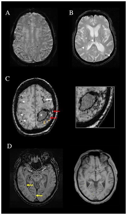
Patterns of cerebral microbleeds (CMB). (A) Multiple strictly lobar CMB on T2*-weighted MRI of a 69-year-old woman who presented with a spontaneous lobar intracerebral hemorrhage. Brain autopsy showed advanced CAA. (B) Mixed CMB (arrowheads) affecting the right thalamus, a deep hemispheric territory, as well as lobar brain regions and therefore not fulfilling Boston criteria for probable CAA. (C) Subacute left frontoparietal lobar hemorrhage (asterisk), numerous strictly lobar CMB (white arrowheads), and a left frontal focus of cortical superficial siderosis (white arrow) on susceptibility-weighted imaging (SWI) MRI of a 78-year-old woman. The image additionally shows foci of CMB (orange arrowheads) and cortical superficial siderosis (red arrows, also seen in magnified image) that are immediately adjacent to the lobar hemorrhage and therefore not counted as separate lesions in determining the number of lobar hemorrhagic foci. (D) SWI from a 71-year-old man with memory loss and CAA on brain biopsy. The yellow arrows point to CMB in the right temporal and right occipital lobes that might appear distant from the brain surface, but would be counted as lobar microbleeds. The aligned T1-weighted slice shows that their positions (asterisks on the right panel) are within or very close to the cortical ribbon.
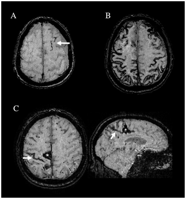
Patterns of cortical superficial siderosis (cSS) in CAA patients. (A) Susceptibility-weighted imaging (SWI) MRI showing a single sulcus with cSS (arrow), classified as focal cSS. (B) SWI showing cSS affecting multiple cortical sulci, classified as disseminated (>3 affected sulci). (C) SWI images from a 68-year-old man with a right parasagittal CAA-related spontaneous lobar intracerebral hemorrhage (asterisk). The arrow points to an area of cSS close to the hematoma on the axial slice. The corresponding sagittal slice (right panel) shows that this cSS focus connects to the lobar hemorrhage and thus would not be counted as an independent hemorrhagic lesion. There are multiple lobar cerebral microbleeds in the left hemisphere.
References
-
- Vinters HV. Cerebral amyloid angiopathy. A critical review. Stroke. 1987;18:311–324. - PubMed
-
- Greenberg SM, Rebeck GW, Vonsattel JP, Gomez-Isla T, Hyman BT. Apolipoprotein e epsilon 4 and cerebral hemorrhage associated with amyloid angiopathy. Ann Neurol. 1995;38:254–259. - PubMed
-
- Greenberg SM, Edgar MA. Case records of the massachusetts general hospital, case 22–1996. N Engl J Med. 1996;335:189–196. - PubMed
-
- Charidimou A, Fox Z, Werring DJ, Song M. Mapping the landscape of cerebral amyloid angiopathy research: An informetric analysis perspective. J Neurol Neurosurg Psychiatry. 2016;87:252–259. - PubMed
-
- McKhann G, Drachman D, Folstein M, Katzman R, Price D, Stadlan EM. Clinical diagnosis of alzheimer’s disease: Report of the nincds-adrda work group under the auspices of department of health and human services task force on alzheimer’s disease. Neurology. 1984;34:939–944. - PubMed
Publication types
MeSH terms
Grants and funding
LinkOut - more resources
Full Text Sources
Other Literature Sources
Medical
