. 2018 Jan 15;8(1):787.
doi: 10.1038/s41598-017-18899-2.

RFA Guardian: Comprehensive Simulation of Radiofrequency Ablation Treatment of Liver Tumors

Affiliations

RFA Guardian: Comprehensive Simulation of Radiofrequency Ablation Treatment of Liver Tumors

Philip Voglreiter et al. Sci Rep. .

Abstract

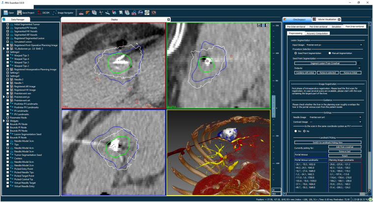
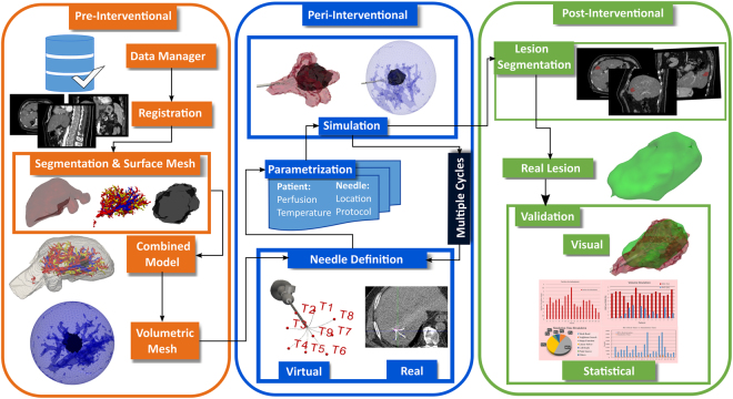
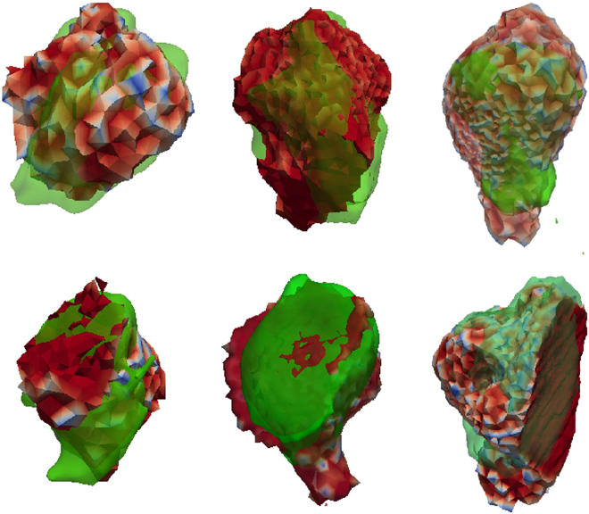
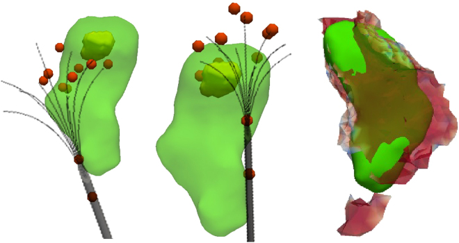
The RFA Guardian is a comprehensive application for high-performance patient-specific simulation of radiofrequency ablation of liver tumors. We address a wide range of usage scenarios. These include pre-interventional planning, sampling of the parameter space for uncertainty estimation, treatment evaluation and, in the worst case, failure analysis. The RFA Guardian is the first of its kind that exhibits sufficient performance for simulating treatment outcomes during the intervention. We achieve this by combining a large number of high-performance image processing, biomechanical simulation and visualization techniques into a generalized technical workflow. Further, we wrap the feature set into a single, integrated application, which exploits all available resources of standard consumer hardware, including massively parallel computing on graphics processing units. This allows us to predict or reproduce treatment outcomes on a single personal computer with high computational performance and high accuracy. The resulting low demand for infrastructure enables easy and cost-efficient integration into the clinical routine. We present a number of evaluation cases from the clinical practice where users performed the whole technical workflow from patient-specific modeling to final validation and highlight the opportunities arising from our fast, accurate prediction techniques.


Conflict of interest statement

The authors declare that they have no competing interests.

Figures

Figure 1
Figure 1
The RFA Guardian interface, showing a case from the pre-clinical evaluation. The left-most part is the data manager, a collection of all data loaded and generated during the workflow. The large central area is reserved for visualization, containing three orthogonal slice viewers and a 3D representation. The right part contains the control elements for navigating through the technical wokflow.
Figure 2
Figure 2
The technical workflow of simulation in the RFA Guardian. When considering the clinical procedure, three distinct phases assert themselves: In the pre-interventional stage, diagnostic scans contribute to generating a patient-specific anatomical model and potentially allow inference of parameters relevant for the simulation. This model allows for high-performance simulation in the peri-interventional phase with user-defined parameterization of the needle and generator. Finally, the post-interventional phase serves for validation purposes and potential analysis tasks for optimizing the treatment in advance.
Figure 3
Figure 3
A simulation ensemble generated from the parameter space sampling routine. This particular set contains 25 simulation results while varying perfusion of tumor and tissue concurrently, with 5 linear variations each. Even small uncertainties in measuring the parameters can already have a considerable impact on the outcome. However, the parameter space sampling allows the user to estimate the expected range of results.
Figure 4
Figure 4
The same ensemble as in Fig. 3, but visualized with Contour Boxplots. The simplification and consecutive condensation of information into a median (dark blue) and respective variance (medium blue and light blue), in conjunction with outliers (orange) and vessels (red), provides more insight into the behaviour of the lesion under varying parameters. Using this technique, the user can reason whether, e.g. the vessel is likely to be coagulated or survive. Factors like this can be critical in treatment planning, but are often overlooked in planning applications.
Figure 5
Figure 5
Visual comparison of six distinct simulation results (color-coded opaque mesh) and the respective segmented real lesions (green transparent surface). Despite non-optimal imaging conditions and protocols, the accuracy achieved during pre-clinical evaluation of the RFA Guardian is very promising.
Figure 6
Figure 6
Visual comparison of an unsuccessful case with a tumor at the liver capsule. This particular case was treated with two consecutive ablation cycles with needle repositioning inbetween (left and center images). While the registration between real induced lesion (green) and tumor (yellow) seems appropriate, the registration of the needle geometry was not accurate enough due to severe tissue deformation at the liver capsule. The left and central figures shows an idealized needle model in gray, while the red spheres visualize the actual simulation input. These single tips resulted from segmentation and registration of the patient images for this case. The deviation from the optimal umbrella shape consecutively leads to a mismatch between simulated and real treatment (right), is below the necessary accuracy. However, such registration mismatches are typically easy to identify during the workflow and, consecutively, do not pose a risk for patients if observed carefully.

References

    1. Forner A, Gilabert M, Bruix J, Raoul J-L. Treatment of intermediate-stage hepatocellular carcinoma. Nature Reviews Clinical Oncology. 2014;11:525–535. doi: 10.1038/nrclinonc.2014.122. - DOI - PubMed
    1. Otto G, et al. Radiofrequency ablation as first-line treatment in patients with early colorectal liver metastases amenable to surgery. Annals of surgery. 2010;251:796–803. doi: 10.1097/SLA.0b013e3181bc9fae. - DOI - PubMed
    1. Bahn DK, et al. Targeted cryoablation of the prostate: 7-year outcomes in the primary treatment of prostate cancer. Urology. 2002;60:3–11. doi: 10.1016/S0090-4295(02)01678-3. - DOI - PubMed
    1. Onik G, Mikus P, Rubinsky B. Irreversible electroporation: implications for prostate ablation. Technology in cancer research & treatment. 2007;6:295–300. doi: 10.1177/153303460700600405. - DOI - PubMed
    1. Paulides MM, et al. Simulation techniques in hyperthermia treatment planning. International Journal of Hyperthermia. 2013;29:346–357. doi: 10.3109/02656736.2013.790092. - DOI - PMC - PubMed

Publication types