. 2018 Jan 15;8(1):727.
doi: 10.1038/s41598-017-18905-7.

Synaptic localisation of SRF coactivators, MKL1 and MKL2, and their role in dendritic spine morphology

Affiliations

Synaptic localisation of SRF coactivators, MKL1 and MKL2, and their role in dendritic spine morphology

Marisa Kaneda et al. Sci Rep. .

Abstract

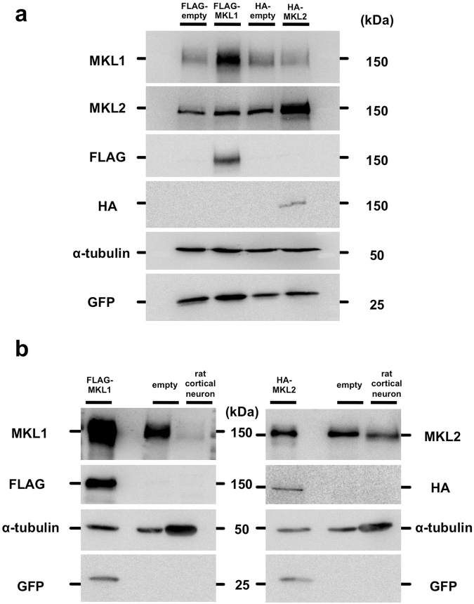
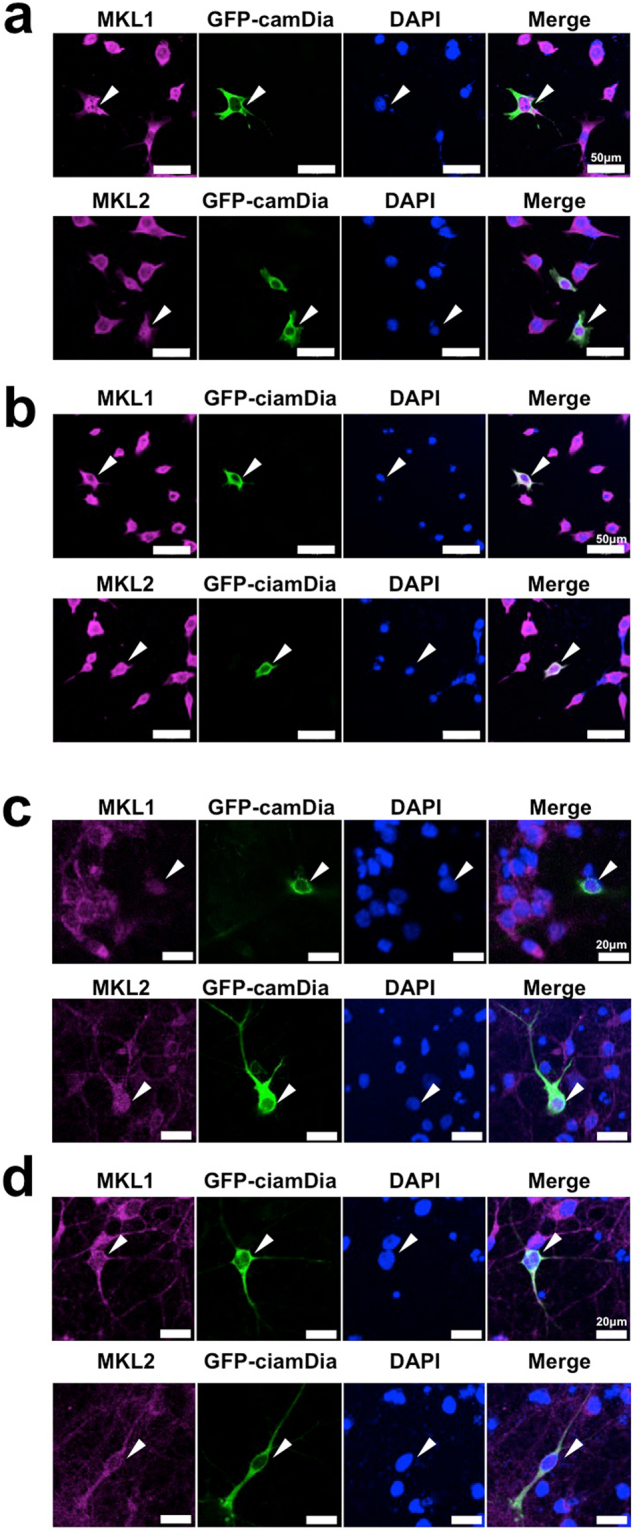
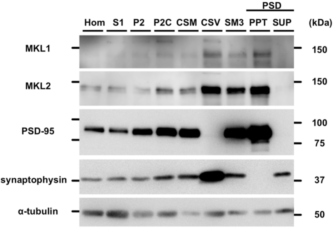
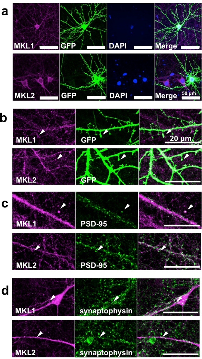
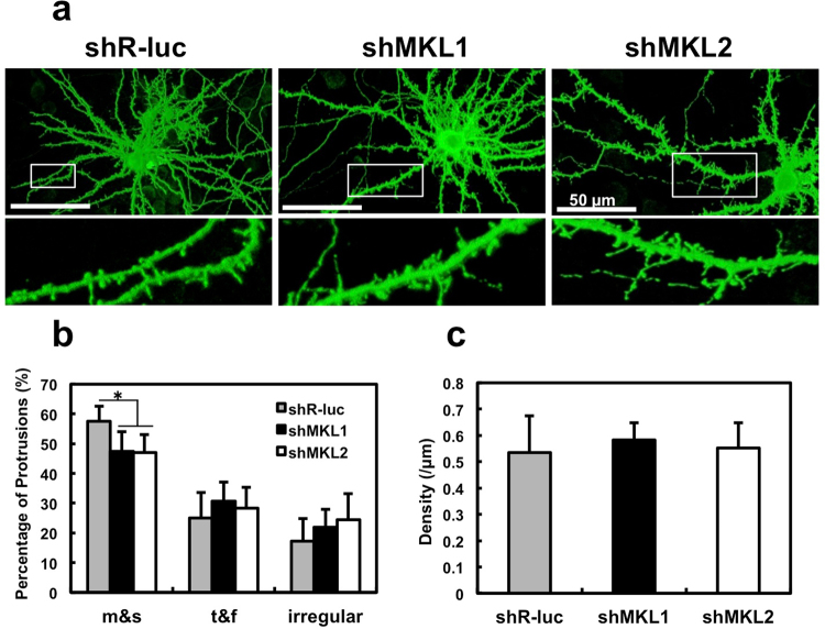
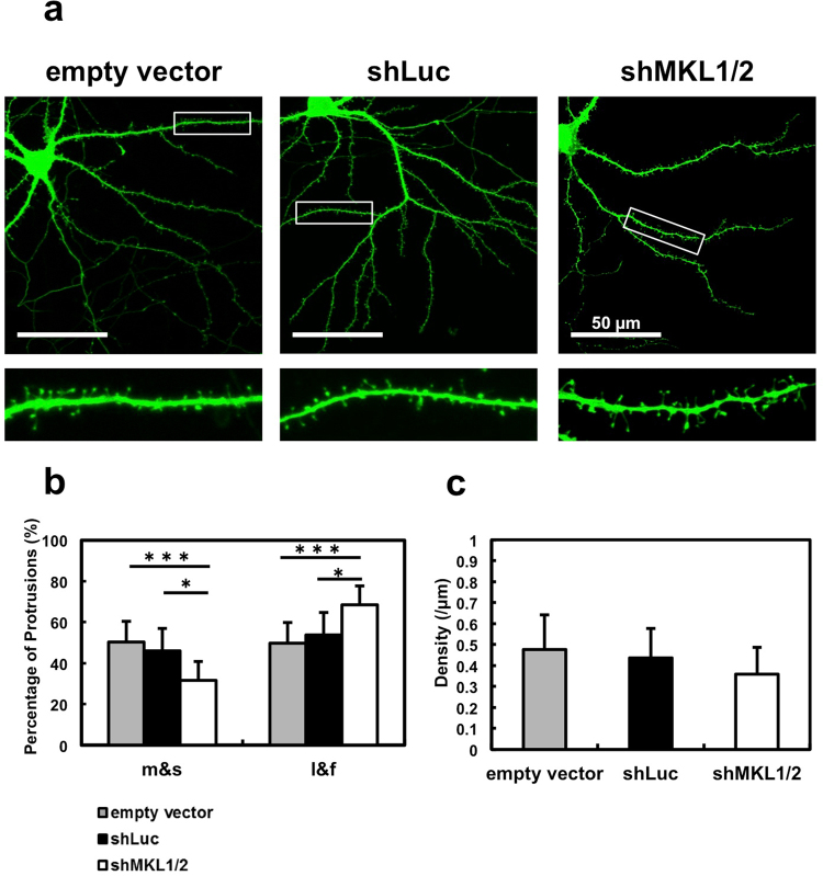
The megakaryoblastic leukaemia (MKL) family are serum response factor (SRF) coactivators, which are highly expressed in the brain. Accordingly, MKL plays important roles in dendritic morphology, neuronal migration, and brain development. Further, nucleotide substitutions in the MKL1 and MKL2 genes are found in patients with schizophrenia and autism spectrum disorder, respectively. Thus, studies on the precise synaptic localisation and function of MKL in neurons are warranted. In this study, we generated and tested new antibodies that specifically recognise endogenously expressed MKL1 and MKL2 proteins in neurons. Using these reagents, we biochemically and immunocytochemically show that MKL1 and MKL2 are localised at synapses. Furthermore, shRNA experiments revealed that postsynaptic deletion of MKL1 or MKL2 reduced the percentage of mushroom- or stubby-type spines in cultured neurons. Taken together, our findings suggest that MKL1 and MKL2 are present at synapses and involved in dendritic spine maturation. This study may, at least in part, contribute to better understanding of the molecular mechanisms underlying MKL-mediated synaptic plasticity and neurological disorders.


Conflict of interest statement

The authors declare that they have no competing interests.

Figures

Figure 1
Figure 1
Specific detection of MKL1 and MKL2 by anti-MKL1 and anti-MKL2 antibodies. (a) pEGFP-C1 vector and pFLAG-CMV2 vector (FLAG-empty) or pFLAG-CMV2-MKL1 vector (FLAG-MKL1), pCMV-HA vector (HA-empty) or pCMV-HA-MKL2 vector (HA-MKL2) were co-transfected into NIH3T3 cells. Cell lysates were lysed 24 hours after transfection. Anti-MKL1 (MKL1) and anti-MKL2 (MKL2) antibody were used for detection of exogenous and endogenous MKL1 and MKL2, respectively. Anti-FLAG and anti-HA antibodies were used for detection of exogenous MKL1 and MKL2, respectively. Anti-α-tubulin and anti-GFP antibodies were used as internal (loading) controls. (b) Detection of endogenous MKL1 and MKL2 in cortical neurons. Left-most lanes: cell lysates from NIH3T3 cells overexpressing FLAG-MKL1 and HA-MKL2. Lane 2: cell lysates from untransfected NIH3T3 cells. Lane 3: cell lysates from rat cortical neurons at 7 days in culture. Full-length blots are presented in Supplementary Figure S5.
Figure 2
Figure 2
Nucleocytoplasmic staining of MKL in NIH3T3 cells and cortical neurons expressing a Rho effector, mDia. NIH3T3 cells were transfected with GFP-tagged constitutively active (ca) mDia (a) or constitutively inactive (cia) mDia (b). Twenty-four hours later, cells were immunostained with anti-GFP and anti-MKL1 or anti-MKL2 antibodies. Cortical neurons (7 days in culture) were transfected with camDia (4 μg/well, c) or ciamDia (4 μg/well, d). Twenty-four hours later, cells were immunostained with anti-GFP and anti-MKL1 or anti-MKL2 antibodies. Arrowheads indicate GFP-mDia-positive cells. Data regarding the cellular localisation of MKL are presented in Supplementary Figure S3.
Figure 3
Figure 3
Detection of endogenous MKL1 and MKL2 protein in subcellular fractions of rat brains. Subcellular fractionation was performed from homogenates of 6-week-old male rat brain. Each protein (5 μg) was analysed by western blotting using anti-MKL1, anti-MKL2, anti-PSD-95 (postsynaptic marker), anti-synaptophysin (presynaptic marker), and anti-α-tubulin antibodies. Hom: homogenate; S1: crude synaptosomal fraction; P2: crude membrane fraction; P2C: synaptosomal fraction; CSM: crude synaptic membrane fraction; CSV: crude synaptic vesicle fraction; SM3: synaptic membrane fraction 3; PSD: postsynaptic density fraction; PPT: 1% Triton X-100-insoluble fraction of PSD; SUP: 1% Triton X-100-soluble fraction of PSD. Full-length blots are presented in Supplementary Figure S6.
Figure 4
Figure 4
Immunocytochemical detection of MKL1 and MKL2 protein in cortical neurons with synapses. (a) Immunostaining of mature, cortical neuronal cultures. pEGFP-C1 vector (4 μg/well) was transfected into cortical neurons at 18 days in culture. Three days later, cells were subjected to immunostaining using anti-GFP and anti-MKL1 or anti-MKL2 antibodies. (b) High magnification of GFP-positive cortical neurons at 21 days in culture. Arrowheads indicate dendritic spines. (c,d) Synaptic staining of MKL1 and MKL2 in mature, cortical neurons. Cortical neurons at 21 days in culture were immunostained using anti-MKL1, anti-MKL2, anti-postsynaptic density (PSD)-95, and anti-synaptophysin antibodies. MKL and PSD-95 signals merged. MKL and synaptophysin signals also merged.
Figure 5
Figure 5
Effect of MKL1 or MKL2 knock-down on dendritic spine morphology and density in cortical neurons. (a) Spine morphology of cortical neurons transfected with pEGFP-C1 (1 μg/well) and shR-luc, shMKL1, or shMKL2 (1 μg/well). Transfection was performed at 16 days in culture. Neurons (21 days in culture) were immunostained using anti-GFP antibody. (b,c) Spine morphology (m and s, mushroom or stubby; t and f, thin or filopodia; irregular) and spine density. Graphs show mean ± S.D. from seven independent experiments. The statistical significance of differences (vs. shR-luc) was analysed by ANOVA with the Tukey–Kramer test. *p < 0.05.
Figure 6
Figure 6
Effect of MKL knock-down on dendritic spine morphology and density in hippocampal neurons. (a) Spine morphology of hippocampal neurons transfected with pSynapsinGFP (0.5 μg/well) and 1 μg/well of pSuper (empty vector), shLuc, or shMKL1/2. Transfection was performed at 9 or 10 days in vitro (DIV). Neurons (DIV 16) were immunostained using anti-GFP antibody. Dendritic spine morphology was analysed using the semi-automated SpineMagick software. (b) The percentage of protrusions clustered into four categories: mushroom, stubby, long, and filopodia (m and s: mushroom or stubby; l and f: long or filopodia). (c) Dendritic spine density. Graphs show mean ± S.D. from four independent experiments. The statistical significance of differences was analysed by ANOVA with Dunn’s multiple comparisons test. *p < 0.05, ***p < 0.001.

References

    1. Ramanan N, et al. SRF mediates activity-induced gene expression and synaptic plasticity but not neuronal viability. Nat. Neurosci. 2005;8:759–767. doi: 10.1038/nn1462. - DOI - PubMed
    1. Kawashima T, et al. Synaptic activity-responsive element in the Arc/Arg3.1 promoter essential for synapse-to-nucleus signaling in activated neurons. Proc. Natl. Acad. Sci. USA. 2009;106:316–321. doi: 10.1073/pnas.0806518106. - DOI - PMC - PubMed
    1. Knöll B, Nordheim A. Functional versatility of transcription factors in the nervous system: the SRF paradigm. Trends Neurosci. 2009;32:432–442. doi: 10.1016/j.tins.2009.05.004. - DOI - PubMed
    1. Miano JM. Serum response factor: toggling between disparate programs of gene expression. J. Mol. Cell. Cardiol. 2003;35(6):577–593. doi: 10.1016/S0022-2828(03)00110-X. - DOI - PubMed
    1. Etkin A, et al. A role in learning for SRF: deletion in the adult forebrain disrupts LTD and the formation of an immediate memory of a novel context. Neuron. 2006;50:127–143. doi: 10.1016/j.neuron.2006.03.013. - DOI - PubMed

Publication types