. 2018 Oct;36(5):743-754.
doi: 10.1007/s10637-017-0558-5. Epub 2018 Jan 16.

Sphingadienes show therapeutic efficacy in neuroblastoma in vitro and in vivo by targeting the AKT signaling pathway

Affiliations

Sphingadienes show therapeutic efficacy in neuroblastoma in vitro and in vivo by targeting the AKT signaling pathway

Piming Zhao et al. Invest New Drugs. 2018 Oct.

Erratum in

Abstract

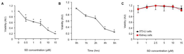
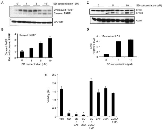
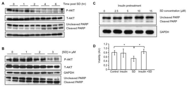
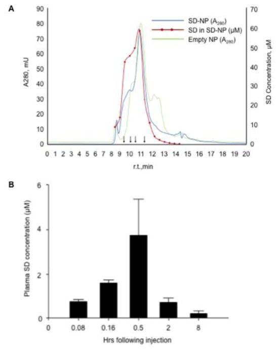
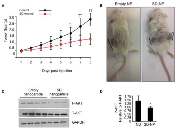
Neuroblastoma is a childhood malignancy that accounts for approximately 15% of childhood cancer deaths. Only 20-35% of children with metastatic neuroblastoma survive with standard therapy. Identification of more effective therapies is essential to improving the outcome of children with high-stage disease. Sphingadienes (SD) are growth-inhibitory sphingolipids found in natural sources including soy. They exhibit chemopreventive activity in mouse models of colon cancer, where they mediate cytotoxicity by inhibiting key pro-carcinogenic signaling pathways. In this study, the effect of SD on neuroblastoma was analyzed. Low micromolar concentrations of SD were cytotoxic to transformed and primary neuroblastoma cells independently of N-Myc amplification status. SD induced both caspase-dependent apoptosis and autophagy in neuroblastoma cells. However, only inhibition of caspase-dependent apoptosis protected neuroblastoma cells from SD-mediated cytotoxicity. SD also inhibited AKT activation in neuroblastoma cells as shown by reduced phosphorylated AKT levels. Pre-treatment with insulin attenuated SD-mediated cytotoxicity in vitro. SD-loaded nanoparticles (NP) administered parenterally to immunodeficient mice carrying neuroblastoma xenografts resulted in cytotoxic levels of SD in the circulation and significantly reduced tumor growth compared to vehicle-treated controls. Analysis of tumor extracts demonstrated reduced AKT activation in tumors of mice treated with SD-NP compared to controls treated with empty NP. Our findings indicate SD are novel potential chemotherapeutic agents that promote neuroblastoma cell death and reduce tumorigenicity in vivo.

Keywords: AKT; Nanoparticle; Neuroblastoma; PI3K; Sphingadienes; Sphingolipids.


Figures

Figure 1
Figure 1. SD are cytotoxic to neuroblastoma cells but not non-malignant cells
(A) Dose response of SD-mediated cytotoxicity. Exponentially growing Kelly cells were exposed to a range of SD concentrations (0–15 μM). After 24 h incubation, cell viability was assessed by MTS assay. Results are presented as arbitrary units (AU). * P values: p < 0.001 for 0.5 μM; p = 0.02 for 1 μM; p < 0.001 for 5 μM; p = 2 x10⁻⁶ for 10 μM; p < 2 x 10⁻⁶ for 15 μM. The results shown are representative of three similar experiments; n = 3 for each point. (B) Time-dependent inhibition of viability in response to SD treatment. Exponentially growing Kelly cells were treated with 10 μM SD or vehicle and incubated for 0–6 h, and viability was determined by MTS assay. * P values: p < 0.02 for 2h; p = 1 x 10⁻⁵ for 4h; p = 3 x 10⁻⁷ for 6h. The results shown are representative of three similar experiments; n = 3 for each point. (C) NIH 3T3 fibroblasts and primary human kidney cells established as described in Methods were exposed to a range of SD concentrations from 0–15 μM. After 24 h, cell viability was assessed by the MTS assay. The results shown are representative of three similar experiments; n = 3 for each point. There is no significant difference in SD-treated vs. vehicle-treated NIH3T3 or kidney cells at any concentration.
Figure 2
Figure 2. SD mediate cytotoxicity by inducing apoptosis in neuroblastoma cells
Kelly cells were grown to 70% confluence in DMEM plus 10% FCS, then changed to serum-free medium and treated with vehicle or varying concentrations of SD. Cells were harvested at 6 h, and whole cell extracts were analysed by immunoblotting to detect (A–B) uncleaved and cleaved PARP as an indicator of apoptosis, or (C–D) unprocessed and processed LC3 as an indicator of autophagy. Actin is used as a loading control. (B) and (D) represent image quantification of immunoblots shown in (A) and (C), respectively. In each case, the ratio of cleaved PARP to uncleaved PARP or LC3II to LC3I in the vehicle sample was arbitrarily set at one. These experiments were repeated at least three times with similar results. (E) Kelly neuroblastoma cells were pre-treated with either autophagy inhibitors bafilomycin (BAF, 1 μM) or 3-methyladenine (3MA, 1 mM), or with the pan-caspase inhibitor of apoptosis Z-VAD-FMK (100 μM) for 2 h, followed by treatment with either 10 μM SD or vehicle. Cells were incubated for an additional 12 h, harvested and evaluated for viability by MTS assay. This experiment was repeated at least three times with similar results.
Figure 3
Figure 3. SD mediate cytotoxicity in neuroblastoma cells by inhibiting AKT signaling
(A) Kelly cells were grown to 70% confluence in the preferred medium plus 10% FCS, switched to serum-free medium, treated with 10 μM SD and incubated for an additional 0, 1, 2, 4 or 6 h. Cells were harvested at the designated time point, and whole cell extracts were analyzed by immunoblotting for total AKT (T-AKT) and phosphorylated AKT (P-AKT) and cleaved and uncleaved PARP. (B) Kelly cells were grown as described in (A) and after incubation in serum-free medium for 8 h were then treated with 0–3 μM SD for 20 h. Cells were harvested and whole cell extracts were analyzed by immunoblotting for T-AKT, P-AKT and cleaved and uncleaved PARP. (C) Kelly cells were serum starved for 12 h, pre-treated with insulin (2 μg/ml) for 1 h, followed by treatment with SD (0–15 μM). After 6 h of incubation, cells were harvested, and cleaved and uncleaved PARP were measured by immunoblotting of whole cell extracts. GAPDH was used as loading control. (D) Kelly cells received either no pre-treatment (control), insulin (2 μg/ml) pre-treatment for 1 h to activate AKT signaling, 10 μM SD without pre-treatment, or insulin pre-treatment followed by SD treatment. Cells were incubated for an additional 6 h, and cell viability was determined by the MTS assay. * p < 0.05. These experiments were repeated at least three times with similar results.
Figure 4
Figure 4. Characterization and kinetics SD-NP
(A) SD-NP were subjected to HPLC on a GF-250 size-exclusion column with continuous monitoring of protein absorbance at 280 nm (blue line). The red line is a plot of the SD content of specified fractions (filled red circles) determined by LC/MS/MS analysis. The SD concentration in μM is shown on the right hand Y axis. The chromatograph depicted in green corresponds to the 280 nm absorbance profile of control ND lacking SD while the black arrows depict the elution position of molecular weight standards (330, 199.5, 133 and 66.5 kDa). (B) Plasma SD levels determined by LC/MS after delivery to wild type mice by i.p. route as described in Methods.
Figure 5
Figure 5. SD delivered via SD-NP inhibit tumor growth and AKT signaling in neuroblastoma xenografts in vivo
(A) Adult NOD/SCID mice were injected subcutaneously with 2 million Kelly cells suspended 1:1 (v/v) in matrigel. Xenograft tumor growth was assessed daily by calipers. Mice were randomly assigned to groups and received either SD-NP (10 mg/kg/d) or vehicle by i.p. injection twice daily starting when tumors were 100 mm3 in size and continuing for approximately 8 d. Average tumor size in SD-NP treated mice (n = 17) are shown in red circles, and vehicle-treated mice (n = 3) are shown in black circles. Statistical analysis was performed using two-way ANOVA followed by Tukey’s post-test. †, p < 0.05 for control vs. SD-NP; ††, p < 0.0001 for control vs. SD-NP. (B) Shown are representative tumors from one control mouse (in this case harboring a single xenograft in one flank) treated with empty NP and one SD-NP treated-mouse (harboring xenograft tumors in both flanks). Brackets show longest diameter of each tumor. (C) Adult NOD/SCID mice were implanted with Kelly xenografts as described in (A). When tumors were 100 mm3 in size, mice either received a single dose of empty NP (n = 4) or SD-NP (n = 3) by i.p. injection. Mice were euthanized by CO2 inhalation 8 h after administration of SD-NP or empty NP. Tumors were excised and flash frozen for molecular analysis. Whole tumor extracts were analyzed by immunoblotting to detect total AKT (T-AKT) or phosphorylated AKT (P-AKT). GAPDH was used as a loading control. (D) Image quantification of P-AKT/ T-AKT in immunoblot shown in (C).

Similar articles

Cited by

References

    1. Matthay KK, George RE, Yu AL. Promising therapeutic targets in neuroblastoma. Clin Cancer Res. 2012;18(10):2740–53. - PMC - PubMed
    1. Ries LA, et al. Cancer incidence and survival among children and adolescents: United States SEER Program 1975–1995. Bethesda, MD: National Cancer Institute; 1999. SEER Program, NIH Pub. No. 99 4649 192.
    1. Young JL, Jr, Miller RW. Incidence of malignant tumors in U. S. children. Journal of Pediatrics. 1975;86(2):254–8. - PubMed
    1. Cheung NK, Dyer MA. Neuroblastoma: developmental biology, cancer genomics and immunotherapy. Nat Rev Cancer. 2013;13(6):397–411. - PMC - PubMed
    1. Frappaz D, et al. LMCE3 treatment strategy: results in 99 consecutively diagnosed stage 4 neuroblastomas in children older than 1 year at diagnosis. Journal of Clinical Oncology. 2000;18(3):468–76. - PubMed

Publication types

MeSH terms