

Comparison of gray matter volume between migraine and "strict-criteria" tension-type headache
- PMID: 29335889
- PMCID: PMC5768588
- DOI: 10.1186/s10194-018-0834-6
Comparison of gray matter volume between migraine and "strict-criteria" tension-type headache
Abstract
Background: Despite evidently distinct symptoms, tension-type headache (TTH) and migraine are highly comorbid and exhibit many similarities in clinical practice. The purpose of this study was to investigate whether both types of headaches are similar in brain morphology.
Methods: Consecutive patients with TTH and age- and sex-matched patients with migraine and healthy controls were enrolled for brain magnetic resonance imaging examination. Patients with TTH were excluded if they reported any headache features or associated symptoms of migraine. Changes in gray matter (GM) volume associated with headache diagnosis (TTH vs. migraine) and frequency (episodic vs. chronic) were examined using voxel-based morphometry. The correlation with headache profile and the discriminative ability between TTH and migraine were also investigated for these GM changes.
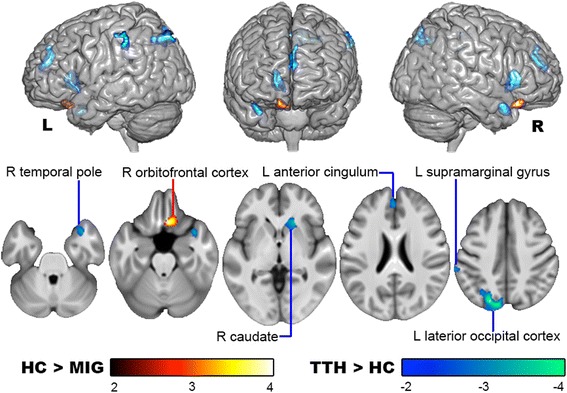
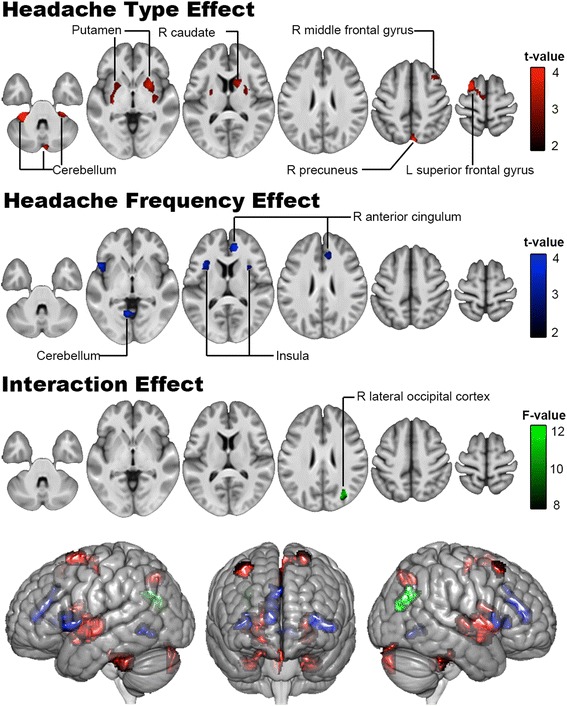
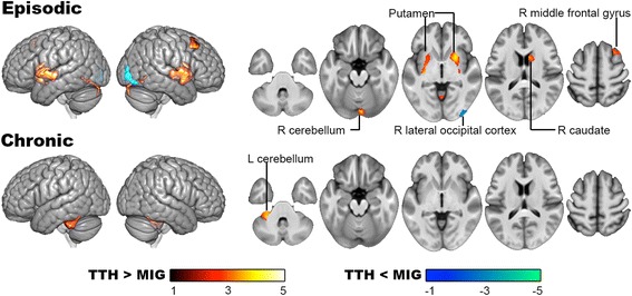
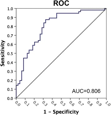
Results: In comparison with controls (n = 43), the patients with TTH (25 episodic and 24 chronic) exhibited a GM volume increase in the anterior cingulate cortex, supramarginal gyrus, temporal pole, lateral occipital cortex, and caudate. The patients with migraine (31 episodic and 25 chronic) conversely exhibited a GM volume decrease in the orbitofrontal cortex. These GM changes did not correlate with any headache profile. A voxel-wise 2 × 2 factorial analysis further revealed the substantial effects of headache types and frequency in the comparison of GM volume between TTH and migraine. Specifically, the migraine group (vs. TTH) had a GM decrease in the superior and middle frontal gyri, cerebellum, dorsal striatum, and precuneus. The chronic group (vs. episodic group) otherwise demonstrated a GM decrease in the bilateral insula and anterior cingulate cortex. In receiver operating characteristic analysis, the GM volumes of the left superior frontal gyrus and right cerebellum V combined had good discriminative ability for distinguishing TTH and migraine (area under the curve = 0.806).
Conclusions: TTH and migraine are separate headache disorders with different characteristics in relation to GM changes. The major morphological difference between the two types of headaches is the relative GM decrease of the prefrontal and cerebellar regions in migraine, which may reflect a higher allostatic load associated with this disabling headache.
Keywords: Gray matter; Magnetic resonance imaging (MRI); Migraine; Tension-type headache; Voxel-based morphometry.
Conflict of interest statement
Authors’ information
None
Ethics approval and consent to participate
The Institutional Review Board of Taipei Veterans General Hospital approved the study protocol (Reference No. 2012–07-013 AC) and each participant provided written informed consent.
Consent for publication
Not applicable.
Competing interests
WT Chen, KH Chou, PL Lee, FJ Hsiao, DM Niddam, KL Lai, and CP Lin report no competing interests. SJ Wang has served on the advisory boards of Allergan, and Eli Lilly Taiwan. He has received honoraria as a moderator from local companies (Taiwan branches) of Pfizer, Eli Lilly and Esai. JL Fuh is a member of the scientific advisory board of Novartis, and has received research support from the Taiwan National Science Council and Taipei-Veterans General Hospital.
Publisher’s Note
Springer Nature remains neutral with regard to jurisdictional claims in published maps and institutional affiliations.
Figures
References
-
- Disease GBD, Injury I, Prevalence C. Global, regional, and national incidence, prevalence, and years lived with disability for 328 diseases and injuries for 195 countries, 1990-2016: a systematic analysis for the global burden of disease study 2016. Lancet. 2017;390:1211–1259. doi: 10.1016/S0140-6736(17)32154-2. - DOI - PMC - PubMed
MeSH terms
Grants and funding
- NSC101-2314-B-075-068-MY2, MOST 106-2321-B-010-009-, and MOST 106-2314-B-075-026-/Ministry of Science and Technology, Taiwan
- 103-2321-B-010-017, 102-2321-B-010-030, 100-2314-B-010-018-MY3, 104-2745-B-010-003 and MOST 106-2321-B-010-009/Ministry of Science and Technology, Taiwan
- V105C-092, V105E9-005-MY2-1, and VGHUST104-G7-1-3/Taipei Veterans General Hospital
- VGHUST105-G7-1-1, V105C-127, V105E9-001-MY2-1, and VTA105-V1-1-1/Taipei Veterans General Hospital
LinkOut - more resources
Full Text Sources
Other Literature Sources
Medical
