

Inter- and intracellular colonization of Arabidopsis roots by endophytic actinobacteria and the impact of plant hormones on their antimicrobial activity
- PMID: 29335919
- PMCID: PMC5913384
- DOI: 10.1007/s10482-018-1014-z
Inter- and intracellular colonization of Arabidopsis roots by endophytic actinobacteria and the impact of plant hormones on their antimicrobial activity
Abstract
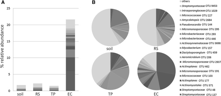
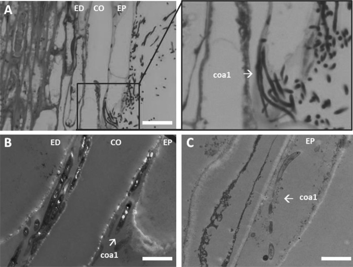
Many actinobacteria live in close association with eukaryotes such as fungi, insects, animals and plants. Plant-associated actinobacteria display (endo)symbiotic, saprophytic or pathogenic life styles, and can make up a substantial part of the endophytic community. Here, we characterised endophytic actinobacteria isolated from root tissue of Arabidopsis thaliana (Arabidopsis) plants grown in soil from a natural ecosystem. Many of these actinobacteria belong to the family of Streptomycetaceae with Streptomyces olivochromogenes and Streptomyces clavifer as well represented species. When seeds of Arabidopsis were inoculated with spores of Streptomyces strain coa1, which shows high similarity to S. olivochromogenes, roots were colonised intercellularly and, unexpectedly, also intracellularly. Subsequent exposure of endophytic isolates to plant hormones typically found in root and shoot tissues of Arabidopsis led to altered antibiotic production against Escherichia coli and Bacillus subtilis. Taken together, our work reveals remarkable colonization patterns of endophytic streptomycetes with specific traits that may allow a competitive advantage inside root tissue.
Keywords: Cryptic antibiotics; Electron microscopy; Plant hormone; Plant–microbe interactions; Streptomyces.
Conflict of interest statement
The authors declare no conflict of interests.
Figures


Similar articles
-
Revealing structure and assembly cues for Arabidopsis root-inhabiting bacterial microbiota.Nature. 2012 Aug 2;488(7409):91-5. doi: 10.1038/nature11336. Nature. 2012. PMID: 22859207
-
Defining the core Arabidopsis thaliana root microbiome.Nature. 2012 Aug 2;488(7409):86-90. doi: 10.1038/nature11237. Nature. 2012. PMID: 22859206 Free PMC article.
-
Morphological, Physiological, and Taxonomic Characterization of Actinobacterial Isolates Living as Endophytes of Cacao Pods and Cacao Seeds.Microbes Environ. 2016;31(1):56-62. doi: 10.1264/jsme2.ME15146. Epub 2016 Mar 5. Microbes Environ. 2016. PMID: 26947442 Free PMC article.
-
Endophytic actinobacteria of medicinal plants: diversity and bioactivity.Antonie Van Leeuwenhoek. 2015 Aug;108(2):267-89. doi: 10.1007/s10482-015-0502-7. Epub 2015 Jun 21. Antonie Van Leeuwenhoek. 2015. PMID: 26093915 Free PMC article. Review.
-
Diverse endophytic Streptomyces species with dynamic metabolites and their meritorious applications: a critical review.Crit Rev Microbiol. 2020 Nov;46(6):750-758. doi: 10.1080/1040841X.2020.1828816. Epub 2020 Oct 12. Crit Rev Microbiol. 2020. PMID: 33044894 Review.
Cited by
-
Drought Drives Spatial Variation in the Millet Root Microbiome.Front Plant Sci. 2020 May 28;11:599. doi: 10.3389/fpls.2020.00599. eCollection 2020. Front Plant Sci. 2020. PMID: 32547572 Free PMC article.
-
Sewage sludge fertilization affects microbial community structure and its resistome in agricultural soils.Sci Rep. 2024 Sep 9;14(1):21034. doi: 10.1038/s41598-024-71656-0. Sci Rep. 2024. PMID: 39251745 Free PMC article.
-
Diversity and Bioactivity of Endophytic Actinobacteria Associated with the Roots of Artemisia herba-alba Asso from Algeria.Curr Microbiol. 2024 Oct 11;81(12):402. doi: 10.1007/s00284-024-03932-1. Curr Microbiol. 2024. PMID: 39392504
-
The Arabidopsis thaliana-Streptomyces Interaction Is Controlled by the Metabolic Status of the Holobiont.Int J Mol Sci. 2022 Oct 26;23(21):12952. doi: 10.3390/ijms232112952. Int J Mol Sci. 2022. PMID: 36361736 Free PMC article.
-
Tapping into the maize root microbiome to identify bacteria that promote growth under chilling conditions.Microbiome. 2020 Apr 18;8(1):54. doi: 10.1186/s40168-020-00833-w. Microbiome. 2020. PMID: 32305066 Free PMC article.
References
MeSH terms
Substances
Grants and funding
LinkOut - more resources
Full Text Sources
Other Literature Sources
