. 2018 Jun 15;27(12):819-830.
doi: 10.1089/scd.2017.0265. Epub 2018 Mar 20.

Simulated Microgravity Impairs Cardiac Autonomic Neurogenesis from Neural Crest Cells

Affiliations

Simulated Microgravity Impairs Cardiac Autonomic Neurogenesis from Neural Crest Cells

Konstantinos E Hatzistergos et al. Stem Cells Dev. .

Abstract

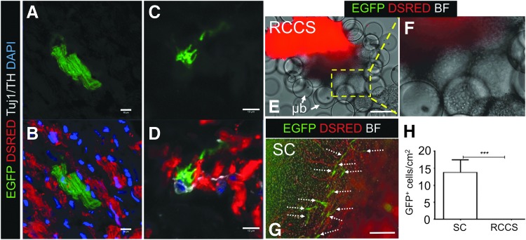
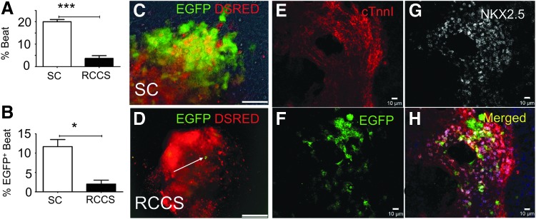
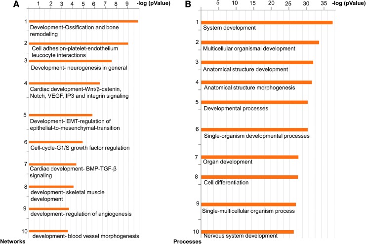
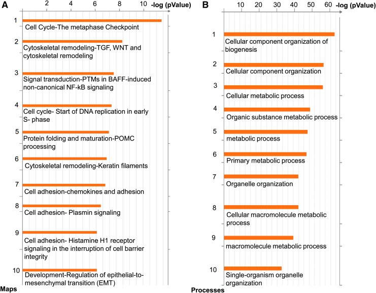
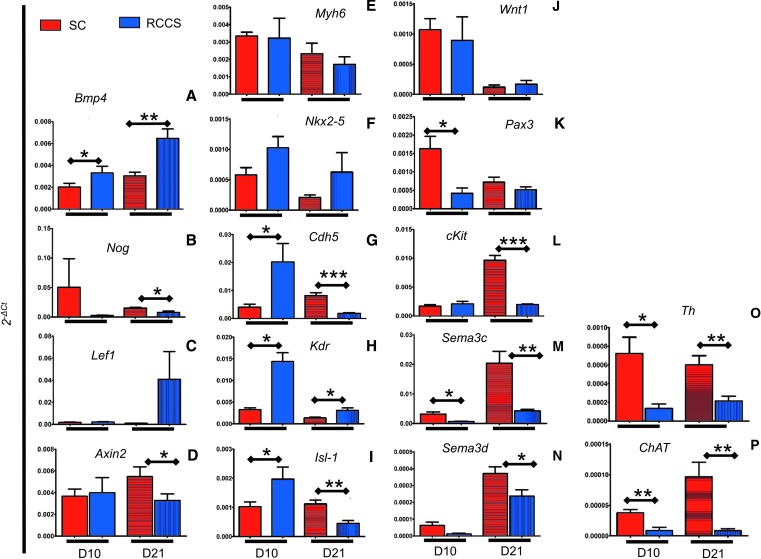
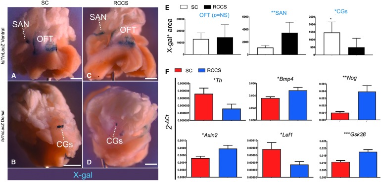
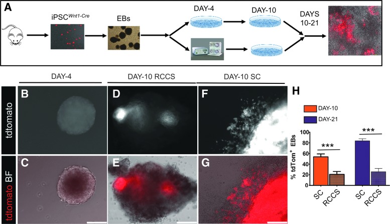
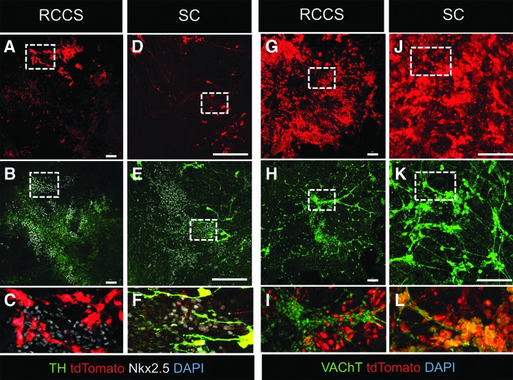
Microgravity-induced alterations in the autonomic nervous system (ANS) contribute to derangements in both the mechanical and electrophysiological function of the cardiovascular system, leading to severe symptoms in humans following space travel. Because the ANS forms embryonically from neural crest (NC) progenitors, we hypothesized that microgravity can impair NC-derived cardiac structures. Accordingly, we conducted in vitro simulated microgravity experiments employing NC genetic lineage tracing in mice with cKitCreERT2/+, Isl1nLacZ, and Wnt1-Cre reporter alleles. Inducible fate mapping in adult mouse hearts and pluripotent stem cells (iPSCs) demonstrated reduced cKitCreERT2/+-mediated labeling of both NC-derived cardiomyocytes and autonomic neurons (P < 0.0005 vs. controls). Whole transcriptome analysis, suggested that this effect was associated with repressed cardiac NC- and upregulated mesoderm-related gene expression profiles, coupled with abnormal bone morphogenetic protein (BMP)/transforming growth factor beta (TGF-β) and Wnt/β-catenin signaling. To separate the manifestations of simulated microgravity on NC versus mesodermal-cardiac derivatives, we conducted Isl1nLacZ lineage analyses, which indicated an approximately 3-fold expansion (P < 0.05) in mesoderm-derived Isl-1+ pacemaker sinoatrial nodal cells; and an approximately 3-fold reduction (P < 0.05) in cardiac NC-derived ANS cells, including sympathetic nerves and Isl-1+ cardiac ganglia. Finally, NC-specific fate mapping with a Wnt1-Cre reporter iPSC model of murine NC development confirmed that simulated microgravity directly impacted the in vitro development of cardiac NC progenitors and their contribution to the sympathetic and parasympathetic innervation of the iPSC-derived myocardium. Altogether, these findings reveal an important role for gravity in the development of NCs and their postnatal derivatives, and have important therapeutic implications for human space exploration, providing insights into cellular and molecular mechanisms of microgravity-induced cardiomyopathies/channelopathies.

Keywords: cardiac autonomic nervous system; cardiomyopathy; microgravity; neural crest cells; pacemaker cells; space travel.


Conflict of interest statement

Dr. Hare discloses a relationship with Vestion Inc. that includes equity, board membership, and consulting. Dr. Joshua Hare is the Chief Scientific Officer, a compensated consultant, and advisory board member for Longeveron and holds equity in Longeveron. Dr. Hare is also the coinventor of intellectual property licensed to Longeveron. Dr. Hatzistergos and Mrs. Valasaki disclose a relationship with Vestion Inc. that includes equity. Vestion did not contribute funding to this study. The other authors report no conflicts.

Figures

<b>FIG. 1.</b>
FIG. 1.
Rotary Cell Culture System (RCCS) impairs cKit+ cells in the adult heart. (A–D) An enhanced green fluorescent protein (EGFP)+ cardiomyocyte (A–B) and Tuj1+/TH+ neuron (C, D) following 2-week cKitCreERT2/+;IRG lineage fate mapping (n = 3), indicates contribution of adult cKit+ cells to postnatal cardiomyocyte and ANS renewal. (E, F) Live tissue imaging of EGFP and Discosoma sea anemone-derived red fluorescent protein (DSRED) in cardiac explants from a cKitCreERT2/+;IRG mouse heart, cultured for 24 days in RCCS in the presence of 4-hydroxytamoxifen, illustrates loss of the cKit-CreERT2 lineage, as indicated by the lack of EGFP expression in the explant as well as in explant-derived cells (boxed region) migrating onto the MEF-coated microbeads (μb). (F) Is a higher magnification of the boxed region in (E). (G) In contrast to RCCS, culture under static (SC) conditions promotes the outgrowth of EGFP+ cardiac explant-derived cells (arrows). (H) Quantification of EGFP+ cells under SC and RCCS (n = 5 mice/group). Values are mean ± SEM. ***P < 0.0005. Scale bars 10 μm (A–D), and 150 μm (E, F). ANS, autonomic nervous system; BF, brightfield; MEF, mouse embryonic fibroblast; RCCS, rotary cell suspension culture system; TH, tyrosine hydroxylase.
<b>FIG. 2.</b>
FIG. 2.
RCCS impairs embryonic development of cKit+ cells in an iPSCkit-Cre model of cardiogenesis. (A, B) Quantification of spontaneously beating EBs (A) and spontaneously beating EBs expressing enhanced green fluorescent protein (EGFP) (B) under SC or RCCS (n = 3/group). (C, D) Live tissue imaging of EGFP and Discosoma sea anemone-derived red fluorescent protein (DSRED) in SC (C) and RCCS-grown (D) iPSCkit-Cre-derived EBs, following NC differentiation and induction of Cre-mediated recombination with 4-hydroxytamoxifen. (E–H) Confocal immunofluorescence showing colocalization of EGFP, NKX2.5, and cardiac troponin I in iPSCkit-Cre-derived cKit+ NCs, grown under SC. Two-tailed t-test, *P < 0.05, ***P < 0.0005. Values are mean ± SEM. Scale bars 150 μm (C, D) and 10 μm (E–H). EBs, embryoid bodies; iPSC, induced pluripotent stem cells; NC, neural crest.
<b>FIG. 3.</b>
FIG. 3.
RCCS affects cardiogenic and neurogenic programs by altering the Wnt and bone morphogenetic protein (BMP) signaling pathways. (A) The 10 process networks most significantly affected (statistically significant), based on microarray enrichment analysis of day 10 iPSCkit-Cre-derived EBs, grown under SC or RCCS. (B) The 10 GO processes most significantly affected, based on microarray enrichment analysis of day 10 iPSCkit-Cre-derived EBs, grown under SC or RCCS. (n = 2 biological replicates for day 10 RCCS, and n = 3 biological replicates for day 10 SC). GO, gene ontology.
<b>FIG. 4.</b>
FIG. 4.
Abnormal BMP/TGF-β and Wnt/β-catenin following transition from RCCS to SC. (A) The 10 signaling pathways most significantly affected based on microarray enrichment analysis of day 21 iPSCkit-Cre-derived EBs, grown continuously under SC or following transfer from RCCS to SC on day 10. (B) The 10 GO processes most significantly affected, based on microarray enrichment analysis of day 21 iPSCkit-Cre-derived EBs, grown continuously under SC or following transfer from RCCS to SC on day 10 (n = 3 biological replicates per group).
<b>FIG. 5.</b>
FIG. 5.
Impact of RCCS on NC- and cardiogenic mesoderm-related genes. qPCR in NC and cardiogenic mesoderm-related genes found to be differentially expressed between SC and RCCS, at EB days 10 and 21. (A, B) RCCS-mediated activation of bone morphogenetic protein (BMP) signaling indicated by upregulated Bmp4 and downregulated Noggin (Nog) expression. (C, D) RCCS-mediated activation of Wnt signaling indicated by upregulated Lef1 and downregulated Axin2 expression. (E, F) Comparison of cardiomyogenic genes Myh6 and Nkx2-5 between SC and RCCS, before and after transition to SC. (G, H) Comparison of vasculogenic genes Cdh5 and Kdr between SC and RCCS, before and after transition to SC. (I) Expression of Isl-1 is upregulated in RCCS at day 10, and downregulated after transition to SC. (J–N) Comparison of NC-related genes Wnt1, Pax3, cKit, Sema3c, and Sema3d between SC and RCCS, before and after transition to SC. (O, P) Expression of Th and ChAT on day 10 and 21 iPSCKit-Cre EBs. (n = 2 biological replicates for day 10 RCCS, and n = 3 for all other groups). Two-tailed t-test, *P < 0.05, **P < 0.005, ***P < 0.0005. Values are mean ± SEM. qPCR, quantitative polymerase chain reaction.
<b>FIG. 6.</b>
FIG. 6.
RCCS expands the pool of Isl1nLacZ+ pacemaker cells and reduces the pool of Isl1nLacZ+ ANS cells. (A, B) X-gal analysis in Isl1nLacZ adult heart tissues following 2 weeks in SC, indicates that Isl-1 is abundantly expressed in sinoatrial node pacemaker cells (A, SAN) and the proximal outflow tract (A, OFT). Dorsally, Isl-1 is abundantly expressed in cardiac ganglia (B, CGs). (C, D) In contrast, tissues cultured for 2 weeks in RCCS exhibit an expansion of X-gal staining in the SAN (C) and diminished X-gal staining in the CGs (D). No differences are noted in the proximal outflow tract (C). (E) Quantification of X-gal+area in the OFT, SAN, and CGs, between groups. (F) qPCR analysis of Isl1nLacZ adult heart tissues indicates that compared with SC, the RCCS group exhibits reduced sympathetic neurogenesis, as indicated by Th expression. Furthermore, these differences are accompanied by abnormal bone morphogenetic protein (BMP) signaling, as indicated by the expression of Bmp4 and Noggin (Nog); as well as abnormal canonical Wnt signaling, as indicated by the expression of Axin2, Lef1, and Gsk3β. n = 5/group. *P < 0.05; **P < 0.005; ***P < 0.0005, two-tailed T-test. Values are mean ± SEM. Scale bars 5 mm.
<b>FIG. 7.</b>
FIG. 7.
RCCS specifically compromises NC lineage development. (A) Schematic of the iPSCsWnt1-Cre model of microgravity-induced cardiomyopathy. (B, C) Live epifluorescence imaging demonstrating absence of tdTomato expression in day 4 iPSCWnt1-Cre EBs, immediately before transfer to RCCS, indicating that NC differentiation has not commenced. (D–G) tdTomato expression in day 10 iPSCWnt1-Cre EBs, indicative of NC differentiation. Expression of tdTomato is less abundant in RCCS- (D, E) than SC-grown (F, G) EBs. (H) Quantification of tdTomato epifluorescence under RCCS and SC on day 10; and day 21, after transition on day 10 EBs from SC to RCCS (n = 17/group at day 10; n = 7 for RCCS on day 21; n = 12 for SC at day 21). ***P < 0.0001, two-tailed T-test. Values are mean ± SEM. Scale bars 150 μm.
<b>FIG. 8.</b>
FIG. 8.
RCCS compromises NC differentiation into cardiac ANS. (A–F) Confocal immunofluorescence of TH, Nkx2.5, and tdTomato on day 19 iPSCWnt1-Cre EBs, transitioned from RCCS on day 10 (A–C) or continuously grown in SC (D–F). Exposure to RCCS supports differentiation of EBs into tdTomato+/Nkx2.5+ myocardium, which however lacks sympathetic innervation, as indicated by the complete lack of TH+ neurons (A–C). In contrast, SC-differentiated Nkx2.5+ myocardium is innervated with tdTomato+/TH+ sympathetic neurons (D–F). (C, F) Are blownup images of the areas depicted in insets in (A, B, D, E), respectively. (G, H) Confocal immunofluorescence of vesicular acetylcholine transporter (VAChT) and tdTomato on day 19 iPSCWnt1-Cre EBs, transitioned from RCCS on day 10 (G–I) or continuously grown in SC (J–L). Exposure to RCCS results in EB differentiation into VAChT+/tdTomato-negative parasympathetic neurons (G–I). In contrast, parasympathetic neurons in SC-differentiated EBs are derived from NCs, as indicated by colocalization of tdTomato+/VAChT+ (J–L). (I, L) Are higher magnification images of the areas depicted in insets in (G, H, J, K), respectively. Scale bars 100 μm.

Similar articles

Cited by

References

    1. Delp MD, Charvat JM, Limoli CL, Globus RK. and Ghosh P. (2016). Apollo lunar astronauts show higher cardiovascular disease mortality: possible deep space radiation effects on the vascular endothelium. Sci Rep 6:29901. - PMC - PubMed
    1. Norsk P, Asmar A, Damgaard M. and Christensen NJ. (2015). Fluid shifts, vasodilatation and ambulatory blood pressure reduction during long duration spaceflight. J Physiol 593:573–584 - PMC - PubMed
    1. Perhonen MA, Franco F, Lane LD, Buckey JC, Blomqvist CG, Zerwekh JE, Peshock RM, Weatherall PT. and Levine BD. (2001). Cardiac atrophy after bed rest and spaceflight. J Appl Physiol (1985) 91:645–653 - PubMed
    1. Liu J, Li Y, Verheyden B, Chen S, Chen Z, Gai Y, Liu J, Gao J, Xie Q, et al. (2015). Is autonomic modulation different between European and Chinese astronauts? PLoS One 10:e0120920. - PMC - PubMed
    1. Vandeput S, Widjaja D, Aubert AE. and Van Huffel S. (2013). Adaptation of autonomic heart rate regulation in astronauts after spaceflight. Med Sci Monit 19:9–17 - PMC - PubMed

Publication types

LinkOut - more resources